Which Imaging Study Is Best for Nontraumatic Brachial Plexopathy Without Malignancy?
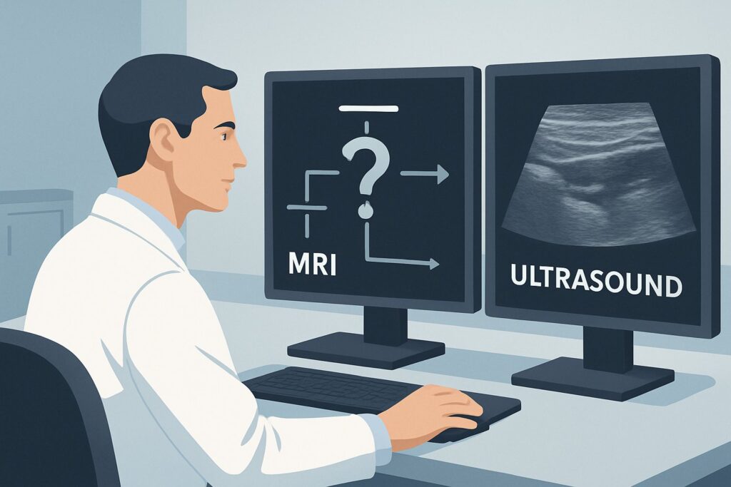
A 45-year-old patient presents to your clinic with a three-month history of progressive weakness in their right hand and forearm, accompanied by sensory changes along the ulnar aspect. There is no history of trauma, and a review of systems is negative for constitutional symptoms. The physical exam localizes the deficit to the lower trunk of the brachial plexus. You suspect a nontraumatic plexopathy, but the differential is broad, ranging from inflammatory conditions to subtle compression. The immediate clinical question is which imaging study will provide the most diagnostic clarity without unnecessary radiation or contrast. For this specific scenario, the American College of Radiology (ACR) finds that MRI brachial plexus without IV contrast is Usually appropriate. This article details the clinical workflow and rationale behind that recommendation.
Who Fits This Clinical Scenario?
This guidance applies to a specific patient population: adults or children presenting with acute or chronic symptoms localizing to the brachial plexus where there is no history of a precipitating traumatic event or a known malignancy. Symptoms may include motor weakness, sensory deficits (paresthesia, numbness), or pain that follows the distribution of the brachial plexus nerve roots, trunks, divisions, cords, or terminal branches.
This workflow is not intended for patients where the clinical picture is different. Key exclusions include:
- Traumatic Injury: If the symptoms began after a motor vehicle collision, fall, penetrating injury, or a difficult childbirth (in the case of perinatal injury), the workup follows the traumatic brachial plexopathy guidelines, as the pre-test probability of nerve root avulsion, transection, or hematoma is much higher.
- Known or Suspected Malignancy: For a patient with a history of lung cancer, breast cancer, lymphoma, or a Pancoast tumor, the primary concern is neoplastic invasion or compression of the plexus. This requires a different imaging approach, often involving contrast-enhanced studies or PET/CT, as detailed in the plexopathy with known malignancy scenario.
- Clear Cervical Spine Origin: If symptoms are purely dermatomal, associated with neck pain, and exacerbated by neck movements, the primary diagnosis may be cervical radiculopathy, which would prioritize a cervical spine MRI.
What Diagnoses Are You Working Up in This Scenario?
In the absence of trauma or malignancy, the differential diagnosis for brachial plexopathy is driven by inflammatory, compressive, and idiopathic causes. The choice of imaging is designed to differentiate among these possibilities.
Parsonage-Turner Syndrome (Brachial Neuritis): This is a common cause of nontraumatic brachial plexopathy. It is an idiopathic, presumed autoimmune, inflammatory condition that typically presents with the sudden onset of severe shoulder and arm pain, followed days to weeks later by profound, patchy weakness and muscle atrophy. MRI is highly sensitive for detecting the characteristic nerve edema (T2 hyperintensity) and thickening associated with this condition.
Thoracic Outlet Syndrome (TOS): This diagnosis refers to the compression of the neurovascular bundle (brachial plexus, subclavian artery, and vein) as it passes through the thoracic outlet. Neurogenic TOS, caused by compression of the brachial plexus, is the most common form. Imaging aims to identify potential compressive structures, such as a cervical rib, anomalous fibrous bands, or hypertrophy of the scalene muscles.
Chronic Inflammatory Demyelinating Polyneuropathy (CIDP): While often presenting as a more generalized polyneuropathy, CIDP can manifest as a plexopathy. It is an immune-mediated disorder causing demyelination of peripheral nerves. MRI may reveal diffuse thickening and enhancement of the nerve roots and plexus, though this finding is not always present.
Benign Nerve Sheath Tumors: Less commonly, a benign tumor such as a schwannoma or neurofibroma can arise from the nerve roots or trunks of the brachial plexus. These masses are typically slow-growing and present with progressive neurological deficits. MRI is the gold standard for identifying and characterizing these lesions.
Why Is MRI of the Brachial Plexus Without IV Contrast Usually Appropriate for This Presentation?
The ACR designates MRI brachial plexus without IV contrast as Usually appropriate because it provides superior soft-tissue resolution to directly visualize the nerves of the plexus, their surrounding structures, and any intrinsic signal abnormalities, all without ionizing radiation (0 mSv).
The rationale for this choice is rooted in its ability to address the key differential diagnoses. High-resolution T2-weighted and STIR (Short Tau Inversion Recovery) sequences are exceptionally sensitive for detecting nerve edema, which is the hallmark of inflammatory conditions like Parsonage-Turner syndrome. These sequences cause fluid and inflamed tissue to appear bright, making abnormal nerves stand out against suppressed background fat and muscle. Furthermore, MRI can clearly delineate the anatomical relationships in the thoracic outlet, identifying compressive structures like cervical ribs or fibrous bands that cause TOS.
It is important to note that the ACR also rates MRI brachial plexus without and with IV contrast as Usually appropriate. The decision to add gadolinium-based contrast depends on the specific clinical question. While non-contrast sequences are often sufficient to diagnose inflammatory or compressive etiologies, contrast may be valuable if a benign tumor (e.g., schwannoma) or an unexpected neoplastic or infectious process is suspected, as these lesions typically demonstrate avid enhancement.
Why Other Studies Are Rated Lower
- CT Neck with IV Contrast: This study is rated May be appropriate. While CT excels at evaluating osseous structures and can identify a cervical rib, its ability to visualize the nerves of the plexus is significantly inferior to MRI. It exposes the patient to ionizing radiation (☢☢☢ 1-10 mSv) and is generally reserved for patients with contraindications to MRI or when bony detail is the primary question.
- Ultrasound (US) Neck: Rated Usually not appropriate, ultrasound is limited in this context. It is highly operator-dependent and cannot visualize the entire course of the brachial plexus, particularly the nerve roots as they exit the neural foramina and the portions of the plexus deep to the clavicle. While it can identify focal abnormalities in the supraclavicular region, it does not provide the comprehensive anatomical overview required for an initial diagnostic workup.
What’s Next After MRI of the Brachial Plexus? Downstream Workflow
The results of the brachial plexus MRI will guide the subsequent clinical pathway. The downstream workflow is contingent on whether the findings are positive, negative, or indeterminate.
- Positive for a Definitive Cause: If the MRI reveals clear evidence of brachial neuritis (Parsonage-Turner syndrome), the diagnosis is confirmed, and management typically involves pain control, physical therapy, and observation, often in consultation with a neurologist. If a compressive lesion like a benign tumor or a structural cause of TOS is found, the next step is typically a referral to neurology and/or neurosurgery for further evaluation, which may include electrodiagnostic studies (EMG/NCS) and consideration for surgical intervention.
- Negative or Non-diagnostic MRI: A normal brachial plexus MRI is a valuable finding that effectively rules out structural, compressive, and significant inflammatory causes within the plexus itself. In this case, the clinical focus should shift to mimics of plexopathy. The most common next step is to evaluate for cervical radiculopathy. Ordering an MRI of the cervical spine (rated May be appropriate for the initial workup) becomes the logical next investigation to assess for nerve root compression from disc herniation or foraminal stenosis.
- Indeterminate Findings: If the MRI shows subtle or nonspecific signal changes, the next step is often correlation with electrodiagnostic testing. EMG and nerve conduction studies can help confirm the presence of a plexopathy, localize the lesion, assess its severity and chronicity, and differentiate it from radiculopathy or mononeuropathy, providing crucial functional information to complement the anatomical data from the MRI.
Pitfalls to Avoid (and When to Get Help)
Navigating the workup for nontraumatic brachial plexopathy requires careful attention to the clinical details to avoid common diagnostic errors.
- Ordering the Wrong MRI: A “cervical spine MRI” is not the same as a “brachial plexus MRI.” The latter requires specific high-resolution sequences and a dedicated field of view tailored to the plexus anatomy. Be explicit in your order to ensure the correct protocol is performed.
- Overlooking Mimics: Do not anchor on a plexopathy diagnosis too early. A negative plexus MRI should immediately prompt consideration of cervical radiculopathy, mononeuropathies (e.g., ulnar neuropathy at the elbow), or even central causes.
- Ignoring Red Flags: The absence of trauma and known malignancy is a key assumption of this scenario. If a patient develops new constitutional symptoms (weight loss, fevers) or has a remote history of cancer, the possibility of an occult malignancy must be reconsidered, which may warrant a contrast-enhanced study or further investigation.
If the clinical picture is complex, the MRI findings are ambiguous, or the patient fails to improve with conservative management, consultation with a neurologist or a musculoskeletal radiologist is the appropriate next step.
Related ACR Topics and Tools
For a comprehensive overview of imaging for all types of plexopathy, including traumatic and malignancy-related scenarios, please see our parent guide. For tools to help with ordering, protocoling, and patient communication, see the resources below.
- For breadth across all scenarios in Plexopathy, see our parent guide: Plexopathy: ACR Appropriateness Decoded.
- To explore other clinical presentations, use the Imaging Appropriateness Selector.
- To see detailed imaging techniques, visit the Imaging Protocol Library.
- To discuss radiation exposure with patients, use the Radiation Dose Calculator.
Frequently Asked Questions
Why is MRI without contrast preferred over MRI with contrast for the initial study?
For many common causes of nontraumatic brachial plexopathy, such as Parsonage-Turner syndrome (brachial neuritis) or most cases of thoracic outlet syndrome, non-contrast MRI sequences like T2/STIR are highly sensitive for detecting the relevant pathology (nerve edema or anatomical compression). The ACR rates both non-contrast and contrast-enhanced MRI as ‘Usually appropriate,’ but starting without contrast avoids the administration of gadolinium unless a tumor or infection is specifically suspected, aligning with the principle of using contrast only when necessary.
If my patient has a pacemaker, can I still order an MRI for brachial plexopathy?
Many modern pacemakers and implantable defibrillators are MRI-conditional. However, this requires a specific protocol and coordination with the radiology department and cardiology to ensure patient safety. If a patient has a non-conditional device or if MRI is otherwise contraindicated, a CT of the neck with IV contrast becomes a reasonable alternative, though it provides less detail of the nerves themselves. It is rated ‘May be appropriate’ by the ACR for this scenario.
How do I differentiate clinically between a C8/T1 radiculopathy and a lower trunk plexopathy?
This can be challenging as both can cause weakness and sensory loss in the hand and ulnar forearm. However, a lower trunk plexopathy (affecting C8 and T1 fibers after they’ve joined) may also involve the median-innervated thenar muscles (via the T1 root contribution) and often presents with a classic Horner’s syndrome (ptosis, miosis, anhidrosis) if the T1 sympathetic fibers are involved pre-ganglionically. A pure C8 radiculopathy would not cause Horner’s syndrome. A negative cervical spine MRI with a positive brachial plexus MRI can definitively distinguish the two.
What specific information should I include in my order for a brachial plexus MRI?
To ensure the radiologist performs the correct protocol, your order should specify ‘MRI Brachial Plexus’ and include the laterality (right, left, or bilateral). Briefly describe the clinical history, including the location of symptoms (e.g., ‘weakness in C8/T1 distribution’), chronicity, and the primary clinical question (e.g., ‘r/o brachial neuritis vs. thoracic outlet syndrome’). This context allows the radiologist to tailor the imaging sequences for the highest diagnostic yield.
When should I consider electrodiagnostic studies (EMG/NCS) in this workup?
Electrodiagnostic studies are complementary to imaging. They are particularly useful when the MRI is negative or inconclusive, as they can confirm a neurogenic process, localize the lesion functionally, and assess its severity and chronicity (e.g., acute denervation vs. chronic reinnervation). Many clinicians order EMG/NCS in parallel with or following MRI to get a complete picture of both anatomy and function.
Reviewed by Pouyan Golshani, MD, Interventional Radiologist — May 26, 2026