Should You Order X-Ray or MRI for Acute Cervical Radiculopathy in Adults?
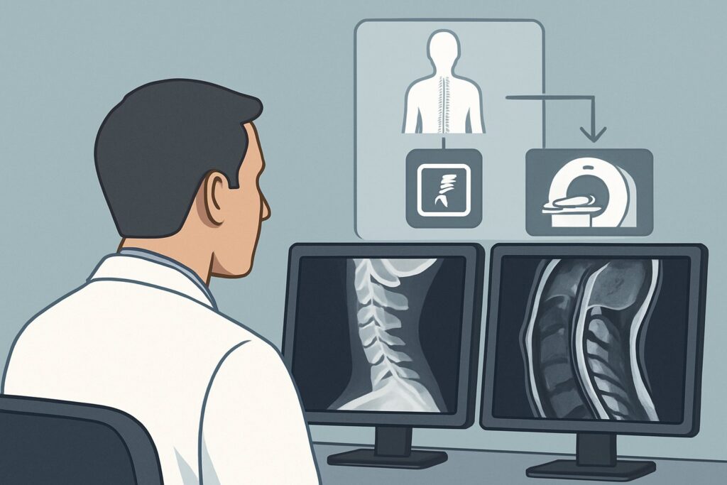
A 45-year-old patient presents to your clinic with a two-week history of neck pain that now radiates down their left arm with associated paresthesias in the thumb and index finger. The neurologic exam reveals 4/5 strength in the biceps and a diminished biceps reflex, but is otherwise normal. There is no history of trauma, fever, weight loss, or malignancy. You suspect a C6 radiculopathy, likely from a disc herniation, and are considering the initial imaging workup. This common scenario requires a thoughtful approach to avoid unnecessary tests while ensuring an accurate diagnosis. This article details the American College of Radiology (ACR) Appropriateness Criteria workflow for an adult with acute or increasing cervical pain with radiculopathy, without trauma or other “red flags.” For this presentation, both Radiography of the cervical spine and Magnetic Resonance Imaging (MRI) of the cervical spine without contrast are rated as May be appropriate.
Who Fits This Clinical Scenario?
This guidance is specifically for adult patients presenting with acute (less than 6 weeks) or subacute, increasing cervical pain accompanied by radicular symptoms. Radiculopathy is defined by signs and symptoms—such as pain, numbness, tingling, or weakness—that follow a specific nerve root distribution into the shoulder, arm, or hand.
Inclusion Criteria:
- Patient: Adult.
- Symptoms: Neck pain with radiating symptoms consistent with a dermatomal or myotomal pattern (radiculopathy).
- Acuity: Acute or progressively increasing symptoms.
- Context: No history of significant trauma and no clinical “red flags.”
Exclusion Criteria (These Route to Different Workflows): This workflow is not appropriate if the patient has:
- Significant Trauma: Any recent injury that could cause a fracture or ligamentous injury requires a different imaging protocol, often starting with CT.
- “Red Flags”: Clinical findings suggesting a more sinister underlying cause, such as fever, unexplained weight loss, night sweats, history of malignancy, immunosuppression, or intravenous drug use. These raise suspicion for infection or tumor and typically warrant more advanced imaging, often with intravenous contrast. See our guide for a patient with suspected infection or known malignancy.
- Myelopathy: Signs of spinal cord compression, such as gait instability, balance problems, hyperreflexia, or bowel/bladder dysfunction, which necessitate urgent MRI.
- Prior Cervical Spine Surgery: Post-operative anatomy changes the imaging approach. See the specific ACR variant for patients with prior surgery.
What Diagnoses Are You Working Up in This Scenario?
When a patient presents with atraumatic cervical radiculopathy, the differential diagnosis is focused on causes of nerve root compression. The goal of initial imaging is to identify the anatomical cause that correlates with the patient’s clinical findings.
The most common cause in this patient population is cervical disc herniation. An intervertebral disc can protrude or extrude, most often in a posterolateral direction, directly impinging on the exiting nerve root in the neural foramen. This is a frequent cause of acute radicular symptoms in younger to middle-aged adults.
A close second, particularly in older adults, is cervical spondylosis. This degenerative process involves osteophyte (bone spur) formation, disc height loss, and facet joint arthropathy. Together, these changes can lead to foraminal stenosis, a narrowing of the bony canal through which the nerve root exits the spinal column. While often a chronic process, an acute inflammatory event or minor disc bulge can trigger radicular symptoms in a pre-existing narrowed foramen.
Less common causes include synovial cysts arising from the facet joints or other non-neoplastic masses that can cause nerve root compression. By definition, this clinical scenario has screened out more urgent diagnoses like epidural abscess, metastasis, or primary bone tumors, which would be accompanied by red flag symptoms.
Why Are Radiography and MRI Both Appropriate Initial Options?
For an adult with acute cervical radiculopathy and no red flags, the ACR designates both Radiography cervical spine and MRI cervical spine without IV contrast as May be appropriate. The choice between them depends on clinical severity, treatment plans, and local practice patterns. Neither is wrong, but they answer different questions.
Radiography cervical spine
A standard set of cervical spine radiographs is a reasonable first step. It is inexpensive, widely available, and provides a good overview of bony anatomy. It can effectively identify degenerative changes like spondylosis, disc space narrowing, and uncover any gross malalignment. While it cannot directly visualize discs or nerves, identifying significant foraminal stenosis from osteophytes may be sufficient to explain the patient’s symptoms and guide initial conservative management. The associated radiation dose is low (ACR Relative Radiation Level ☢☢, 0.1-1 mSv).
MRI cervical spine without IV contrast
MRI is the gold standard for visualizing soft tissues, including the spinal cord, nerve roots, and intervertebral discs. It is the most sensitive and specific non-invasive test for diagnosing a disc herniation and evaluating the degree of nerve root or spinal cord compression. Because it uses no ionizing radiation (ACR Relative Radiation Level O, 0 mSv), it is an excellent choice, particularly if symptoms are severe, persistent despite initial conservative therapy, or if a more invasive treatment like an epidural steroid injection or surgery is being considered.
Why Alternatives Are Rated Lower
- CT cervical spine without IV contrast is rated Usually not appropriate. While excellent for bone detail, it exposes the patient to significantly more radiation than radiographs (ACR RRL ☢☢☢, 1-10 mSv) and provides substantially less information about the nerve roots and discs than an MRI.
- MRI cervical spine with IV contrast is also rated Usually not appropriate. In the absence of red flags for tumor, infection, or prior surgery, gadolinium-based contrast adds cost and potential risk (e.g., nephrogenic systemic fibrosis in patients with renal impairment, allergic reactions) without providing additional diagnostic information for a standard degenerative radiculopathy.
The decision often comes down to a stepped-care approach versus a direct-to-definitive-imaging approach. Starting with radiography is a valid, resource-conscious strategy. Proceeding directly to MRI is also valid, especially when the clinical suspicion for a surgically-amenable lesion is high.
What’s Next After Imaging? Downstream Workflow
The results of your initial imaging study will guide the next steps in management, which often involve a combination of physical therapy, medication, and potentially interventional procedures.
If you started with Radiography:
- Positive Result: If radiographs show severe degenerative changes (e.g., advanced spondylosis, significant foraminal stenosis) that correlate with the patient’s symptoms, this may be sufficient to confirm the diagnosis. You can proceed with a course of conservative management (4-6 weeks). If the patient fails to improve, the next step is an MRI without contrast to better define the soft-tissue component of the nerve compression.
- Negative or Non-diagnostic Result: A normal radiograph does not rule out the most common cause of acute radiculopathy: a soft-tissue disc herniation. If symptoms are significant or persistent, the clear next step is to obtain an MRI of the cervical spine without contrast.
If you ordered an MRI (either initially or after radiography):
- Positive Result: If the MRI identifies a disc herniation or foraminal stenosis that clinically correlates with the patient’s radiculopathy, the findings can be used to guide specific treatments. This may include targeted physical therapy, oral medications, referral for an epidural steroid injection, or a consultation with a spine surgeon if symptoms are severe or progressive.
- Negative Result: A completely normal MRI of the cervical spine is a powerful finding. It largely rules out a structural cause in the neck. In this case, you should reconsider the differential diagnosis to include non-spinal causes of arm pain, such as brachial plexopathy, thoracic outlet syndrome, or peripheral nerve entrapment (e.g., carpal tunnel syndrome). Further workup may include electromyography (EMG) and nerve conduction studies.
Pitfalls to Avoid (and When to Get Help)
Navigating this common clinical scenario involves being mindful of a few potential missteps.
- Stopping at a Normal X-ray: Do not assume a normal radiograph excludes a clinically significant, treatable cause of radiculopathy. Soft disc herniations are invisible on X-ray and are a primary cause of symptoms.
- Ordering Unnecessary Contrast: For routine, atraumatic radiculopathy, ordering an MRI with contrast is rarely indicated. This adds unnecessary cost and potential risk to the patient.
- Ignoring Progressive Deficits: This workflow is for stable or slowly progressing symptoms. If a patient develops rapidly worsening weakness or any signs of myelopathy (e.g., new-onset clumsiness, gait difficulty, urinary retention), this constitutes a clinical emergency.
- Misclassifying the Patient: Ensure the patient truly has no red flags. A cursory history can miss a subtle history of cancer or recent infection that would completely change the imaging workup.
Escalation: If any red flags are present or if the patient develops signs of myelopathy, escalate care immediately. This typically involves an urgent, non-contrast MRI of the cervical spine and a consultation with a neurosurgeon or spine specialist.
Related ACR Topics and Tools
For a comprehensive overview of all clinical variants related to neck pain, and for tools to help you apply these criteria in your practice, please see the following resources.
- For breadth across all scenarios in Cervical Pain or Cervical Radiculopathy, see our parent guide: Cervical Pain or Cervical Radiculopathy: ACR Appropriateness Decoded.
- ACR Appropriateness Criteria Lookup — for adjacent scenarios
- Imaging Protocol Library — for technique on the recommended study
- Radiation Dose Calculator — for cumulative dose conversations
Frequently Asked Questions
Why not just order an MRI for every patient with cervical radiculopathy?
While MRI is the most definitive test, many cases of acute cervical radiculopathy resolve with 4-6 weeks of conservative therapy. A stepped-care approach starting with radiography can be a more cost-effective strategy, confirming a degenerative cause and reserving MRI for patients who fail to improve or have severe symptoms. This avoids the cost and access issues of MRI for a self-limited condition.
What specific ‘red flags’ would change this imaging recommendation?
Red flags suggesting a more serious condition like tumor, infection, or fracture would change the recommendation. Key red flags include: fever, chills, unexplained weight loss, history of cancer, immunosuppression, intravenous drug use, recent significant trauma, and progressive or profound neurologic deficits (myelopathy).
Is a CT myelogram ever appropriate for this condition?
As an initial imaging study for this scenario, CT myelography is rated ‘Usually not appropriate’ by the ACR. It is an invasive procedure with higher radiation exposure. Its primary role is as a problem-solving tool for patients with symptoms of nerve root compression who have a contraindication to MRI (e.g., an incompatible pacemaker or cochlear implant).
How long should I wait before ordering imaging for acute cervical radiculopathy?
For patients without red flags, it is often reasonable to trial a course of conservative management (e.g., physical therapy, anti-inflammatory medications) for 4-6 weeks before ordering imaging. However, imaging should be considered earlier if symptoms are severe, debilitating, or associated with any motor weakness.
Are flexion-extension radiographs needed for this initial workup?
No. For the initial evaluation of atraumatic cervical radiculopathy, the ACR rates flexion-extension views as ‘Usually not appropriate.’ These specialized views are used to assess for ligamentous instability, which is not the primary concern in this clinical scenario. They are more relevant in the setting of trauma or certain rheumatologic conditions.
Reviewed by Pouyan Golshani, MD, Interventional Radiologist — May 29, 2026