Why Is Imaging Not Recommended for Acute Low Back Pain Without Red Flags?
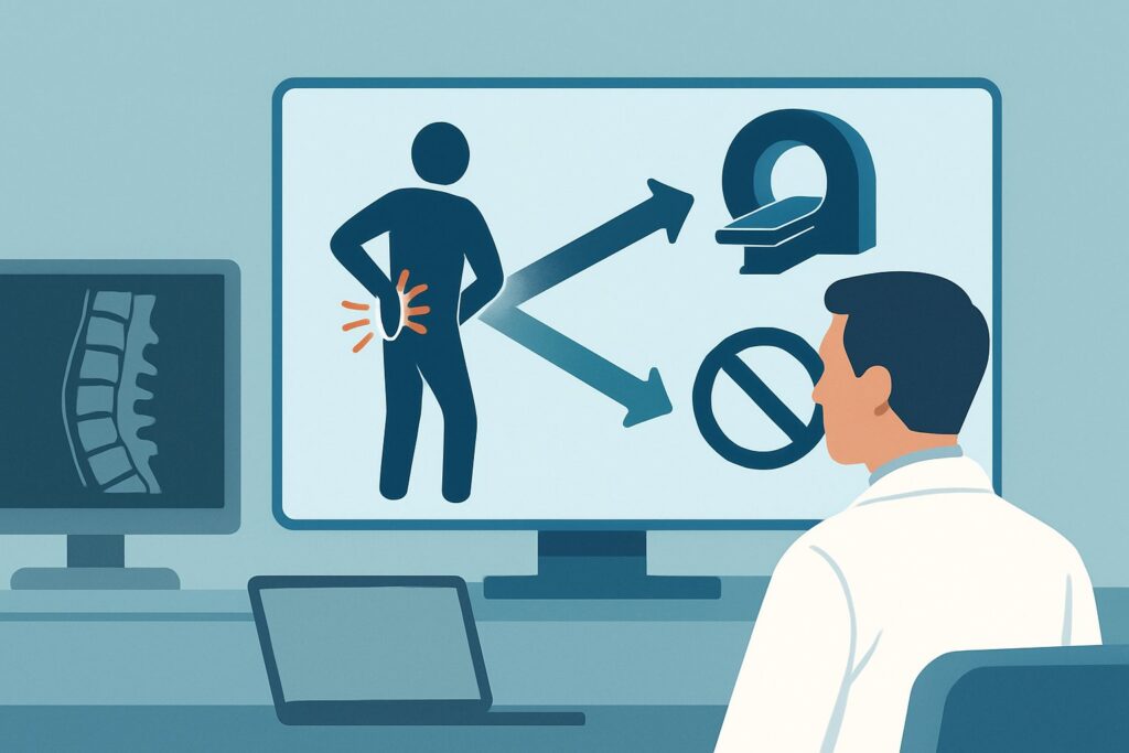
A 45-year-old software engineer presents to your clinic on a Tuesday morning with three days of low back pain after helping a friend move over the weekend. The pain is sharp, radiates down his posterior left thigh to the knee, and is worse with sitting. He has no weakness, numbness, or bowel or bladder changes. You perform a straight leg raise, which is positive on the left. Your clinical suspicion is high for acute lumbar radiculopathy, and you consider ordering a lumbar spine X-ray to “see what’s going on.” For this exact presentation—acute low back pain with or without radiculopathy, no red flags, and no prior treatment—what is the most appropriate next step?
This article provides a detailed clinical workflow for this specific scenario, grounded in the American College of Radiology (ACR) Appropriateness Criteria. For this presentation, the evidence-based consensus is clear: nearly all forms of initial imaging, including `Radiography lumbar spine`, are rated Usually not appropriate. We will explore the rationale behind this recommendation, the appropriate non-imaging workflow, and the pitfalls to avoid.
Who Fits This Clinical Scenario?
This guidance applies to a very specific and common patient population. Correctly identifying these patients is key to avoiding unnecessary imaging and focusing on effective initial management.
Inclusion Criteria for This Workflow:
- Acute Onset: The low back pain began less than six weeks ago.
- With or Without Radiculopathy: The patient may have simple axial back pain or pain that radiates along a nerve root distribution (e.g., sciatica), but without progressive or profound neurologic deficits.
- No Red Flags: A thorough history and physical exam reveal no warning signs of a serious underlying condition. Key red flags include a history of cancer, unexplained weight loss, fever, recent infection, immunosuppression, significant trauma, or symptoms of cauda equina syndrome (e.g., saddle anesthesia, new-onset bowel or bladder incontinence).
- No Prior Management: This is the patient’s initial presentation for this episode of back pain, and they have not yet undergone a course of conservative therapy.
Exclusion Criteria (These Patients Require a Different Workflow):
This advice does not apply if the clinical picture is different. If your patient presents with one of the following, they fit a different ACR variant that may warrant imaging:
- Suspicion of Cauda Equina Syndrome: This is a neurologic emergency requiring immediate imaging.
- Presence of Red Flags: Suspicion of cancer, infection, or fracture from significant trauma.
- Chronic Pain: The pain has persisted for more than 12 weeks.
- Prior Lumbar Surgery: New or progressing symptoms in a post-operative patient.
What Diagnoses Are You Working Up in This Scenario?
In the absence of red flags, the differential diagnosis for acute low back pain is narrow and dominated by benign, self-limiting conditions. The decision to defer imaging is based on the high probability that the cause is one of the following, for which initial management is conservative regardless of imaging findings.
Mechanical Low Back Pain (Lumbar Strain/Sprain)
This is the most common diagnosis, representing injury to the muscles, ligaments, or tendons of the lower back. It is a diagnosis of exclusion. The pain is typically axial, exacerbated by movement, and resolves with time and conservative care. Imaging is almost always normal or shows non-specific degenerative changes unrelated to the acute symptoms.
Acute Herniated Nucleus Pulposus (HNP) with Radiculopathy
This is the classic “slipped disc” causing sciatica. A fragment of the intervertebral disc protrudes and irritates or compresses a nearby nerve root. While this can be intensely painful, the natural history is favorable for most patients. The vast majority of cases of acute radiculopathy from an HNP improve or resolve completely within weeks to months with non-operative management. Initial imaging does not alter this conservative treatment plan.
Symptomatic Degenerative Disc or Facet Disease
Underlying degenerative changes are ubiquitous in adults and often asymptomatic. An acute flare can occur, but it is clinically indistinguishable from a simple lumbar strain. Importantly, imaging will almost certainly show these chronic changes, creating a significant risk of misattributing the acute pain to a chronic finding. This can lead to patient anxiety, iatrogenic harm from unnecessary interventions, and a focus on structural abnormalities that are not the true pain generator.
Why Is Imaging Usually Not Appropriate for Acute Low Back Pain Without Red Flags?
The ACR panel’s consensus reflects a large body of evidence demonstrating that for this specific patient group, the potential harms of routine initial imaging outweigh the benefits. The core rationale is that imaging findings do not change the initial management plan and can lead to a cascade of negative consequences.
All imaging modalities for this scenario are rated `Usually not appropriate`. Let’s examine the two most commonly considered studies:
- Radiography lumbar spine: This study is rated `Usually not appropriate`. While it can assess alignment and bone integrity, it has very low sensitivity for the most common causes of acute low back pain, such as disc herniation or muscle strain. It cannot visualize soft tissues or nerves. Furthermore, it exposes the patient to ionizing radiation (an effective dose of 1-10 mSv, designated ☢☢☢ by the ACR) with minimal to no diagnostic benefit in this context.
- MRI lumbar spine without IV contrast: This is also rated `Usually not appropriate` for initial imaging. Although MRI is the gold standard for visualizing disc herniations and nerve root compression, obtaining it at the first visit is premature. The findings, whether positive or negative for a disc herniation, do not alter the initial 4-to-6-week course of conservative therapy. Reserving MRI for patients who fail this initial management period ensures the test is used when it can genuinely guide the next steps, such as considering epidural steroid injections or surgical consultation.
The primary risks of premature imaging are not physical but diagnostic and psychological. The high prevalence of asymptomatic “abnormalities” (e.g., disc bulges are seen on MRI in a substantial portion of asymptomatic adults) can lead to a focus on non-causative findings, increased patient fear and anxiety, and a higher likelihood of receiving unnecessary, costly, and potentially harmful interventions.
What Is the Recommended Workflow Instead of Initial Imaging?
With imaging off the table, the focus shifts entirely to a clinical workflow centered on conservative management, patient education, and scheduled re-evaluation.
Initial Management (First 4-6 Weeks):
1. Patient Education and Reassurance: Explain the benign nature of most acute low back pain. Reassure the patient that serious conditions are unlikely and that significant improvement is expected within a few weeks.
2. Activity Modification: Advise the patient to remain active as tolerated. Prolonged bed rest is counterproductive. Encourage gentle movement and avoidance of activities that significantly worsen the pain.
3. Non-pharmacologic Therapy: Recommend superficial heat for comfort. If available, referral to a physical therapist can be highly effective for guided exercise and manual therapy.
4. Pharmacologic Therapy: Non-steroidal anti-inflammatory drugs (NSAIDs) or acetaminophen are appropriate first-line agents. Muscle relaxants may be considered for short-term use if significant muscle spasm is present.
Downstream Decision Point (at 4-6 Weeks):
- If Symptoms Are Improving or Resolved: No further action or imaging is needed. The patient can be advised on secondary prevention strategies like core strengthening and proper lifting mechanics.
- If Symptoms Are Unchanged or Worsening: This is the critical juncture where the patient’s condition may be reclassified. They may now fit the ACR scenario for Subacute or chronic low back pain. At this point, advanced imaging, typically an MRI of the lumbar spine without contrast, may become appropriate to guide further treatment, such as injections or surgical evaluation.
Pitfalls to Avoid (and When to Re-evaluate)
Navigating this common scenario effectively requires avoiding several potential missteps.
- Succumbing to Patient Pressure for Imaging: Patients often believe an image will provide a definitive answer and a quick fix. A key clinical skill is explaining why imaging is not helpful at this stage and can even be misleading, while confidently outlining the evidence-based plan for recovery.
- Missing Evolving Red Flags: The initial assessment is a snapshot in time. Instruct patients on what new symptoms to watch for (e.g., progressive leg weakness, changes in bowel/bladder function) and to seek immediate re-evaluation if they occur.
- Attributing Pain to Incidental Findings: If an X-ray or MRI was done for an unrelated reason, be extremely cautious about linking chronic degenerative changes to the patient’s acute symptoms. This is a classic attribution error.
When to Escalate: If a red flag emerges at any point during the follow-up period, the “watchful waiting” approach ends. The patient’s clinical scenario has changed, and they should be re-evaluated promptly for a workup that may include immediate imaging, such as for suspected cauda equina syndrome or spinal infection.
Related ACR Topics and Tools
This article covers one specific variant within the broader topic of Low Back Pain. For a comprehensive overview of all related scenarios and for tools to help with clinical decision-making, please see the resources below.
- For breadth across all scenarios in Low Back Pain, see our parent guide: Low Back Pain: ACR Appropriateness Decoded.
- ACR Appropriateness Criteria Lookup — for adjacent scenarios
- Imaging Protocol Library — for technique on the recommended study
- Radiation Dose Calculator — for cumulative dose conversations
Frequently Asked Questions
My patient has classic sciatica. Shouldn’t I get an MRI to confirm a disc herniation?
Even with classic radicular symptoms suggestive of a disc herniation, the ACR guidance is to defer imaging for the first 4-6 weeks in the absence of red flags. The initial management—conservative therapy—is the same whether a disc herniation is confirmed on MRI or not. The MRI becomes appropriate if the patient fails to improve with this initial course of treatment.
What if the patient is over 50? Does that change the recommendation?
Age alone, without other risk factors or red flags, does not automatically trigger imaging for acute low back pain. However, if an elderly patient has additional risk factors, such as a history of osteoporosis or even low-velocity trauma, they may fit a different ACR scenario where initial radiography could be appropriate to rule out a compression fracture.
How long is ‘acute’ low back pain?
In the context of the ACR Appropriateness Criteria, ‘acute’ generally refers to symptoms lasting less than six weeks. Pain lasting from six to twelve weeks is considered ‘subacute,’ and pain lasting longer than twelve weeks is ‘chronic.’ The imaging recommendations may change as the condition becomes subacute or chronic.
Is there any role for a lumbar spine X-ray at all in this specific scenario?
For uncomplicated acute low back pain without red flags, the role is extremely limited, which is why the ACR rates it ‘Usually not appropriate.’ It does not visualize soft tissues like discs or nerves and exposes the patient to radiation for little to no diagnostic benefit. Its primary utility is in different clinical scenarios, such as after significant trauma or when there is a suspicion of fracture or instability.
What are the key ‘red flags’ that would make me order imaging immediately?
The presence of red flags moves the patient into a different, more urgent clinical scenario. Key red flags include: suspicion of cancer (e.g., unexplained weight loss, personal history of cancer), suspicion of infection (e.g., fever, chills, IV drug use), cauda equina syndrome (e.g., saddle anesthesia, new-onset bowel or bladder incontinence), significant trauma, or a progressive or profound neurologic deficit (e.g., foot drop).
Reviewed by Pouyan Golshani, MD, Interventional Radiologist — May 29, 2026