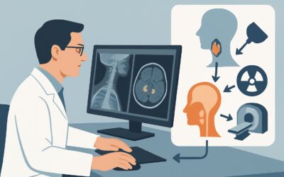
What Is the Best Initial Imaging for New Headache During Pregnancy or Peripartum?

A 32-year-old woman, three days postpartum after an uncomplicated delivery, presents to the emergency department with a severe, throbbing headache that started this morning. She has a history of migraines, but states, “this feels different—more intense and all over.” Her blood pressure is 165/105 mmHg. You are considering the broad and serious differential for a new or changing headache in the peripartum period and must decide on the most appropriate initial imaging study. This article details the clinical workflow for this specific scenario, guiding you through the diagnostic rationale and downstream decisions. For a new or changing headache during pregnancy or the peripartum period, the American College of Radiology (ACR) rates `MRI head without IV contrast` as Usually appropriate.
Who Fits This Clinical Scenario?
This guidance applies to a specific and high-risk patient population: individuals who are currently pregnant or are in the peripartum period (typically defined as up to six weeks postpartum) and present with a headache of new onset or a headache that represents a significant change from their usual pattern. The “change in pattern” is a critical qualifier; a patient with a long history of stable migraines who presents with a typical attack does not fit this scenario. Instead, this workflow is for the patient whose headache has new features, is significantly more severe, is associated with new neurologic symptoms, or fails to respond to standard therapies.
It is equally important to identify who does not fit this scenario to ensure correct diagnostic routing:
- Sudden Onset “Thunderclap” Headache: A patient describing an explosive headache reaching maximal intensity within a minute has a distinct differential (e.g., subarachnoid hemorrhage, reversible cerebral vasoconstriction syndrome) and falls under the ACR variant for sudden onset severe headache.
- Classic Migraine or Tension-Type Headache: A patient with a known primary headache disorder and a normal neurologic exam, presenting with a headache typical for them, generally does not require initial imaging.
- Positional Headache: A headache that is clearly worse when upright and improves when lying flat suggests intracranial hypotension and is addressed by a separate ACR workflow.
What Diagnoses Are You Working Up in This Scenario?
The differential diagnosis for a new headache in pregnancy or the peripartum period includes both common headache types and several life-threatening conditions unique to or more prevalent in this population. The goal of initial imaging is to rapidly identify or exclude these serious etiologies.
Preeclampsia/Eclampsia: This is a primary concern, especially when the headache is accompanied by hypertension, proteinuria, or visual disturbances. The headache can be a manifestation of cerebral edema or vasospasm and may be a premonitory sign of an impending eclamptic seizure. Imaging is crucial to evaluate for complications.
Posterior Reversible Encephalopathy Syndrome (PRES): Often occurring in the setting of preeclampsia, eclampsia, or severe hypertension, PRES is characterized by vasogenic edema, typically affecting the posterior cerebral white matter. Patients present with headache, seizures, altered mental status, and visual changes. It is a critical diagnosis to make, as it is often reversible with prompt blood pressure control.
Cerebral Venous Sinus Thrombosis (CVST): Pregnancy and the postpartum period are prothrombotic states, significantly increasing the risk of CVST. Headache is the most common presenting symptom, reported in up to 90% of patients. The headache can be variable in character and onset, making clinical diagnosis challenging without imaging.
Reversible Cerebral Vasoconstriction Syndrome (RCVS): While often associated with a classic “thunderclap” headache, RCVS can also present with a less abrupt but severe headache. It is characterized by multifocal constriction of cerebral arteries and is more common in the postpartum period.
Pituitary Apoplexy: The pituitary gland undergoes physiologic enlargement during pregnancy, making it more vulnerable to hemorrhage or infarction. This can present with a sudden, severe headache, visual field defects, and cranial nerve palsies.
Why Is MRI Head without IV Contrast the Recommended Study for This Presentation?
The ACR designates `MRI head without IV contrast` as Usually appropriate for the initial evaluation of a new or changing headache pattern during pregnancy or the peripartum period. This recommendation is based on its high diagnostic yield for the key differential diagnoses while avoiding potential risks.
The primary advantage of Magnetic Resonance Imaging (MRI) is its superior soft-tissue contrast without the use of ionizing radiation. This is a critical consideration in a pregnant patient, aligning with the As Low As Reasonably Achievable (ALARA) principle. While the fetal radiation dose from a head CT is negligible, avoiding radiation entirely is preferable when a non-ionizing modality offers equivalent or superior diagnostic information.
For the specific differential in this scenario:
- PRES and Preeclampsia: MRI, particularly with Fluid-Attenuated Inversion Recovery (FLAIR) and T2-weighted sequences, is highly sensitive for detecting the characteristic vasogenic edema of PRES, which may be subtle or entirely missed on a non-contrast CT.
- Cerebral Venous Sinus Thrombosis (CVST): A non-contrast MRI can often suggest the diagnosis by showing abnormal signal intensity or loss of the normal flow void within a dural venous sinus. While a dedicated MR Venography (MRV) is more definitive, the initial brain MRI is an excellent screening tool.
- Pituitary Apoplexy: MRI is the modality of choice for evaluating the pituitary gland, capable of clearly depicting hemorrhage or infarction within an enlarged gland.
Alternative Imaging Considerations:
- `CT head without IV contrast` is also rated Usually appropriate. It is an excellent alternative when MRI is unavailable, contraindicated, or if the clinical need for speed is paramount (e.g., to rapidly exclude a large intracranial hemorrhage). However, its sensitivity for non-hemorrhagic conditions like PRES, early CVST, or ischemia is lower than MRI. It involves a low but non-zero dose of ionizing radiation (ACR Relative Radiation Level ☢☢☢ 1-10 mSv).
- `MRV head without IV contrast` is rated May be appropriate. This is a valuable study but is more focused than a standard brain MRI. It is the best non-contrast test to directly visualize the dural venous sinuses and should be considered as the next step or an initial add-on if CVST is the leading clinical suspicion.
- Contrast-enhanced studies, such as `MRI head with IV contrast`, are rated Usually not appropriate for the initial workup. Gadolinium-based contrast agents cross the placenta and have an unknown, though likely low, risk to the fetus. They are generally avoided during pregnancy unless the potential diagnostic benefit is deemed to substantially outweigh this theoretical risk. Postpartum, concerns are related to excretion in breast milk, though current guidelines suggest breastfeeding can continue safely.
What’s Next After MRI Head without IV Contrast? Downstream Workflow
The results of the initial non-contrast head MRI will guide the subsequent clinical pathway. The goal is to move from broad suspicion to a specific diagnosis and management plan.
- If the MRI is positive for PRES: The immediate focus is aggressive blood pressure management and treatment of the underlying cause, which in this population is often preeclampsia. This typically involves consultation with obstetrics and maternal-fetal medicine, and may necessitate delivery if the patient is still pregnant.
- If the MRI is positive for hemorrhage (e.g., subarachnoid, intraparenchymal, or pituitary apoplexy): This is a medical emergency requiring immediate neurosurgical and/or neurointerventional consultation. Further vascular imaging (such as CTA or MRA) is often required to identify an underlying aneurysm or vascular malformation.
- If the MRI is suggestive of CVST: When the non-contrast study shows findings suspicious for venous thrombosis, the definitive next step is dedicated venography. `MRV head without IV contrast` is an excellent choice. If results are still equivocal, `CTV head with IV contrast` or a contrast-enhanced MRV may be necessary. Once confirmed, treatment with anticoagulation is initiated in consultation with neurology and hematology.
- If the MRI is negative: A normal non-contrast MRI is reassuring and makes many of the most life-threatening conditions much less likely. However, if clinical suspicion remains high for a vascular etiology like RCVS or an occult CVST, further imaging may be warranted. This could include MRA/MRV or CTA/CTV. If the headache resolves and the neurologic examination remains normal, the diagnosis may be a primary headache disorder (e.g., migraine) exacerbated by the physiologic changes of pregnancy or postpartum, and outpatient neurologic follow-up is appropriate.
Pitfalls to Avoid (and When to Get Help)
Navigating this clinical scenario requires vigilance to avoid common diagnostic errors.
- Attribution Error: Do not automatically attribute a severe headache in a patient with a known migraine history to “just another migraine.” A change in character, severity, or associated symptoms in the peripartum period warrants a full workup.
- Ignoring Vital Signs: A new headache in the setting of elevated blood pressure is a major red flag for preeclampsia and PRES until proven otherwise. This combination demands urgent evaluation.
- Hesitation to Image: Fear of imaging a pregnant patient can lead to dangerous delays in diagnosing time-sensitive conditions like CVST or hemorrhage. A non-contrast head MRI carries no known risk to the fetus.
- Incomplete Workup: A negative non-contrast head CT does not fully exclude serious pathology like PRES or CVST. If clinical suspicion is high, do not stop the workup; proceed to MRI.
If the patient presents with a focal neurologic deficit, seizure, or rapidly declining mental status, escalate immediately with neurologic and obstetric consultation, and proceed to the most rapidly available imaging, which is often a non-contrast head CT.
Related ACR Topics and Tools
For a comprehensive overview of imaging for all headache presentations, refer to the parent topic article. For additional tools to help refine your imaging orders and discuss them with patients, see the resources below.
- For breadth across all scenarios in Headache, see our parent guide: Headache: ACR Appropriateness Decoded.
- ACR Appropriateness Criteria Lookup: Look up adjacent scenarios or other clinical questions.
- Imaging Protocol Library: Review detailed imaging techniques for recommended studies.
- Radiation Dose Calculator: Quantify and discuss radiation exposure for CT and other modalities.
Frequently Asked Questions
Is a non-contrast head CT ever the first choice in a pregnant patient with a new headache?
Yes. While MRI is generally preferred for its lack of radiation and superior soft-tissue detail, a non-contrast head CT is also rated ‘Usually appropriate’ by the ACR. It is often faster and more readily available, making it the first choice in an unstable patient or when there is high suspicion for acute hemorrhage and MRI is not immediately accessible.
If the non-contrast MRI is normal, have I ruled out Cerebral Venous Sinus Thrombosis (CVST)?
Not definitively. A standard non-contrast MRI can show indirect signs of CVST, but it can also be normal, especially in the early stages. If your clinical suspicion for CVST remains high despite a normal MRI, the next step is a dedicated venography study, such as a non-contrast MRV (rated ‘May be appropriate’ as an initial study) or a CTV.
What are the concerns with gadolinium contrast in a postpartum, breastfeeding patient?
The amount of gadolinium-based contrast agent excreted into breast milk is extremely small (less than 0.04% of the administered dose), and the amount absorbed by the infant from the milk is even smaller. The American College of Radiology and other professional bodies state that it is safe for the mother and infant to continue breastfeeding without interruption after receiving gadolinium.
My patient has a new headache and hypertension. Should I wait for imaging before starting blood pressure medication?
No. Severe hypertension in this setting is a medical emergency. You should initiate antihypertensive therapy concurrently with the diagnostic workup, including imaging. Controlling the blood pressure is a key part of treatment for conditions like preeclampsia and PRES and can prevent further neurologic injury.
How does the workup change if the patient is 5 months pregnant versus 5 days postpartum?
The core diagnostic algorithm remains the same, as the differential diagnoses (PRES, CVST, etc.) are relevant throughout this entire period. The primary difference is the management of pregnancy-specific conditions. A diagnosis of severe preeclampsia or eclampsia in a pregnant patient may necessitate delivery, whereas in a postpartum patient, the focus is solely on maternal treatment.
Reviewed by Pouyan Golshani, MD, Interventional Radiologist — June 27, 2026