Which Imaging Study Is Best for Altered Mental Status with a Suspected Medical Cause?
A 78-year-old woman with a history of diabetes and hypertension is brought to the emergency department by her family for worsening confusion over the past 48 hours. She is lethargic but arousable, oriented only to person, and her neurologic exam is non-focal. Her fingerstick glucose is 450 mg/dL, and initial labs suggest diabetic ketoacidosis. As you begin fluid resuscitation and insulin therapy, you consider whether to order brain imaging. Is this purely a metabolic encephalopathy, or could there be a concurrent intracranial process? This article details the American College of Radiology (ACR) imaging recommendations for this specific scenario: an adult with altered mental status where a medical illness or toxic-metabolic cause is strongly suspected. For this presentation, both MRI head without IV contrast and CT head without IV contrast are rated as May be appropriate (Disagreement), reflecting the clinical nuance of when to image.
Who Fits This Clinical Scenario?
This guidance applies to adult patients presenting with altered mental status—be it delirium, lethargy, or coma—where the initial clinical assessment points strongly toward a systemic, non-neurologic cause. The key inclusion criteria are:
- Altered Mental Status: A global change in consciousness or cognition.
- Suspected Systemic Cause: The history and initial laboratory findings suggest a medical illness (e.g., sepsis, uremia, hepatic encephalopathy, severe electrolyte disturbance, diabetic ketoacidosis) or a toxic exposure (e.g., drug overdose, alcohol withdrawal).
- Absence of Focal Neurologic Deficits: The physical examination does not reveal signs like unilateral weakness, aphasia, or visual field cuts that would point to a specific brain lesion.
It is critical to distinguish this scenario from similar presentations that require a different imaging pathway. This guidance does not apply if:
- Focal Neurologic Deficits Are Present: A patient with confusion and a new facial droop falls under the ACR variant for suspected intracranial pathology, which prioritizes a more urgent stroke or mass workup.
- The Patient Fails to Improve: If the suspected metabolic derangement is corrected (e.g., glucose is normalized, ammonia levels decrease) but the patient’s mental status does not improve, they transition to a different clinical scenario requiring a new imaging evaluation.
- The Presentation is New Onset Psychosis: A patient with hallucinations or delusions without a clear medical trigger fits the new onset psychosis variant, which has its own distinct workup.
What Diagnoses Are You Working Up in This Scenario?
In this context, imaging is primarily a tool to exclude structural mimics and to identify specific brain complications of systemic disease. While the most likely cause of the patient’s altered mental status is the metabolic derangement itself, which often has no specific imaging correlate, imaging can reveal critical, treatable conditions.
Posterior Reversible Encephalopathy Syndrome (PRES): Often associated with severe hypertension, renal failure, or certain medications, PRES can present with altered mental status, seizures, and visual changes. It has a characteristic pattern of vasogenic edema, typically in the posterior cerebral white matter, that is best seen on MRI.
Anoxic or Hypoxic Brain Injury: Severe systemic insults like profound hypoglycemia, shock, or cardiac arrest can cause diffuse brain injury. Imaging, particularly MRI with diffusion-weighted imaging (DWI), is highly sensitive for detecting the resulting cytotoxic edema in vulnerable areas like the basal ganglia, cortex, and hippocampus.
Osmotic Demyelination Syndrome (ODS): Previously known as central pontine myelinolysis, this is a feared complication of the rapid correction of chronic hyponatremia. While the clinical signs may be delayed, imaging can show characteristic non-enhancing signal abnormalities in the pons and other susceptible white matter tracts, for which MRI is far more sensitive than CT.
Occult Structural Lesions: Less commonly, a patient with a known metabolic issue may have a concurrent, unrelated brain process. Imaging serves as a screen to rule out a large ischemic stroke, hemorrhage, hydrocephalus, or a brain tumor that could be masquerading as or complicated by a metabolic encephalopathy.
Why MRI or CT Without Contrast Are Considered for This Presentation
The ACR panel rates both MRI head without IV contrast and CT head without IV contrast as May be appropriate (Disagreement). This rating reflects a lack of consensus and highlights the central clinical question: is imaging necessary at all in the initial phase? The rationale for this nuanced stance is rooted in balancing diagnostic yield against resource utilization and potential risks.
Many clinicians reasonably argue that if the clinical picture and lab work strongly support a metabolic cause, the most appropriate next step is to treat that cause and defer imaging. The pre-test probability of finding a clinically significant, unexpected structural lesion is low. However, others argue for initial imaging to definitively exclude a catastrophic mimic, especially in elderly patients or those with an unclear history.
When imaging is pursued, the choice between CT and MRI depends on the specific clinical suspicion and practical considerations:
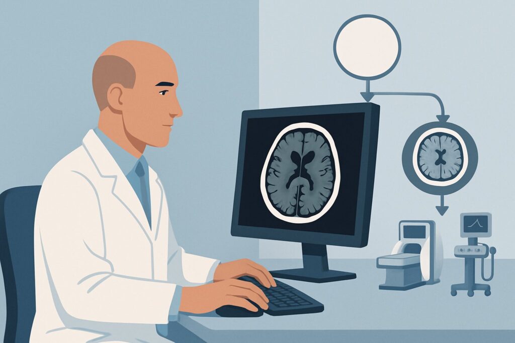
- MRI head without IV contrast (Radiation Dose: 0 mSv): This is the more sensitive study for the key differential diagnoses in this scenario. It excels at detecting the subtle edema of PRES, the early cytotoxic changes of anoxic injury, and the white matter abnormalities of ODS. Its superiority in tissue characterization makes it the preferred modality if a specific complication of metabolic disease is suspected.
- CT head without IV contrast (Radiation Dose: ☢☢☢ 1-10 mSv): The primary advantages of CT are speed and accessibility. It is excellent for rapidly excluding large, emergent conditions like hemorrhage, hydrocephalus, or a significant mass. In an unstable, agitated, or uncooperative patient, a quick non-contrast CT is often the most pragmatic and safest initial choice. However, it is insensitive to the early or subtle findings of PRES, anoxic injury, or ODS.
Studies involving intravenous contrast, such as CT head with IV contrast or MRI head with and without IV contrast, are rated Usually not appropriate. In the initial evaluation of a suspected metabolic derangement, contrast adds little diagnostic information and introduces unnecessary risk, particularly in patients with acute kidney injury, a common contributor to metabolic encephalopathy.
What’s Next After Imaging? Downstream Workflow
The results of the initial imaging study will guide the subsequent clinical pathway. The most common outcome is a negative or non-specific study, which reinforces the initial diagnosis of a toxic-metabolic encephalopathy.
- If the study is negative: The focus remains squarely on managing the underlying systemic illness. Continue aggressive treatment of the sepsis, DKA, uremia, or other identified cause. If the patient’s mental status fails to improve as expected with correction of the metabolic parameters, this constitutes a new clinical question. The patient may now fit the ACR scenario for “Altered mental status despite clinical management,” which often warrants a more advanced imaging study, such as an MRI if a CT was done initially, or a contrast-enhanced study if infection or inflammation is suspected.
- If the study is positive for a specific diagnosis (e.g., PRES, anoxic injury): The management plan must be expanded to address the neurologic diagnosis. This typically involves a neurology consultation. For PRES, this means aggressive blood pressure control; for anoxic injury, it involves supportive care and prognostication. The systemic illness must still be managed concurrently.
- If the study shows an unexpected finding (e.g., tumor, chronic subdural hematoma): This discovery fundamentally changes the diagnostic landscape. The workflow pivots to address the new finding, which will almost always require a neurosurgery or neurology consultation and potentially further, more specific imaging.
Pitfalls to Avoid (and When to Get Help)
Navigating the workup for altered mental status requires careful clinical judgment to avoid common missteps.
- Over-reliance on imaging: Do not let the imaging workflow delay fundamental treatment. Correcting severe hypoglycemia or starting antibiotics for sepsis should happen concurrently with, or even before, sending the patient for a scan.
- Choosing the wrong initial study: Ordering a non-contrast CT to “rule out” PRES or early anoxic injury is a common pitfall; CT is not sensitive enough for these conditions. If those are high on the differential, MRI is the appropriate study if the patient is stable enough.
- Ignoring patient stability: Attempting to obtain a 45-minute MRI on an agitated, delirious, or hemodynamically unstable patient is often impractical and unsafe. A rapid non-contrast CT is a more appropriate first step in such cases to rule out immediate life threats.
If the patient develops any new focal neurologic deficits, experiences a seizure, or fails to show any cognitive improvement despite correction of their metabolic abnormalities, it is critical to escalate care with an urgent neurology consultation.
Related ACR Topics and Tools
This article covers one specific variant within the broader ACR topic of Altered Mental Status. For a comprehensive overview of all related scenarios, from suspected intracranial pathology to new onset psychosis, see our parent guide: Altered Mental Status, Coma, Delirium, and Psychosis: ACR Appropriateness Decoded.
For additional decision support and technical details, the following GigHz resources are available:
- ACR Appropriateness Criteria Lookup — for exploring adjacent clinical scenarios.
- Imaging Protocol Library — for detailed techniques of the recommended studies.
- Radiation Dose Calculator — for discussing cumulative radiation exposure with patients and families.
Frequently Asked Questions
Why is there ‘Disagreement’ in the ACR rating for this scenario?
The ‘Disagreement’ reflects the valid clinical debate on whether imaging is needed at all for this specific presentation. Many cases of toxic-metabolic encephalopathy resolve with medical treatment alone, making initial imaging low-yield. The rating acknowledges that both imaging upfront (to exclude mimics) and deferring imaging (while treating the underlying cause) can be reasonable approaches depending on the clinical context.
If my patient has acute kidney injury, which study is safer?
Both non-contrast CT and non-contrast MRI are safe in patients with acute kidney injury, as neither uses intravenous contrast. Non-contrast MRI is often preferred because it provides more detailed information about potential brain complications of uremia (like PRES) without any radiation exposure. The key is to avoid gadolinium-based contrast agents in this setting unless absolutely necessary and after careful risk-benefit analysis.
Should I just get a non-contrast head CT on every patient with altered mental status?
No. While a non-contrast CT is a fast and widely available screening tool, it should be used judiciously. For this specific scenario (suspected metabolic cause, no focal deficits), the diagnostic yield is low. Furthermore, CT exposes the patient to ionizing radiation and is insensitive to key diagnoses like PRES, osmotic demyelination, or early ischemic injury. The decision to image, and with which modality, should be based on the full clinical picture.
What specific MRI sequences are most important in this workup?
For a non-contrast MRI in this setting, the most critical sequences are Diffusion-Weighted Imaging (DWI) and Apparent Diffusion Coefficient (ADC) maps, which are extremely sensitive for cytotoxic edema seen in anoxic injury and acute stroke. T2-weighted and FLAIR (Fluid-Attenuated Inversion Recovery) sequences are essential for identifying the vasogenic edema characteristic of PRES and the signal changes of osmotic demyelination.
If the initial non-contrast CT is negative but the patient doesn’t improve, what is the next step?
A negative initial CT in a patient who fails to improve after correction of their metabolic derangement is a strong indication for further investigation. The patient now fits a different clinical scenario. The next logical step is typically a brain MRI without and with IV contrast to look for more subtle pathology that the CT may have missed, such as encephalitis, small strokes, or an underlying inflammatory or infectious process.
Reviewed by Pouyan Golshani, MD, Interventional Radiologist — May 29, 2026