Which Imaging Study Is Best for Mild Head Trauma (GCS 13-15) After a Positive Clinical Rule?
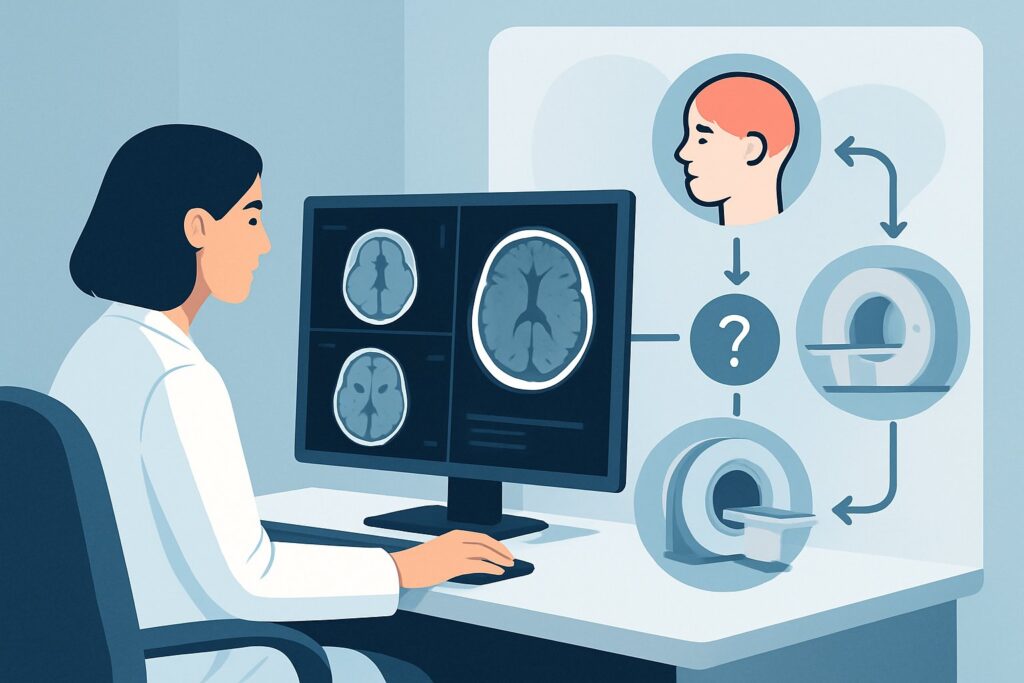
It’s 2 a.m. in the emergency department, and you’re evaluating a 52-year-old who fell from a stepladder while changing a lightbulb. He had a brief loss of consciousness, per his spouse, and now complains of a persistent headache. His Glasgow Coma Scale (GCS) is 15, but he has a significant scalp hematoma and vomited once in the waiting room. The Canadian CT Head Rule is positive, clearly indicating a need for imaging to rule out a clinically important brain injury. The question is no longer if you should image, but what you should order first. For this specific scenario—acute, mild head trauma in a patient who meets criteria for imaging—the American College of Radiology (ACR) designates one study as the clear first choice. Per the ACR Appropriateness Criteria, a CT head without IV contrast is Usually Appropriate.
Who Fits This Clinical Scenario for Mild Head Trauma?
This imaging workflow is designed for a precise patient population: adults presenting with acute, mild traumatic brain injury (TBI). To apply this guidance correctly, the patient must meet all of the following criteria:
- Acute Presentation: The injury occurred recently, typically within the last 24 to 48 hours.
- Mild TBI Classification: The patient has a Glasgow Coma Scale (GCS) score of 13, 14, or 15 on evaluation.
- Imaging Indicated by a Clinical Decision Rule (CDR): A validated rule, such as the Canadian CT Head Rule (CCHR) or the New Orleans Criteria (NOC), has been applied and found to be positive. This means the patient has one or more high-risk features (e.g., age ≥65, dangerous mechanism of injury, vomiting, signs of a basilar skull fracture, severe headache, amnesia) that place them at a higher-than-baseline risk for a significant intracranial injury.
It is critical to distinguish this scenario from similar presentations. This guidance does not apply to:
- Patients with a negative CDR: If a patient with mild head trauma does not meet imaging criteria per a validated rule, imaging is generally not indicated. This falls under a different ACR variant: Acute head trauma, mild (GCS 13–15), imaging not indicated by clinical decision rule.
- Patients with moderate or severe TBI: Any patient with a GCS score of 12 or below, or those with penetrating trauma, requires a different and more urgent diagnostic approach.
- Patients presenting with subacute or chronic symptoms: If the injury occurred weeks or months ago and the patient now has new or persistent neurologic deficits, the differential diagnosis and imaging strategy change significantly.
What Diagnoses Are You Working Up in This Scenario?
When you order imaging for mild TBI, your primary goal is to identify acute, space-occupying lesions that may require immediate neurosurgical intervention. The differential diagnosis is focused on traumatic injuries that are readily visible on the recommended imaging study.
Epidural Hematoma (EDH): This is a classic neurosurgical emergency. It involves an arterial bleed, most often from the middle meningeal artery, that collects between the dura mater and the skull. Patients may experience a “lucid interval” before rapid neurologic decline. On imaging, it appears as a hyperdense, biconvex (lens-shaped) collection.
Subdural Hematoma (SDH): More common than EDH, especially in older adults with brain atrophy, an SDH results from the tearing of bridging veins. The venous bleed collects between the dura and arachnoid mater. It typically appears as a hyperdense, crescent-shaped collection that conforms to the surface of the brain.
Traumatic Subarachnoid Hemorrhage (tSAH): This is one of the most common findings after TBI. Blood from injured surface vessels tracks into the subarachnoid space, appearing as hyperdensity within the cerebral sulci, cisterns, and fissures. While often managed non-operatively, it indicates significant force was applied to the head.
Intraparenchymal Hemorrhage and Contusion: These are essentially bruises of the brain tissue itself, often occurring at the site of direct impact (a “coup” injury) or on the opposite side (a “contrecoup” injury). They appear as ill-defined areas of hyperdensity within the brain parenchyma.
Skull Fracture: The imaging study must also be able to clearly identify fractures, particularly those that are depressed (pushed inward) or involve the skull base, as these are associated with a higher risk of underlying brain injury, vascular injury, or cerebrospinal fluid leak.
Why Is CT Head Without IV Contrast the Recommended Study for This Presentation?
The ACR rates CT head without IV contrast as Usually Appropriate because it optimally balances speed, accessibility, and diagnostic accuracy for the critical questions in this scenario. Its primary purpose is to rapidly and reliably detect acute hemorrhage and skull fractures.
The rationale is threefold:
- Excellent Sensitivity for Acute Blood: Acute hemorrhage is hyperdense (appears bright white) on a non-contrast CT scan. This makes it an extremely sensitive tool for identifying epidural, subdural, subarachnoid, and intraparenchymal bleeding—the most immediate life-threats.
- Speed and Availability: A non-contrast head CT can be performed in under a minute of scan time and is available 24/7 in virtually every emergency department. This speed is critical when evaluating a potentially unstable trauma patient.
- Superior Bone Detail: CT provides exquisite detail of the skull, making it the gold standard for identifying and characterizing fractures, which can have important prognostic and management implications.
Alternative studies are rated lower for specific, important reasons in this initial evaluation:
- MRI head without IV contrast is rated Usually not appropriate. While MRI is more sensitive for certain subacute findings like diffuse axonal injury, it is significantly slower, less available in an emergency setting, and more susceptible to motion artifact. It fails the primary test of being a rapid screening tool for emergent, surgically-correctable bleeds.
- Radiography skull is also rated Usually not appropriate. A skull x-ray can identify a fracture but provides zero information about the brain parenchyma. A patient can have a life-threatening intracranial hemorrhage with no skull fracture, making a normal x-ray dangerously reassuring.
- CT head with IV contrast is Usually not appropriate as the initial study. Intravenous contrast is not necessary to visualize acute blood; in fact, the enhancement of normal vascular structures can sometimes obscure a small hemorrhage. It adds time, cost, and the risks of contrast allergy and nephropathy without improving the detection of the key traumatic injuries.
The radiation dose for a non-contrast head CT in an adult is categorized as ☢☢☢ (1-10 mSv), a level considered justified given the high stakes of missing an intracranial bleed. Once you’ve decided on CT head without IV contrast, our protocol guide covers the technique, key imaging planes, and reading principles in detail: CT Brain Without Contrast.
What’s Next After CT Head Without IV Contrast? Downstream Workflow
The results of the initial non-contrast head CT will dictate your next steps, creating a clear decision tree for patient management.
- If the study is positive for a significant, space-occupying hemorrhage (e.g., a large epidural or subdural hematoma with midline shift), the immediate next step is an urgent neurosurgical consultation. The patient will require admission, likely to an intensive care unit, for close neurologic monitoring and potential surgical intervention like a craniotomy for hematoma evacuation.
- If the study is positive for less critical findings (e.g., a small traumatic subarachnoid hemorrhage, a non-displaced skull fracture, or a small cerebral contusion), the patient typically requires admission for a period of observation (often 24 hours). Serial neurologic examinations are key. If the patient’s clinical status worsens—for example, they develop a new neurologic deficit—this may trigger a new workflow for Acute head trauma with new or progressive neurologic deficit(s), often involving repeat imaging.
- If the study is negative for any acute traumatic finding, management depends on the patient’s clinical picture. If symptoms are controlled, the neurologic exam is non-focal, and they have reliable support at home, the patient can often be safely discharged with strict head injury precautions and concussion education. They should be instructed to return immediately for any worsening symptoms.
A negative initial CT provides strong reassurance against an immediate, life-threatening injury. However, it does not rule out delayed complications or the clinical syndrome of concussion.
Pitfalls to Avoid (and When to Get Help)
Even in this seemingly straightforward scenario, several clinical pitfalls can lead to poor outcomes. Be mindful of the following:
- Over-relying on GCS alone: A patient with a GCS of 15 can still harbor a lethal intracranial hematoma. This is precisely why validated clinical decision rules are so important—they identify high-risk features even in patients who appear well.
- Ignoring anticoagulation status: A patient on any anticoagulant or antiplatelet agent is at a substantially higher risk for intracranial bleeding, even from minor trauma. This factor should lower your threshold for imaging and may warrant a period of observation or even repeat imaging regardless of initial CT findings.
- Dismissing clinical signs of basilar skull fracture: Physical exam findings like hemotympanum (blood behind the eardrum), “raccoon eyes” (periorbital ecchymosis), or Battle’s sign (postauricular ecchymosis) are high-risk indicators and strongly support the need for imaging.
- Providing inadequate discharge instructions: A patient with a negative CT still has a head injury. Clear, written instructions on when to return to the emergency department are essential for patient safety.
If a patient develops a worsening headache, new focal weakness, seizure, or any decline in their GCS after the initial evaluation, escalate immediately for repeat imaging and neurosurgical consultation.
Related ACR Topics and Tools
This article covers one specific scenario in depth. For a broader view of imaging across all head trauma presentations, from mild to severe, please consult our parent guide. For help with adjacent scenarios or calculating radiation dose, the following GigHz resources are available.
- For breadth across all scenarios in Head Trauma, see our parent guide: Head Trauma: ACR Appropriateness Decoded.
- Imaging Appropriateness Selector — for adjacent scenarios
- Imaging Protocol Library — for technique on the recommended study
- Radiation Dose Calculator — for cumulative dose conversations
Frequently Asked Questions
What if the patient is on anticoagulants like warfarin or a DOAC? Does that change the imaging choice?
The choice of imaging study remains the same: a CT head without IV contrast is still the best initial test to look for hemorrhage. However, anticoagulation status dramatically lowers the threshold to order the scan in the first place and may influence the decision for repeat imaging or a longer period of observation, even after a negative initial CT, due to the increased risk of delayed bleeding.
Why not just get an MRI to be more thorough?
In the acute setting, the primary goal is to quickly rule out a life-threatening bleed that requires surgery. A non-contrast CT is the fastest, most available, and most sensitive tool for this specific task. MRI is slower, less accessible, and more prone to motion artifact. It is a valuable second-line tool for evaluating subacute injuries or when there is concern for non-hemorrhagic injury like diffuse axonal injury, but it is not the appropriate first test for an acute trauma workup.
The Canadian CT Head Rule was negative, but I’m still clinically concerned. Can I order a CT anyway?
Clinical decision rules are highly sensitive tools designed to reduce unnecessary imaging, but they do not replace clinical judgment. If you have a compelling reason to suspect an intracranial injury despite a negative CDR (e.g., a subtle but persistent neurologic finding, a very frail patient), you can still order the scan. It is crucial to document your reasoning clearly, as this clinical situation falls into a different ACR scenario where imaging is rated ‘Usually Not Appropriate.’
Does a normal non-contrast head CT rule out a concussion?
No. A concussion is a clinical diagnosis of a functional brain disturbance, not a structural one. A non-contrast head CT is performed to rule out structural injuries like bleeding, fractures, and swelling. In most concussions, the CT scan is expected to be normal. The diagnosis of concussion is based on the history and symptoms, not the imaging results.
How does this guidance apply to children with mild head trauma?
This workflow is intended for adult patients. Pediatric head trauma is managed differently, using specific pediatric clinical decision rules like the PECARN (Pediatric Emergency Care Applied Research Network) rule. These rules are designed to be even more selective about using CT scans due to children’s increased sensitivity to ionizing radiation. The ACR provides separate appropriateness criteria for pediatric head trauma.
Reviewed by Pouyan Golshani, MD, Interventional Radiologist — May 26, 2026