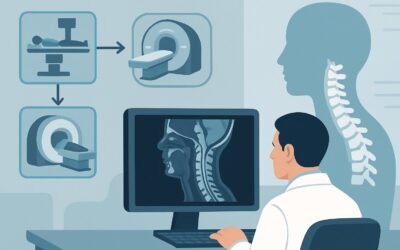
What Is the Best Initial Staging Imaging for Suspected Nasopharynx Cancer?

A 52-year-old man presents to your otolaryngology clinic with a three-month history of unilateral hearing loss, a new neck lump, and occasional epistaxis. On examination, you note a right-sided cervical lymph node and serous fluid behind the tympanic membrane. You suspect nasopharyngeal carcinoma (NPC), a diagnosis often linked to Epstein-Barr virus (EBV). Your next step is to confirm the diagnosis and, critically, to accurately stage the disease to guide therapy. This raises an immediate imaging question: which study provides the most detailed and comprehensive initial assessment? This article details the clinical workflow for this specific scenario, explaining why the American College of Radiology (ACR) finds MRI orbits face neck without and with IV contrast to be Usually appropriate for the initial staging of suspected nasopharynx cancer.
Who Fits This Clinical Scenario?
This guidance applies to patients undergoing initial evaluation for suspected or newly diagnosed nasopharyngeal carcinoma. It is also the correct pathway for patients presenting with an unknown primary head and neck cancer where serology is positive for Epstein-Barr virus (EBV), a strong indicator that the nasopharynx is the primary site. The key elements are that this is for initial staging, not for post-treatment surveillance or assessment of treatment response.
This workflow should be distinguished from similar, but distinct, clinical presentations:
- Other Head and Neck Cancers: Patients with suspected primary tumors of the oral cavity, oropharynx, larynx, or salivary glands follow different imaging algorithms. These sites have different patterns of spread and require tailored imaging protocols.
- Post-Treatment Surveillance: A patient with a history of treated NPC who presents for routine follow-up or with new symptoms of recurrence falls under a separate ACR variant. Surveillance imaging has different goals and considerations.
- Sinonasal Tumors: While anatomically adjacent, cancers originating in the paranasal sinuses or nasal cavity have unique staging requirements, often with a greater initial emphasis on bony invasion, which may alter the choice or ranking of imaging modalities.
Applying this workflow is appropriate when the clinical suspicion is squarely focused on the nasopharynx as the primary site of malignancy.
What Diagnoses Are You Working Up in This Scenario?
When ordering initial staging imaging for a suspected nasopharyngeal mass, the primary goal is to confirm and stage the most likely malignancy, but several other possibilities must be considered. The imaging findings will help differentiate among these potential diagnoses.
Nasopharyngeal Carcinoma (NPC) is the most common and critical diagnosis to confirm. It is an aggressive malignancy strongly associated with EBV infection, particularly in endemic regions like Southern China, Southeast Asia, and North Africa. Imaging is essential to define the tumor’s local extent (T stage), including invasion of the skull base, parapharyngeal spaces, and cranial nerves, as well as regional lymph node involvement (N stage).
Nasopharyngeal Lymphoma is another important consideration, as it can present with a bulky nasopharyngeal mass and extensive cervical lymphadenopathy, mimicking NPC. While biopsy is definitive, certain imaging features, such as a lack of skull base erosion despite large size and homogeneous enhancement, may suggest lymphoma over carcinoma.
Benign Lesions, while less common in the context of a suspicious neck mass, can also occur. A Thornwaldt cyst is a benign midline developmental cyst that is typically an incidental finding but can become large or infected. In adolescent males, a juvenile nasopharyngeal angiofibroma is a highly vascular benign tumor that must be considered, though it is rare in the typical adult NPC population.
Metastatic Disease to the nasopharynx from a distant primary is rare but remains in the differential. Similarly, other rare malignancies like rhabdomyosarcoma (more common in children) or minor salivary gland tumors can arise in this location.
Why Is MRI of the Orbits, Face, and Neck the Recommended Study?
For the initial local and regional staging of suspected nasopharyngeal carcinoma, the ACR designates MRI orbits face neck without and with IV contrast as Usually appropriate. This recommendation is based on MRI’s superior ability to delineate the primary tumor and its relationship to critical adjacent structures, which is paramount for accurate T-staging.
The key advantage of MRI is its exceptional soft-tissue contrast resolution. This allows for precise evaluation of:
- Tumor Extent: Differentiating tumor from surrounding normal mucosa, muscle, and parapharyngeal fat.
- Intracranial Extension: Detecting subtle invasion through the skull base foramina into the cavernous sinus or other intracranial compartments.
- Perineural Spread: Identifying tumor tracking along cranial nerves, particularly branches of the trigeminal nerve (V) and the abducens nerve (VI), which is a common and prognostically significant pathway of spread for NPC.
While other advanced imaging modalities are also rated Usually appropriate, they serve different primary roles.
CT neck with IV contrast is excellent for assessing cortical bone erosion of the skull base and is often faster and more accessible than MRI. However, its soft-tissue resolution is inferior, making it less sensitive for early perineural or intracranial spread. It is a valid alternative but may not provide the same level of detail for T-staging as MRI.
FDG-PET/CT skull base to mid-thigh is also Usually appropriate and is the standard for detecting distant metastatic disease (M-staging) and assessing the full burden of nodal disease. While it provides crucial whole-body information, its anatomic resolution of the primary tumor site is lower than that of a dedicated MRI. Therefore, PET/CT is typically performed to complete the staging process, but not to replace the detailed local assessment provided by MRI.
A significant benefit of MRI is the absence of ionizing radiation (0 mSv), which is an important consideration, especially in younger patients. This contrasts with the radiation dose from CT (1-10 mSv, ☢☢☢) and PET/CT (10-30 mSv, ☢☢☢☢).
What’s Next After MRI? Downstream Workflow
The results of the initial staging MRI will guide the subsequent diagnostic and therapeutic pathway. The workflow branches based on the findings, leading to biopsy, further imaging, or multidisciplinary tumor board discussion.
If the MRI is positive for a suspicious nasopharyngeal mass: The next step is typically an endoscopic examination with a targeted biopsy of the lesion to obtain a histopathologic diagnosis. The MRI findings are crucial for guiding the endoscopist to the most representative area of the tumor. Once NPC is confirmed, the MRI data (T and N stage) are combined with the results of a whole-body staging study, usually an FDG-PET/CT, to establish the final TNM stage. This comprehensive stage dictates the treatment plan, which for most non-metastatic NPC involves definitive chemoradiation.
If the MRI is negative or shows only benign findings: If a high clinical suspicion for malignancy remains despite a negative MRI (e.g., in a patient with a neck mass that is EBV-positive), the focus may shift. An FDG-PET/CT may be performed to search for an occult primary tumor elsewhere or to guide a biopsy of the most metabolically active lymph node. The patient should be referred for a comprehensive endoscopic evaluation, including examination under anesthesia with directed biopsies of the nasopharynx (even if it appears normal on MRI) and other high-risk sites.
If the MRI is indeterminate: In cases where findings are ambiguous, a multidisciplinary discussion involving otolaryngology, radiology, radiation oncology, and medical oncology is essential. A short-interval follow-up MRI or a PET/CT may be recommended to assess for change or to provide functional information that can help clarify the nature of the finding.
Pitfalls to Avoid (and When to Get Help)
Navigating the initial workup for suspected nasopharyngeal cancer requires careful attention to detail to avoid common errors that can delay diagnosis or lead to inaccurate staging.
- Inadequate MRI Field of View: Ordering a standard “MRI Brain” or “MRI Neck” may not be sufficient. The protocol must cover the entire region from the orbits through the skull base and down to the clavicles to fully assess the primary tumor and cervical lymph nodes.
- Ignoring Cranial Nerve Deficits: A new cranial nerve palsy (e.g., diplopia from CN VI palsy) in a patient at risk for NPC is a red flag for perineural spread and skull base invasion. This finding should expedite the imaging workup with MRI.
- Over-reliance on CT for T-staging: While CT is useful, relying on it alone can lead to underestimation of the tumor’s extent, particularly regarding subtle intracranial or perineural invasion.
- Delaying Biopsy: Imaging provides the stage, but tissue provides the diagnosis. Imaging should not unduly delay obtaining a biopsy, which is required before initiating treatment.
If the clinical picture and imaging findings are discordant, or if the case is complex, escalation to a multidisciplinary head and neck tumor board is the most appropriate next step.
Related ACR Topics and Tools
For a comprehensive overview of imaging for all head and neck cancer scenarios, this article is best used alongside our broader topic guide. The following resources provide additional context for evidence-based imaging decisions.
- For breadth across all scenarios in Staging and Post-Therapy Assessment of Head and Neck Cancer, see our parent guide: Staging and Post-Therapy Assessment of Head and Neck Cancer: ACR Appropriateness Decoded.
- To explore other clinical presentations and their recommended imaging studies, use the ACR Appropriateness Criteria Lookup.
- For detailed technical parameters of the recommended study, consult the Imaging Protocol Library.
- To discuss radiation exposure with patients, the Radiation Dose Calculator can help quantify and compare study doses.
Frequently Asked Questions
Why is MRI preferred over CT for initial staging of nasopharynx cancer?
MRI is preferred for its superior soft-tissue contrast, which provides a more detailed evaluation of the primary tumor’s extent, including subtle invasion into adjacent soft tissues, the skull base, and along cranial nerves. This is critical for accurate T-staging. While CT is excellent for assessing bone erosion, it can miss early soft tissue or perineural spread.
If PET/CT is also ‘Usually Appropriate’, can I just order that instead of an MRI?
While FDG-PET/CT is essential for overall staging (especially for detecting distant metastases and nodal disease), it should not replace a dedicated high-resolution MRI of the nasopharynx and neck. The anatomic detail from MRI for the primary tumor (T-stage) is superior to that of PET/CT. The standard of care is typically to perform both: an MRI for local-regional staging and a PET/CT for distant staging.
Does the patient need contrast for the MRI?
Yes, both non-contrast and post-contrast sequences are essential. Non-contrast images, particularly fat-suppressed T2-weighted sequences, are excellent for detecting the tumor. Post-contrast images are crucial for assessing the pattern of enhancement, delineating the tumor margins, and identifying perineural spread, where abnormal nerve enhancement is a key finding.
What if the patient has a pacemaker or other contraindication to MRI?
In cases where MRI is contraindicated, a contrast-enhanced CT of the neck is the best alternative and is also rated ‘Usually Appropriate’ by the ACR. While it has limitations in soft-tissue assessment, it provides excellent detail regarding bone invasion and cervical lymph nodes. This would be combined with an FDG-PET/CT for complete staging.
The patient has an enlarged cervical lymph node, but the nasopharynx looks normal on endoscopy. What is the role of imaging?
This presentation is highly suspicious for an unknown primary tumor, and if EBV serology is positive, the nasopharynx is the most likely source. An MRI is critical in this scenario, as it can often detect a small, submucosal primary tumor that is not visible on endoscopy. If the MRI is also negative, an FDG-PET/CT is the next step to identify the primary site or guide a biopsy of the most metabolically active node.
Reviewed by Pouyan Golshani, MD, Interventional Radiologist — May 30, 2026