How Should You Image Suspected Cerebral Vasospasm? An ACR-Guided Workflow
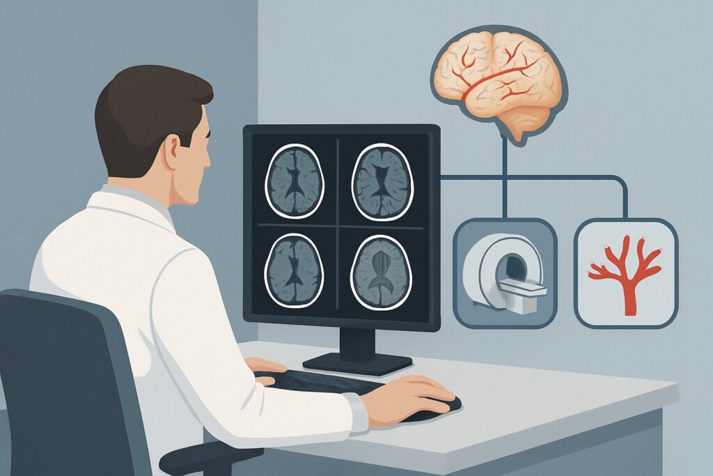
It’s day eight in the neuro-intensive care unit for a 58-year-old patient recovering from a coiled anterior communicating artery aneurysm rupture. The initial post-procedure course was smooth, but this morning, the patient develops a new left-sided hemiparesis and fluctuating aphasia. The clinical team’s leading concern is delayed cerebral ischemia secondary to vasospasm, a common and dangerous complication following subarachnoid hemorrhage. The immediate question is how to confirm this diagnosis and assess its severity to guide intervention. This article provides a detailed clinical workflow for the initial imaging of suspected cerebral vasospasm, focusing on the American College of Radiology (ACR) Appropriateness Criteria. For this specific scenario, the ACR rates `Arteriography cervicocerebral` as `Usually appropriate`.
Who Fits This Clinical Scenario?
This guidance applies to patients with a new or worsening neurologic deficit where cerebral vasospasm is a primary clinical consideration. The classic context is a patient who is 4 to 14 days post-aneurysmal subarachnoid hemorrhage (SAH), as this is the peak window for this complication. However, the scenario also includes patients with suspected vasospasm from other causes, such as traumatic brain injury, post-operative states, or conditions like reversible cerebral vasoconstriction syndrome (RCVS).
This workflow is specifically for the initial diagnostic imaging when suspicion arises. It is distinct from related but separate clinical questions:
- Initial workup of SAH: This scenario assumes SAH is already known. The workup for a “thunderclap headache” to diagnose the initial hemorrhage follows a different pathway, detailed in the parent topic on cerebrovascular diseases.
- Screening or surveillance: This is not for routine, asymptomatic screening for vasospasm, which often involves non-invasive methods. This guidance is for a symptomatic patient requiring a definitive diagnosis.
- Suspected CNS vasculitis: While vasculitis can present with arterial narrowing, its workup involves a broader differential and often different imaging sequences. This scenario focuses on the more acute presentation of vasospasm.
What Diagnoses Are You Working Up in This Scenario?
When ordering imaging for suspected vasospasm, you are primarily evaluating the caliber of the cerebral arteries to identify narrowing that explains the patient’s new symptoms. The differential diagnosis is focused but includes critical mimics that imaging can help differentiate.
Cerebral Vasospasm Secondary to Subarachnoid Hemorrhage: This is the most common and anticipated cause in the post-SAH setting. Irritation from blood products in the subarachnoid space triggers prolonged, diffuse, or focal arterial constriction. This narrowing reduces blood flow, leading to delayed cerebral ischemia (DCI) and infarction if not treated promptly. Imaging aims to confirm the presence, location, and severity of the vasospasm.
Reversible Cerebral Vasoconstriction Syndrome (RCVS): A key mimic of post-SAH vasospasm, RCVS is characterized by multifocal arterial constriction that typically resolves within three months. It often presents with recurrent thunderclap headaches and can be associated with neurologic deficits. While it can be idiopathic, it is often linked to vasoactive drugs or the postpartum state. Imaging findings of diffuse vasoconstriction can be indistinguishable from post-SAH vasospasm.
Central Nervous System (CNS) Vasculitis: A less common but important consideration, CNS vasculitis is an inflammatory condition of the vessel walls that causes stenosis. Unlike the more acute onset of vasospasm or RCVS, vasculitis may have a more subacute or chronic course. Imaging may show “beading” or irregular narrowing of multiple vessels, often in different vascular territories, and may be associated with parenchymal enhancement or small infarcts in a pattern atypical for large-vessel vasospasm.
Why Is Arteriography the Recommended Study for This Presentation?
The ACR designates both `Arteriography cervicocerebral` (also known as digital subtraction angiography, or DSA) and `CTA head with IV contrast` as `Usually appropriate` for the initial imaging of suspected cerebral vasospasm. However, DSA is often considered the gold standard for diagnosis and offers a direct therapeutic pathway.
Arteriography cervicocerebral (DSA): This invasive procedure provides the highest spatial and temporal resolution for evaluating the entire cervicocerebral vasculature. It is exceptionally sensitive for detecting subtle to severe arterial narrowing and can precisely delineate the affected segments. Its primary advantage is that it is both diagnostic and therapeutic; if significant vasospasm is confirmed, intra-arterial therapies like vasodilator infusion (e.g., verapamil, nicardipine) or angioplasty can be performed during the same session. This “one-stop” capability is critical in a time-sensitive condition where preventing permanent infarction is the goal. The main tradeoffs are its invasive nature, risk of procedural complications (e.g., stroke, dissection), and radiation exposure (adult RRL ☢☢☢ 1-10 mSv).
CTA head with IV contrast: As a non-invasive alternative, CTA is fast, widely available, and highly effective at visualizing the major intracranial arteries. It can reliably demonstrate moderate to severe vasospasm in the circle of Willis and its proximal branches. It is an excellent first-line diagnostic tool, especially at centers where immediate access to interventional neuroradiology is limited or when the clinical suspicion is moderate. The radiation dose is comparable to DSA (adult RRL ☢☢☢ 1-10 mSv).
Alternatives rated lower for this specific scenario include:
- US duplex Doppler transcranial (TCD): Rated `May be appropriate`, TCD is a non-invasive, radiation-free bedside tool used for monitoring trends in blood flow velocities. Elevated velocities can suggest vasospasm. However, it is operator-dependent, has limited acoustic windows in some patients, and is less sensitive for distal vessel vasospasm. It is primarily a screening or monitoring tool, not the definitive diagnostic study for a new, severe deficit.
- MRA head without IV contrast: Rated `Usually not appropriate`, non-contrast MRA techniques often lack the resolution to reliably diagnose vasospasm and are prone to artifacts that can mimic or obscure stenosis. They are generally not considered adequate for this acute, high-stakes clinical question.
What’s Next After Imaging? Downstream Workflow
The results of the initial imaging study will directly guide the subsequent clinical management, which is focused on restoring cerebral perfusion and preventing permanent neurologic injury.
- If the study is positive for significant vasospasm: If DSA was the initial study, intra-arterial therapy can be administered immediately. If CTA confirms vasospasm, the patient may be taken for DSA for intervention. Medical management is also intensified, often involving induced hypertension (permissive hypertension), euvolemia, and potentially oral nimodipine to improve collateral flow and reduce the risk of infarction.
- If the study is negative for vasospasm: A negative high-quality DSA or CTA effectively rules out hemodynamically significant large-vessel vasospasm as the cause of the patient’s symptoms. The clinical team must then aggressively pursue other causes for the neurologic decline. This includes investigating for hydrocephalus, seizures (requiring EEG), metabolic derangements (e.g., hyponatremia), or infection. The focus shifts away from vascular intervention and toward treating the alternative diagnosis.
- If the study is indeterminate or shows only mild/moderate spasm: In cases where imaging shows mild vasospasm that does not fully correlate with the severity of the clinical deficit, a multimodal approach is necessary. This may involve perfusion imaging (`CT head perfusion with IV contrast` or `MRI head perfusion with IV contrast`, both rated `May be appropriate`) to assess the downstream hemodynamic consequences of the narrowing. These studies can reveal areas of oligemia or ischemia, helping to determine if the observed vasospasm is physiologically significant and requires aggressive treatment.
Pitfalls to Avoid (and When to Get Help)
Navigating the workup for suspected vasospasm requires timely and accurate decision-making. Common pitfalls include:
- Delaying definitive imaging: In a patient with a new, significant neurologic deficit post-SAH, time is brain. Over-reliance on indirect measures like TCDs or delaying a CTA or DSA can lead to irreversible infarction.
- Misattributing symptoms: Attributing all neurologic changes in a post-SAH patient to vasospasm without considering mimics like seizures, hydrocephalus, or electrolyte imbalances can lead to missed diagnoses.
- Underestimating CTA limitations: While excellent for proximal vessels, CTA may be less sensitive for vasospasm in smaller, more distal arteries (M2/M3 or A2/A3 segments and beyond). A high clinical suspicion with a negative CTA may still warrant DSA.
If a patient develops a sudden, severe neurologic deficit consistent with a large vessel territory stroke (e.g., dense hemiplegia and aphasia), immediate escalation to the neuro-interventional and neuro-critical care teams is mandatory, often bypassing intermediate steps to proceed directly to DSA.
Related ACR Topics and Tools
This article focuses on a single, specific clinical scenario. For a comprehensive overview of imaging for aneurysms, vascular malformations, and subarachnoid hemorrhage, refer to the parent topic article. The following GigHz tools can also support your clinical workflow:
- For breadth across all scenarios in Cerebrovascular Diseases-Aneurysm, Vascular Malformation, and Subarachnoid Hemorrhage, see our parent guide: Cerebrovascular Diseases-Aneurysm, Vascular Malformation, and Subarachnoid Hemorrhage: ACR Appropriateness Decoded.
- ACR Appropriateness Criteria Lookup — for adjacent scenarios
- Imaging Protocol Library — for technique on the recommended study
- Radiation Dose Calculator — for cumulative dose conversations
Frequently Asked Questions
When should I choose CTA over DSA for suspected vasospasm?
CTA is an excellent non-invasive first choice when it is readily available and can be performed quickly. It is particularly useful for ruling out significant proximal vasospasm. Choose CTA if the patient is relatively stable, if the diagnosis is less certain, or as a fast screening tool before committing to an invasive procedure. DSA is preferred when the clinical suspicion is very high, if a therapeutic intervention is anticipated, or if the CTA is negative but symptoms persist, suggesting more distal or subtle disease.
What is the role of Transcranial Doppler (TCD) in managing vasospasm?
TCD is rated ‘May be appropriate’ by the ACR for diagnosis but is primarily used as a non-invasive daily monitoring tool to detect trends in cerebral blood flow velocities after SAH. A sudden rise in velocities can signal the onset of vasospasm, prompting a confirmatory study like CTA or DSA. It is not a substitute for anatomic imaging when a patient develops a new focal neurologic deficit.
Can a normal non-contrast head CT rule out vasospasm?
No. A non-contrast CT of the head is used to detect hemorrhage, hydrocephalus, or a large territory stroke. It provides no direct visualization of the blood vessels and cannot diagnose or rule out vasospasm. A patient can have severe, life-threatening vasospasm with a non-contrast CT that appears unchanged from baseline.
Does perfusion imaging (CTP/MRP) have a role in the initial workup?
Perfusion imaging is rated ‘May be appropriate’ and is typically a problem-solving tool rather than the initial diagnostic study. It is most useful when anatomic imaging (CTA/DSA) shows mild or moderate vasospasm and you need to determine if that narrowing is causing a significant downstream blood flow deficit (ischemia). It helps bridge the gap between anatomic findings and clinical symptoms.
If the patient has renal insufficiency, is CTA with contrast still an option?
In patients with significant renal dysfunction, the risk of contrast-induced nephropathy must be weighed against the risk of permanent brain injury from untreated vasospasm. In this high-stakes scenario, the diagnostic benefit of a contrast-enhanced study (either CTA or DSA) often outweighs the renal risk, especially with appropriate pre- and post-procedure hydration. A formal risk/benefit discussion with the patient/family and consulting with nephrology is recommended.
Reviewed by Pouyan Golshani, MD, Interventional Radiologist — May 29, 2026