Which Imaging Is Best for Chronic Unilateral Vision Loss with a Suspected Orbital Mass?

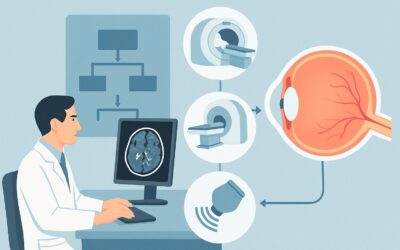
A 62-year-old patient presents to your clinic with six months of slowly worsening vision in his right eye, describing it as a progressive “dimming” and blurriness. On examination, you note subtle proptosis and a relative afferent pupillary defect. You suspect an intraorbital process—perhaps a mass compressing the optic nerve or a slow-flow vascular lesion. Standing at the electronic health record, you face the critical decision: which initial imaging study will provide the clearest answer with the least risk? This article provides a step-by-step clinical workflow for this specific scenario, grounded in the American College of Radiology (ACR) Appropriateness Criteria. For an adult with chronic, progressive unilateral vision loss where an intraorbital mass or vascular lesion is suspected, the ACR rates MRI orbits without and with IV contrast as Usually Appropriate.
Who Fits This Clinical Scenario for Chronic Unilateral Vision Loss?
This guidance is tailored for a specific patient population: adults experiencing a gradual, persistent, and one-sided decline in vision. The key clinical feature is the suspicion of a structural lesion within the orbit—a mass, an enlarged muscle, or a vascular anomaly—based on signs like proptosis (bulging eye), ophthalmoplegia (restricted eye movements), or optic disc swelling or atrophy on fundoscopy.
This workflow is intended for the initial diagnostic imaging step, not for follow-up or post-treatment evaluation.
It is crucial to distinguish this presentation from similar but distinct clinical scenarios that require different imaging pathways:
- Acute Vision Loss: If the vision loss was sudden, the differential shifts towards ischemic, inflammatory, or infectious causes. This would fall under the ACR variant for Acute vision loss, infection or inflammatory disorder suspected.
- Trauma: If the vision loss occurred after an injury, the primary concern is orbital fracture or hemorrhage, routing to the Acute posttraumatic visual defect scenario, where CT is often preferred.
- Bitemporal Hemianopia: Vision loss affecting the outer visual fields of both eyes strongly suggests a lesion at the optic chiasm (e.g., a pituitary adenoma). This presentation aligns with the Sellar or parasellar mass suspected scenario.
Applying this article’s guidance to the wrong clinical context can lead to diagnostic delays or suboptimal imaging.
What Diagnoses Are You Working Up with a Suspected Orbital Mass?
When ordering imaging for a suspected intraorbital lesion, you are investigating a focused differential diagnosis. The goal of the study is to differentiate between these possibilities, as their management pathways vary significantly.
A primary consideration is an optic nerve or sheath tumor. Optic nerve gliomas and optic nerve sheath meningiomas are the most common primary tumors of the optic nerve. They typically cause slow, painless, progressive vision loss. MRI is exceptionally skilled at delineating the optic nerve and its sheath, making it ideal for identifying and characterizing these neoplasms.
Orbital vascular lesions are another key diagnostic category. These include cavernous malformations (often called cavernous hemangiomas), orbital varices, and arteriovenous malformations. These lesions can expand slowly, causing mass effect on the optic nerve or extraocular muscles. Contrast-enhanced imaging is vital to assess their vascular nature and flow characteristics.
Consider inflammatory or infiltrative processes that can mimic a mass. Thyroid eye disease (Graves’ orbitopathy) is a common cause of proptosis and vision loss due to enlargement of the extraocular muscles, which can compress the optic nerve at the orbital apex. Idiopathic orbital inflammation (orbital pseudotumor) is another condition that can present as a discrete, enhancing mass, though it is often associated with pain.
Finally, while less common, metastatic disease to the orbit from primary cancers like breast, lung, or prostate must be considered, particularly in patients with a known history of malignancy.
Why Is MRI of the Orbits the Recommended Study for This Presentation?
The ACR designates MRI orbits without and with IV contrast as Usually Appropriate because it provides the best soft-tissue resolution for evaluating the complex anatomy of the orbit. This is the central rationale for its top rating.
MRI excels at visualizing the optic nerve, its sheath, the extraocular muscles, and the orbital fat—structures that are poorly differentiated on other modalities. The pre-contrast sequences can detect subtle abnormalities, while the post-contrast sequences are critical for characterizing lesions. The pattern and degree of gadolinium enhancement help distinguish between tumor types (e.g., the tram-track sign of a meningioma), vascular malformations, and inflammatory processes. Furthermore, specific sequences like fat suppression are essential to null the bright signal from orbital fat, making enhancing lesions more conspicuous.
Why are other studies rated lower for this specific scenario?
- CT orbits with IV contrast is rated May be appropriate. While excellent for evaluating bony anatomy and detecting calcifications, its soft-tissue contrast is significantly inferior to MRI. It can miss subtle optic nerve pathology or mischaracterize a soft-tissue mass. However, it is a valuable alternative when MRI is contraindicated (e.g., in a patient with an incompatible pacemaker) or unavailable. It involves ionizing radiation (☢☢☢ 1-10 mSv), whereas MRI has a radiation dose of 0 mSv.
- MRA head and neck without and with IV contrast is rated Usually not appropriate for initial imaging. While a vascular lesion is on the differential, a standard orbital MRI with contrast is the correct first step to characterize the anatomy and identify if a vascular lesion is even present. MRA is a specialized functional study designed to visualize arteries, and it lacks the detailed parenchymal and soft-tissue information of a diagnostic MRI. It is typically a downstream, problem-solving tool, not an initial survey.
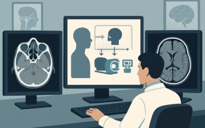
When ordering, be specific: request “MRI orbits without and with contrast” to ensure the protocol includes thin slices through the orbits and appropriate fat-suppression techniques.
What’s Next After an Orbital MRI? Downstream Workflow
The results of the orbital MRI will guide your next steps and necessary referrals. The imaging report is not the end of the workup but a critical branch point in the patient’s care.
- If the MRI identifies a distinct mass (e.g., suspected meningioma, glioma, or schwannoma): The next step is a referral to a multidisciplinary team. This typically includes a neuro-ophthalmologist for functional assessment and specialists in neurosurgery or oculoplastic surgery for consideration of biopsy or resection.
- If the MRI shows findings consistent with a vascular lesion (e.g., cavernous malformation): A neuro-ophthalmology or neurosurgery consult is still warranted. Depending on the findings, further vascular imaging like a formal MRA/MRV or even a digital subtraction angiogram (rated Usually not appropriate for initial workup but valuable for pre-surgical planning) may be considered.
- If the MRI reveals inflammatory changes (e.g., enlarged extraocular muscles in thyroid eye disease or diffuse enhancement in orbital pseudotumor): The workflow shifts from surgical to medical. A referral to ophthalmology is key, often in conjunction with endocrinology (for thyroid disease) or rheumatology (for systemic inflammatory conditions).
- If the MRI is negative or non-diagnostic: Re-evaluate the clinical picture. Could the lesion be further posterior along the visual pathway? This may prompt consideration of an adjacent ACR scenario, such as optic nerve abnormality suspected, and potentially lead to ordering the `May be appropriate` study, MRI head without and with IV contrast, to evaluate the optic chiasm, tracts, and radiations.
Pitfalls to Avoid (and When to Get Help)
Navigating the workup for a suspected orbital mass requires careful attention to detail to avoid common diagnostic errors.
1. Ordering Without Contrast: An MRI of the orbits without IV contrast is rated Usually not appropriate for this scenario. Contrast is essential for lesion detection and characterization. Omitting it can render the study non-diagnostic and necessitate a repeat scan.
2. Defaulting to CT: Unless MRI is absolutely contraindicated, it should be the first-line study. Choosing CT first due to convenience or habit can miss key soft-tissue findings and exposes the patient to unnecessary radiation.
3. Using a Standard “Brain” Protocol: An MRI of the brain is not optimized for the orbits. Ensure the order specifically requests “MRI orbits” to trigger a protocol with thin, high-resolution slices and fat-suppression sequences tailored to this anatomy.
4. Ignoring Red Flags: If a patient presents with rapid vision loss, severe pain, or evidence of acute optic nerve compression, the workup should be expedited. This warrants an urgent referral to a neuro-ophthalmologist or emergency department evaluation, even before imaging is completed.
Related ACR Topics and Tools
This article covers one specific variant within the broader ACR topic of Vision Loss. For a comprehensive overview of all related scenarios, from trauma to suspected inflammatory disease, please see our parent guide. The tools below can help you apply these criteria in your daily practice.
- For breadth across all scenarios in Vision Loss, see our parent guide: Vision Loss: ACR Appropriateness Decoded.
- To look up other clinical scenarios, visit the ACR Appropriateness Criteria Lookup tool.
- For details on imaging techniques, explore the Imaging Protocol Library.
- To discuss radiation exposure with patients, use the Radiation Dose Calculator.
Frequently Asked Questions
My patient has a pacemaker. Can I still order an MRI for a suspected orbital mass?
An MRI may not be possible if the patient has an older, MRI-incompatible pacemaker or other metallic implant. All patients must be screened for MRI safety. If an MRI is contraindicated, the ACR rates ‘CT orbits with IV contrast’ as ‘May be appropriate’. While it has lower soft-tissue resolution, it is the best alternative in this situation.
The MRI report mentions ‘fat suppression’. Why is that important for orbital imaging?
The orbit is filled with fat, which produces a very bright signal on many standard MRI sequences. This bright signal can obscure underlying pathology, especially enhancing lesions. Fat suppression techniques selectively null the signal from fat, making the optic nerve, muscles, and any abnormal tissue stand out more clearly, which is critical for diagnosis.
Is there a role for ultrasound in working up chronic unilateral vision loss?
Ocular ultrasound is a valuable tool, particularly for evaluating the globe itself (e.g., for retinal detachment or intraocular tumors). It can also provide some information about the anterior orbit. However, for evaluating the entire orbit, the optic nerve back to the orbital apex, and the surrounding soft tissues, MRI is far superior. Ultrasound is generally not the primary modality for a suspected retrobulbar mass.
If I suspect thyroid eye disease, should I still order an MRI or just check thyroid labs?
Both are often necessary. Thyroid function tests (TSH, T3, T4) and antibody levels are crucial for the systemic diagnosis. However, imaging is essential to assess the extent of orbital involvement, such as the degree of extraocular muscle enlargement and, most importantly, to check for compressive optic neuropathy at the crowded orbital apex. The MRI provides anatomical information that labs cannot.
The patient’s vision loss is progressive but they also have pain. Does that change the imaging choice?
Pain is an important clinical feature but does not change the initial imaging choice of ‘MRI orbits without and with IV contrast’. The presence of pain may raise suspicion for an inflammatory process like idiopathic orbital inflammation (orbital pseudotumor) or certain aggressive tumors, but MRI remains the best modality to differentiate these possibilities.
Reviewed by Pouyan Golshani, MD, Interventional Radiologist — May 30, 2026