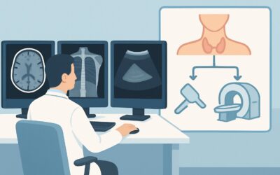
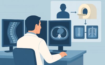
What Imaging Should You Order for Headache with Signs of Intracranial Hypertension?

A 34-year-old woman presents to your clinic with a three-month history of a persistent, dull headache. She describes a “whooshing” sound in her right ear that syncs with her heartbeat, and she notes her vision briefly “greys out” when she stands up quickly or coughs. On fundoscopic examination, you identify bilateral papilledema. You suspect a syndrome of intracranial hypertension, but the key question is whether this is idiopathic or secondary to a dangerous underlying cause. This article details the evidence-based imaging workflow for this specific clinical presentation, guiding you from initial suspicion to a definitive diagnostic plan. For a patient with headache and features of intracranial hypertension, the American College of Radiology (ACR) rates MRI head without and with IV contrast as Usually appropriate.
Who Fits This Clinical Scenario?
This guidance applies to patients presenting with a headache accompanied by specific signs or symptoms suggestive of elevated intracranial pressure (ICP). The classic constellation includes papilledema on fundoscopy, pulsatile tinnitus (a rhythmic “whooshing” sound synchronous with the pulse), and transient visual obscurations, often precipitated by Valsalva maneuvers like coughing, sneezing, or bending over. Other suggestive features can include a sixth cranial nerve (abducens) palsy, leading to horizontal diplopia.
It is critical to distinguish this presentation from similar, but distinct, clinical scenarios that require different workups:
- Sudden, severe “thunderclap” headache: This presentation raises immediate concern for subarachnoid hemorrhage and follows a different imaging pathway, often starting with non-contrast head CT.
- Headache worse when upright, better when supine: This positional feature is the hallmark of intracranial HYPOtension, the clinical opposite of this scenario. Imaging for suspected CSF leak is indicated.
- Headache with fever, neck stiffness, or rapidly progressive neurologic deficits: These are “red flag” features suggesting an urgent process like meningitis, encephalitis, or an evolving stroke, which may alter the initial choice and timing of imaging.
This article is focused solely on the initial imaging workup for the patient whose primary clinical picture points toward chronically elevated ICP.
What Diagnoses Are You Working Up in This Scenario?
The primary goal of imaging in this scenario is not to diagnose idiopathic intracranial hypertension (IIH) directly, but rather to exclude secondary causes of elevated ICP. The differential diagnosis drives the choice of imaging modality.
Idiopathic Intracranial Hypertension (IIH): Also known as pseudotumor cerebri, this is a diagnosis of exclusion and the most common cause of this syndrome, particularly in young women with an elevated body mass index. While the diagnosis is ultimately confirmed with lumbar puncture showing elevated opening pressure and normal cerebrospinal fluid (CSF) composition, imaging is required first to rule out other pathologies that would make a lumbar puncture unsafe or lead to an alternative diagnosis.
Cerebral Venous Sinus Thrombosis (CVST): This is a critical, life-threatening diagnosis that must be excluded. A clot in the dural venous sinuses obstructs venous outflow from the brain, directly causing intracranial hypertension. CVST can present identically to IIH and requires prompt anticoagulation. Imaging must be capable of clearly visualizing the dural sinuses.
Intracranial Mass or Lesion: A primary or metastatic brain tumor, abscess, or other space-occupying lesion can elevate ICP by obstructing CSF pathways (e.g., the cerebral aqueduct) or by compressing venous structures. Imaging must have sufficient soft-tissue resolution to detect and characterize such lesions.
Chronic or Granulomatous Meningitis: Less commonly, chronic inflammation or infection of the meninges (e.g., from tuberculosis or sarcoidosis) can impair CSF absorption at the arachnoid granulations, leading to a communicating hydrocephalus and elevated ICP. Contrast-enhanced imaging is essential to visualize meningeal enhancement.
Why Is MRI Head Without and With IV Contrast the Recommended Study for This Presentation?
The ACR designates MRI head without and with IV contrast as Usually appropriate because it is the single best test to evaluate the entire differential for intracranial hypertension. It provides a comprehensive assessment of the brain parenchyma, ventricular system, and vascular structures without using ionizing radiation.
The non-contrast sequences are excellent for identifying indirect signs that support a diagnosis of IIH, such as:
- Empty sella turcica
- Flattening of the posterior sclera
- Distention of the perioptic nerve sheaths
- Transverse venous sinus stenosis
The administration of intravenous gadolinium-based contrast is crucial. Post-contrast sequences are highly sensitive for detecting enhancing intracranial masses, abnormal meningeal enhancement seen in meningitis, and can help visualize flow-related signal within the dural venous sinuses. An occluded sinus will fail to enhance, a key finding for diagnosing CVST.
How do alternative studies compare?
CT head without IV contrast is also rated Usually appropriate (Radiation Level: ☢☢☢ 1-10 mSv). It is often performed in the emergency setting due to its speed and availability. It is effective at ruling out large masses, significant hydrocephalus, or hemorrhage. However, it lacks the sensitivity of MRI for detecting subtle parenchymal abnormalities, dural sinus thrombosis, and the specific imaging signs of IIH. A negative non-contrast CT does not sufficiently rule out the critical secondary causes of intracranial hypertension.
MR Venography (MRV), either with or without contrast, is rated May be appropriate. An MRV is the gold standard for evaluating the dural venous sinuses and is superior to a standard post-contrast MRI for this specific task. However, it does not provide the detailed parenchymal assessment of a full brain MRI. Therefore, it is often ordered as an additional sequence alongside the primary brain MRI rather than as a standalone initial test.
What’s Next After MRI Head Without and With IV Contrast? Downstream Workflow
The results of the brain MRI will dictate the subsequent clinical pathway. The workflow branches based on whether a secondary cause of intracranial hypertension is identified.
If the MRI is positive for a secondary cause:
- Intracranial Mass: The next step is urgent neurosurgical consultation for biopsy or resection, along with consideration for steroid administration to reduce vasogenic edema.
- Cerebral Venous Sinus Thrombosis: This finding requires immediate consultation with neurology or hematology to initiate anticoagulation therapy. A hypercoagulable workup is often warranted.
- Meningeal Enhancement: If concerning for meningitis, a lumbar puncture is necessary for CSF analysis (cell count, protein, glucose, cultures, cytology) to identify the infectious or inflammatory cause.
If the MRI is negative for a secondary cause:
When the MRI shows no mass, hydrocephalus, or venous thrombosis (and may or may not show supportive signs of IIH), the workup proceeds toward confirming a diagnosis of Idiopathic Intracranial Hypertension. The essential next step is a lumbar puncture (LP) with measurement of the opening pressure. An elevated opening pressure (typically >25 cm H2O in adults) with normal CSF composition confirms the diagnosis of IIH. The patient can then be started on appropriate medical management, such as acetazolamide, and referred to ophthalmology for formal visual field testing and monitoring.
Pitfalls to Avoid (and When to Get Help)
Navigating the workup for suspected intracranial hypertension requires careful attention to detail to avoid common diagnostic errors.
- Accepting a “normal” non-contrast CT: A non-contrast head CT can miss CVST and the subtle signs of IIH. If clinical suspicion is high, a negative CT should not terminate the workup; proceed to contrast-enhanced MRI/MRV.
- Omitting IV contrast: Ordering a non-contrast MRI saves time but can fail to detect enhancing masses, meningitis, or venous sinus thrombosis. For this specific indication, contrast is almost always necessary.
- Performing an LP before imaging: A lumbar puncture in the setting of an obstructive intracranial mass can precipitate catastrophic cerebral herniation. Imaging must always be performed first to ensure it is safe to proceed with an LP.
If a patient presents with rapidly declining vision, severe and intractable headache, or altered mental status, this constitutes a neurologic emergency. Escalate care immediately with an urgent neurology or neurosurgery consultation.
Related ACR Topics and Tools
For a comprehensive overview of imaging for all headache presentations, from migraine to thunderclap headache, please see our parent guide. For tools to help refine your imaging orders, the following resources are available.
- For breadth across all scenarios in Headache, see our parent guide: Headache: ACR Appropriateness Decoded.
- To explore other clinical scenarios and their corresponding ACR ratings, use the ACR Appropriateness Criteria Lookup.
- For detailed technical specifications of imaging studies, consult the Imaging Protocol Library.
- To discuss radiation exposure with patients, especially when considering CT, the Radiation Dose Calculator can be a helpful aid.
Frequently Asked Questions
Why is MRI preferred over CT when CT is also rated ‘Usually appropriate’?
While a non-contrast CT is appropriate for an initial, rapid evaluation to rule out a large mass or hemorrhage, it has significant limitations. MRI offers far superior soft-tissue contrast, making it much more sensitive for detecting the subtle imaging signs of IIH (like optic nerve sheath distention), identifying venous sinus thrombosis, and characterizing smaller enhancing lesions or meningeal inflammation that a CT scan would miss. MRI is the more definitive study for this specific clinical question.
Is a dedicated MR Venogram (MRV) always necessary in addition to the brain MRI?
Not always as the first step. A standard post-contrast brain MRI, particularly with 3D spoiled gradient-echo sequences, can often adequately visualize the major dural venous sinuses. If these images are non-diagnostic or if clinical suspicion for cerebral venous sinus thrombosis (CVST) remains very high despite a normal-appearing MRI, then a dedicated MRV is the appropriate next imaging step. Many institutions include MRV sequences as part of their standard ‘IIH protocol’.
What if my patient has a contraindication to MRI, like a non-compatible pacemaker?
In cases where MRI is contraindicated, the best alternative is a combination of CT studies. The workup would typically involve a non-contrast head CT to assess for mass effect or hydrocephalus, followed by a CT Venogram (CTV) with IV contrast. A CTV is an excellent study for evaluating the dural venous sinuses to rule out thrombosis. This combination provides the most critical information, though with less parenchymal detail than MRI and with the use of ionizing radiation.
Can I make the diagnosis of Idiopathic Intracranial Hypertension (IIH) from the MRI alone?
No. While MRI can show findings highly suggestive of IIH (e.g., empty sella, flattened posterior globes), these signs are not perfectly sensitive or specific. The primary role of MRI is to exclude secondary causes of intracranial hypertension. The definitive diagnosis of IIH requires a lumbar puncture that demonstrates elevated opening pressure with normal CSF composition, performed after imaging has confirmed it is safe to do so.
Does the presence of transverse sinus stenosis on MRI confirm the diagnosis?
Transverse sinus stenosis is a very common finding in patients with IIH, but it can be a ‘chicken-or-the-egg’ problem. The elevated intracranial pressure can cause external compression of the sinuses (secondary stenosis), which in turn can worsen venous outflow and further raise pressure. Conversely, a primary stenosis could potentially cause IIH. Its presence strongly supports the diagnosis but is not independently diagnostic and can also be seen in asymptomatic individuals. The complete clinical and diagnostic picture is required.
Reviewed by Pouyan Golshani, MD, Interventional Radiologist — May 30, 2026