Should You Order MRI for Mild Cognitive Impairment? An ACR-Guided Workflow
It’s a common primary care scenario: a 68-year-old patient, accompanied by their concerned daughter, presents with subjective memory complaints. They mention misplacing keys more often and occasionally struggling for a word. A Montreal Cognitive Assessment (MoCA) score is 24, confirming a mild deficit, but the patient remains fully independent in their daily activities and does not meet the criteria for dementia. You’ve diagnosed mild cognitive impairment (MCI), a crucial juncture where the right workup can clarify prognosis and rule out reversible causes. The immediate question is what, if any, initial imaging to order.
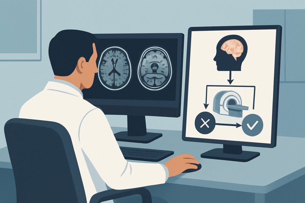
This article provides a deep dive into the American College of Radiology (ACR) Appropriateness Criteria for this specific clinical scenario: an adult with mild cognitive impairment not meeting criteria for dementia, undergoing initial imaging. For this presentation, the ACR rates MRI head without IV contrast as Usually appropriate.
Who Fits This Clinical Scenario for Mild Cognitive Impairment?
This guidance is tailored for a specific patient population. The key inclusion criterion is an adult patient with an objective diagnosis of mild cognitive impairment (MCI). This means there is evidence of cognitive decline, typically in memory, from a previous baseline, confirmed through standardized testing (e.g., MoCA, SLUMS, or formal neuropsychological evaluation). Crucially, this cognitive decline is not severe enough to interfere with the patient’s independence in everyday activities, which is the defining feature that distinguishes MCI from dementia.
This workflow does not apply to patients who already meet the full clinical criteria for a specific dementia syndrome. If your patient’s presentation includes significant functional impairment or specific features suggestive of a particular etiology, a different imaging pathway is indicated. Exclusions from this scenario include:
- Suspected Alzheimer Disease: Patients with memory deficits and functional decline consistent with Alzheimer disease fall under a different ACR variant.
- Suspected Frontotemporal Dementia (FTD): Patients presenting primarily with behavioral changes (e.g., disinhibition, apathy) or progressive aphasia require a workup focused on FTD.
- Suspected Dementia with Lewy Bodies (DLB): Patients with cognitive impairment accompanied by visual hallucinations, parkinsonism, or REM sleep behavior disorder have a distinct diagnostic pathway.
This article focuses exclusively on the initial structural imaging workup for a patient whose symptoms are confined to the MCI stage.
What Diagnoses Are You Working Up With Initial Imaging for MCI?
The primary goal of neuroimaging in mild cognitive impairment is twofold: first, to exclude structural and potentially reversible causes of cognitive symptoms, and second, to identify imaging biomarkers that may suggest an underlying neurodegenerative process and inform prognosis. The differential diagnosis is broad, but imaging helps narrow the possibilities.
Early Neurodegenerative Disease (Prodromal Alzheimer Disease): This is the most common underlying cause of amnestic MCI and the primary concern for most patients and families. While a definitive diagnosis cannot be made on imaging alone, structural MRI can reveal patterns highly suggestive of early Alzheimer’s pathology. The key finding is atrophy of the medial temporal lobes, particularly the hippocampi and entorhinal cortex, which often precedes global brain volume loss.
Vascular Cognitive Impairment: Cerebrovascular disease is a frequent contributor to cognitive decline, either alone or as a co-pathology with neurodegeneration. Imaging is essential for identifying evidence of vascular damage, which can include chronic small vessel ischemic changes (leukoaraiosis), multiple lacunar infarcts, cortical microinfarcts, or evidence of cerebral amyloid angiopathy such as microhemorrhages. Identifying a significant vascular burden can shift management towards aggressive risk factor modification.
Reversible or Treatable Structural Causes: Though less common, it is critical to rule out structural lesions that can mimic or cause cognitive impairment. These include conditions like normal pressure hydrocephalus (NPH), characterized by enlarged ventricles out of proportion to sulcal widening. Other important rule-outs include chronic subdural hematomas, which can present with insidious cognitive decline, and slow-growing brain tumors (e.g., meningiomas) or cysts in strategic locations.
Why Is MRI Head Without IV Contrast the Recommended Study for This Presentation?
For the initial evaluation of mild cognitive impairment, the ACR designates MRI head without IV contrast as Usually appropriate. This recommendation is based on its superior soft tissue resolution and ability to detect the key pathologies in the differential diagnosis without the need for ionizing radiation or intravenous contrast.
MRI provides exquisite detail of brain parenchyma, making it highly sensitive for detecting the subtle volume loss in the medial temporal lobes characteristic of early Alzheimer disease. It is also the most sensitive modality for identifying small vessel ischemic disease, lacunar infarcts, and microhemorrhages (on specific sequences like Susceptibility-Weighted Imaging, or SWI), which are central to diagnosing vascular cognitive impairment. Furthermore, MRI can clearly delineate structural abnormalities like tumors, hydrocephalus, or subdural fluid collections that could be responsible for the patient’s symptoms.
While other modalities are also rated Usually appropriate, they serve different, often more specific, roles:
- CT head without IV contrast: Also rated Usually appropriate, a non-contrast CT is a reasonable alternative if MRI is unavailable, contraindicated (e.g., incompatible pacemaker), or not tolerated by the patient. It is effective at ruling out major structural causes like hydrocephalus, large tumors, or chronic subdural hematomas. However, its sensitivity for early hippocampal atrophy and subtle ischemic changes is significantly lower than MRI. It involves a radiation dose of 1-10 mSv.
- Amyloid PET/CT and FDG-PET/CT brain: These molecular imaging studies are also Usually appropriate but are not typically first-line structural evaluations. Amyloid PET can detect the presence of amyloid plaques, a core feature of Alzheimer’s, while FDG-PET assesses patterns of cerebral metabolism. They are powerful tools for increasing diagnostic certainty, particularly in ambiguous cases, but they are generally considered after initial structural imaging has been performed.
A study rated lower, MRI head without and with IV contrast, is considered May be appropriate. The addition of gadolinium-based contrast is generally not necessary for the initial MCI workup unless there is a specific clinical suspicion for a condition that enhances, such as a primary or metastatic tumor, or an inflammatory/infectious process.
What’s Next After MRI Head Without IV Contrast? Downstream Workflow
The results of the initial non-contrast head MRI will guide the subsequent clinical pathway. The report is not just a “positive” or “negative” result but provides crucial information for counseling, prognosis, and further testing.
- If the MRI shows significant medial temporal lobe atrophy: This finding increases the likelihood that the patient’s MCI is due to an underlying Alzheimer’s disease process. The next steps involve counseling the patient and family about the risk of progression to dementia, initiating lifestyle and risk-factor modifications, and considering more specific biomarker testing (e.g., CSF analysis or Amyloid PET) if a more definitive etiological diagnosis is needed, especially if disease-modifying therapies are being considered.
- If the MRI shows a significant burden of vascular disease: When findings like extensive white matter hyperintensities or multiple lacunar infarcts are prominent, the focus shifts to managing vascular risk factors. This includes aggressive control of hypertension, diabetes, and hyperlipidemia, as well as smoking cessation. This can potentially slow cognitive decline.
- If the MRI is normal or shows only age-expected changes: A normal structural MRI is reassuring but does not rule out an underlying neurodegenerative process. It does, however, lower the probability of a major structural cause. In this case, the next step is typically clinical and cognitive monitoring over time. If the clinical picture remains uncertain or progresses, functional imaging like an FDG-PET scan may be considered to look for metabolic patterns suggestive of neurodegeneration.
- If the MRI reveals an unexpected structural cause (e.g., NPH, tumor): The workflow pivots entirely. The patient should be referred to the appropriate specialist, typically a neurosurgeon, for evaluation and potential treatment of the identified condition.
Pitfalls to Avoid (and When to Get Help)
Navigating the workup for mild cognitive impairment requires careful consideration to avoid common missteps. One pitfall is attributing all findings to “age-related changes” without considering the clinical context; what is normal for an 85-year-old may be abnormal for a 60-year-old. Another common error is ordering a contrast-enhanced MRI by default when it is not indicated, adding unnecessary cost and potential risk. Conversely, failing to order any imaging at all may miss a rare but treatable cause of cognitive decline. Finally, do not overlook the importance of reviewing the patient’s medication list, as polypharmacy and specific drug classes can cause or exacerbate cognitive symptoms. If the clinical picture and imaging findings are discordant or complex, escalation to a neurologist or a specialized memory disorders clinic is the appropriate next step.
Related ACR Topics and Tools
For a comprehensive overview of imaging recommendations across all dementia-related scenarios, from Alzheimer disease to frontotemporal dementia, please see our parent guide. For additional tools to help with ordering decisions and patient communication, the following resources are available.
- For breadth across all scenarios in Dementia, see our parent guide: Dementia: ACR Appropriateness Decoded.
- To explore adjacent clinical presentations, use the ACR Appropriateness Criteria Lookup.
- For details on imaging techniques, consult the Imaging Protocol Library.
- To discuss radiation exposure with patients, use the Radiation Dose Calculator.
Frequently Asked Questions
Why is a non-contrast MRI preferred over a non-contrast CT for initial MCI imaging?
While a non-contrast CT is also rated ‘Usually appropriate’ and is a good alternative if MRI is contraindicated, MRI is generally preferred due to its superior soft tissue contrast. This allows for much better visualization of subtle brain atrophy, particularly in the hippocampi, and is more sensitive for detecting small vessel ischemic changes and microhemorrhages, which are key findings in the MCI workup.
When should I add IV contrast to the MRI for a patient with mild cognitive impairment?
According to the ACR, a non-contrast MRI is sufficient for the initial evaluation. An MRI with IV contrast is rated ‘May be appropriate’ and should be reserved for cases where there is a specific clinical suspicion of a condition that typically enhances, such as a brain tumor, abscess, or certain inflammatory conditions like multiple sclerosis. Routine use of contrast is not recommended.
What if the MRI is completely normal in a patient with confirmed MCI?
A normal structural MRI is a common and somewhat reassuring finding, as it effectively rules out major structural causes like tumors, hydrocephalus, or significant vascular disease. However, it does not exclude an early-stage neurodegenerative process. The next steps typically involve continued clinical follow-up to monitor for progression. If diagnostic uncertainty persists, functional imaging like FDG-PET or biomarker testing (CSF analysis) may be considered.
Should I order an Amyloid PET scan as the first imaging study for MCI?
While Amyloid PET/CT is rated ‘Usually appropriate’, it is generally not the first-line imaging test. The initial step is typically structural imaging (MRI or CT) to rule out reversible causes and assess brain structure. An Amyloid PET scan is a powerful secondary tool used to confirm or exclude the presence of amyloid pathology, which can increase diagnostic certainty for Alzheimer disease, particularly in complex cases or when considering amyloid-targeting therapies.
Does a finding of ‘mild generalized atrophy’ on the MRI report change my management?
The interpretation of ‘mild generalized atrophy’ is highly dependent on the patient’s age. In an older adult (e.g., 80+), this can be a normal age-related finding. In a younger patient (e.g., early 60s), it may be more significant. The key is to look for focal atrophy, especially in the medial temporal lobes, which is more specific for a neurodegenerative process than generalized volume loss. The radiologist’s comparison to age-matched peers is critical.
Reviewed by Pouyan Golshani, MD, Interventional Radiologist — May 30, 2026