What Imaging Is Best for Chronic Vertigo with Unilateral Hearing Loss or Tinnitus?

A 58-year-old patient presents to your clinic with a six-month history of intermittent, spinning vertigo. The episodes are unpredictable, lasting for hours at a time. Over the last two months, he has also noticed a persistent, high-pitched ringing in his left ear and feels his hearing is not as sharp on that side. His neurologic exam is otherwise normal. You suspect a pathology of the vestibulocochlear nerve or inner ear structures. The critical question is which initial imaging study will most effectively evaluate the cerebellopontine angle and internal auditory canals to rule out a structural cause.
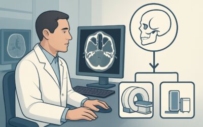
This clinical workflow article addresses this exact scenario: an adult with chronic, recurrent vertigo associated with unilateral auditory symptoms. According to the American College of Radiology (ACR) Appropriateness Criteria, the definitive initial study is MRI head and internal auditory canal without and with IV contrast, which is rated Usually appropriate.
Who Fits This Clinical Scenario?
This guidance is for adult patients presenting with a specific constellation of chronic and focal neurologic symptoms. To apply this workflow, the patient’s history should include:
- Chronic, Recurrent Vertigo: The dizziness is a true spinning sensation (vertigo) that has been occurring intermittently for weeks to months, rather than a single, acute, unremitting episode.
- Unilateral Auditory Symptoms: The key localizing feature is hearing loss or tinnitus affecting only one ear. This strongly suggests a lesion affecting the eighth cranial nerve (vestibulocochlear nerve) or inner ear on that side.
It is crucial to distinguish this presentation from similar, but distinct, clinical scenarios that require different workups:
- Brief, Positional Vertigo: If vertigo lasts seconds to minutes and is reliably triggered by specific head movements (e.g., rolling over in bed), the diagnosis is likely benign paroxysmal positional vertigo (BPPV). Imaging is typically not indicated for this presentation.
- Acute, Persistent Vertigo: A sudden, severe, and continuous vertigo lasting days points toward an acute process like vestibular neuritis or a posterior circulation stroke. The workup for this depends on the HINTS (Head-Impulse, Nystagmus, Test-of-Skew) examination findings.
- Vertigo with Other Brainstem Deficits: If the patient also has diplopia, dysarthria, facial numbness, or significant limb ataxia, the concern for a brainstem or cerebellar lesion is much higher. This represents a different clinical scenario with a broader imaging differential.
What Diagnoses Are You Working Up in This Scenario?
The combination of chronic vertigo and unilateral auditory symptoms directs the diagnostic search toward the internal auditory canal (IAC) and the cerebellopontine angle (CPA). The primary goal of imaging is to identify or exclude a structural lesion in this region.
Vestibular Schwannoma (Acoustic Neuroma): This is the classic, must-not-miss diagnosis. These benign tumors arise from the Schwann cells of the vestibular portion of the eighth cranial nerve. As they grow, they compress the cochlear portion of the nerve, causing tinnitus and sensorineural hearing loss, and affect the vestibular portion, causing vertigo and imbalance. They are the most common tumor of the CPA.
Ménière’s Disease: This is a clinical diagnosis characterized by the triad of episodic vertigo, fluctuating sensorineural hearing loss, and tinnitus. It is caused by endolymphatic hydrops (excess fluid in the inner ear). While imaging is typically normal in Ménière’s disease, it is essential in the initial workup to exclude a structural mimic like a vestibular schwannoma, which can present with identical symptoms.
Other Cerebellopontine Angle (CPA) Masses: Though less common than schwannomas, other tumors can occur in this location and produce the same symptoms. These include meningiomas, epidermoid cysts, and metastatic lesions. Each has characteristic features on MRI that can help differentiate them.
Vascular Loop Compression: In some cases, a loop of an artery (most commonly the anterior inferior cerebellar artery, or AICA) can compress the vestibulocochlear nerve in the IAC, leading to auditory and vestibular symptoms. MRI and MRA can identify these neurovascular conflicts.
Labyrinthitis: While typically an acute condition, chronic or recurrent inflammation of the labyrinth or vestibular nerve can occur. Post-inflammatory changes or active inflammation (which may show enhancement on MRI) can be a source of chronic symptoms.
Why Is MRI Head and Internal Auditory Canal Without and With IV Contrast the Recommended Study?
The ACR rates MRI head and internal auditory canal without and with IV contrast as Usually appropriate because it provides the highest diagnostic yield for the key pathologies in the differential diagnosis for this specific clinical scenario.
The superior soft-tissue contrast of MRI is unmatched for visualizing the cranial nerves, brainstem, and inner ear structures. The protocol must be tailored to this indication:
- High-Resolution Sequences: Thin-slice, high-resolution T2-weighted sequences (like FIESTA, CISS, or SPACE) are critical for delineating the nerves within the fluid-filled internal auditory canal and CPA cistern. This can reveal the nerve of origin for a mass or identify a compressing vascular loop.
- Role of IV Contrast: The administration of gadolinium-based contrast is essential. Vestibular schwannomas are typically avidly enhancing tumors. A small, intracanalicular schwannoma might be invisible on non-contrast sequences but become conspicuous on post-contrast T1-weighted images. This is why a non-contrast-only MRI is rated lower as only May be appropriate—it risks missing the primary diagnosis.
Why Other Studies Are Less Appropriate
CT Temporal Bone Without IV Contrast: While rated Usually appropriate, CT is considered a secondary option. It provides exquisite detail of the bony labyrinth and temporal bone, making it useful for evaluating osseous pathology, but it has poor intrinsic soft-tissue resolution. It cannot reliably visualize the nerves themselves or detect small, non-calcified tumors within the CPA or IAC. CT is an excellent alternative for patients with absolute contraindications to MRI (e.g., incompatible implanted devices). However, it involves ionizing radiation (ACR Relative Radiation Level ☢☢☢, 1-10 mSv), whereas MRI has none.
Routine MRI Brain Without and With IV Contrast: This study is rated Usually not appropriate. A standard brain MRI protocol uses thicker slices and may not include the specific high-resolution sequences focused on the IACs. A small intracanalicular schwannoma can easily be missed on a routine brain protocol. It is critical to specify “Internal Auditory Canals” or “CPA” in the order to ensure the correct protocol is performed.
What’s Next After MRI? Downstream Workflow
The MRI result dictates the subsequent clinical pathway. The findings can be broadly categorized into three outcomes:
Positive for a Structural Lesion (e.g., Vestibular Schwannoma): If the MRI identifies a schwannoma or other CPA mass, the next step is a referral. The patient should be referred to an Otolaryngologist (Neurotologist) and often a Neurosurgeon. Management options depend on tumor size, patient age, and hearing status, and may include observation with serial imaging, stereotactic radiosurgery, or surgical resection.
Negative Study: A normal MRI effectively rules out a structural cause for the patient’s symptoms. This significantly increases the likelihood of a peripheral vestibulopathy like Ménière’s disease or vestibular migraine. The downstream workflow then shifts away from structural diagnosis and toward functional testing and medical management. This typically involves:
- Formal audiogram to quantify and characterize the hearing loss.
- Vestibular function testing (e.g., videonystagmography [VNG]) to assess inner ear function.
- Referral to an Otolaryngologist or Neurologist specializing in vestibular disorders for management, which may include dietary changes, diuretics, or other medical therapies.
Indeterminate or Incidental Finding: Occasionally, the MRI may show non-specific findings, such as subtle nerve enhancement without a discrete mass, or an unrelated incidentaloma. In these cases, a discussion with the interpreting neuroradiologist is invaluable. Further steps might include short-term follow-up imaging or proceeding with the functional testing pathway as described for a negative study.
Pitfalls to Avoid (and When to Get Help)
Navigating this workup requires attention to detail to avoid common missteps:
- Ordering the Wrong Protocol: The most frequent error is ordering a “routine MRI brain.” This protocol lacks the thin-slice sequences necessary to evaluate the IACs and can miss the causative pathology. Always specify “IAC protocol” or “for evaluation of vestibular schwannoma.”
- Omitting IV Contrast: A non-contrast MRI is an incomplete study for this indication. The ACR rates it as only May be appropriate because it cannot reliably exclude a small, enhancing tumor. Unless there is a severe contraindication, contrast is necessary.
- Prematurely Diagnosing Ménière’s Disease: While the clinical picture may strongly suggest Ménière’s, it remains a diagnosis of exclusion. It is a clinical pitfall to anchor on this diagnosis without first performing imaging to rule out a structural mimic.
If a patient with this chronic presentation suddenly develops acute, severe neurologic symptoms—such as diplopia, dense facial palsy, or an inability to walk—this constitutes a neurologic emergency. This suggests a new, superimposed process like a brainstem stroke or hemorrhage into a tumor and requires immediate escalation and evaluation in an emergency setting.
Related ACR Topics and Tools
The ACR Appropriateness Criteria are a comprehensive resource for evidence-based imaging decisions. For breadth across all scenarios in Dizziness and Ataxia, see our parent guide: Dizziness and Ataxia: ACR Appropriateness Decoded.
For additional decision support, the following GigHz tools are available:
- ACR Appropriateness Criteria Lookup — for exploring adjacent clinical scenarios
- Imaging Protocol Library — for technical details on the recommended study
- Radiation Dose Calculator — for discussing cumulative dose with patients when considering CT
Frequently Asked Questions
Why is intravenous contrast so important for the MRI in this scenario?
Intravenous gadolinium-based contrast is critical because the primary diagnosis to exclude, a vestibular schwannoma, is a tumor that typically enhances avidly. A small tumor located entirely within the internal auditory canal might be isointense to the nerves on non-contrast images and could be missed. The bright enhancement after contrast administration makes it conspicuous.
Can I order a CT scan if my patient has a pacemaker or other contraindication to MRI?
Yes. In cases of an absolute contraindication to MRI, a CT of the temporal bones without contrast is rated ‘Usually appropriate’ by the ACR and is the best alternative. While it is less sensitive for soft-tissue tumors, it provides excellent detail of the bone and can sometimes show secondary signs of a mass, such as widening of the internal auditory canal.
What if my patient’s hearing loss and tinnitus are bilateral, not unilateral?
Bilateral hearing loss and tinnitus represent a different clinical scenario with a distinct differential diagnosis. The primary concern in that case would be Neurofibromatosis type 2 (NF2), an inherited disorder characterized by bilateral vestibular schwannomas. This presentation falls outside the scope of this specific ACR variant and requires a dedicated workup.
My patient’s symptoms seem classic for Ménière’s disease. Is imaging still necessary?
Yes. While Ménière’s disease is a clinical diagnosis, its symptoms (vertigo, tinnitus, hearing loss) perfectly mimic those of a vestibular schwannoma. The standard of care is to obtain a high-quality MRI at least once during the initial workup to definitively exclude a structural lesion before settling on a long-term diagnosis of Ménière’s.
What is the difference between this scenario and vertigo with other brainstem deficits?
This scenario is defined by symptoms localizing specifically to the eighth cranial nerve (hearing and balance). The presence of additional brainstem deficits—such as double vision (cranial nerves III, IV, VI), facial numbness (cranial nerve V), or difficulty swallowing (cranial nerves IX, X)—points to a larger lesion or a process affecting the brainstem itself, which is a different and more urgent clinical scenario.
Reviewed by Pouyan Golshani, MD, Interventional Radiologist — May 30, 2026