Which Imaging Study Is Best for Episodic Peripheral Vertigo? An ACR-Guided Workflow
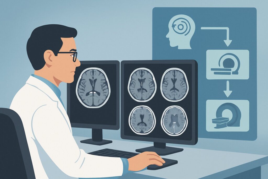
A 48-year-old patient presents to your clinic describing recurrent, spontaneous episodes of intense spinning vertigo lasting about an hour, often accompanied by a feeling of fullness and ringing in her left ear. The physical exam is unremarkable between episodes, but her history is highly suggestive of a peripheral vestibular disorder. The key clinical question is whether imaging is necessary and, if so, which study provides the most diagnostic value without unnecessary radiation or cost. This article provides a detailed workflow for this specific scenario, grounded in the American College of Radiology (ACR) Appropriateness Criteria, which rate an MRI of the head and internal auditory canal without and with IV contrast as Usually Appropriate.
Who Fits This Clinical Scenario for Episodic Peripheral Vertigo?
This guidance applies to patients presenting with episodic vertigo, where the symptoms are intermittent rather than constant. The clinical picture points toward a peripheral etiology, meaning the problem is likely located in the inner ear or the vestibulocochlear nerve. Associated symptoms that strengthen this classification include unilateral hearing loss (which may fluctuate), tinnitus (ringing in the ear), or aural fullness (a sensation of pressure).
It is crucial to distinguish this presentation from others that require a different diagnostic approach. This workflow is NOT for:
- Patients with persistent, unremitting vertigo: A constant sense of dizziness or imbalance, especially when accompanied by other neurological symptoms like double vision, slurred speech, or limb weakness, suggests a central cause (e.g., brainstem or cerebellum). This presentation aligns with the ACR scenario for Persistent vertigo with or without neurological symptoms (central vertigo) and often requires more urgent evaluation.
- Patients with purely conductive hearing loss: If audiometry confirms a conductive deficit and vertigo is absent, the pathology is likely in the external or middle ear (e.g., otosclerosis, ossicular chain disruption). This follows a separate diagnostic pathway.
- Patients with a clear history of trauma: Vertigo following a significant head injury necessitates a different workup, often starting with CT to evaluate for temporal bone fractures.
What Diagnoses Are You Working Up in This Scenario?
When ordering imaging for episodic peripheral vertigo, the goal is primarily to rule out serious structural causes and, in some cases, to confirm a suspected diagnosis. The differential diagnosis guides the choice of imaging modality.
Vestibular Schwannoma (Acoustic Neuroma): This is the most critical “can’t-miss” diagnosis. A benign tumor of the Schwann cells surrounding the vestibulocochlear nerve (Cranial Nerve VIII), it typically grows in the internal auditory canal (IAC). While classic presentation is progressive unilateral hearing loss, it can manifest with episodic vertigo. MRI with contrast is exceptionally sensitive for detecting these tumors, even when they are very small.
Ménière’s Disease: This is a clinical diagnosis characterized by the classic triad of episodic vertigo, fluctuating sensorineural hearing loss, and tinnitus/aural fullness. Imaging is primarily performed to exclude a vestibular schwannoma or other retrocochlear pathology that can mimic its symptoms. Advanced MRI techniques can sometimes visualize endolymphatic hydrops (excess fluid in the inner ear), which is the presumed pathophysiology of Ménière’s, but its primary role remains exclusionary.
Vestibular Neuritis or Labyrinthitis: While typically presenting as a single, severe, and prolonged episode of vertigo, these inflammatory conditions can sometimes cause recurrent or stuttering symptoms. Imaging helps rule out a structural lesion when the clinical course is atypical. On occasion, post-contrast MRI may show enhancement of the vestibular nerve or labyrinth, supporting the inflammatory diagnosis.
Vascular Compression Syndrome: Less commonly, a blood vessel (most often the anterior inferior cerebellar artery, or AICA) can form a loop that compresses the vestibulocochlear nerve, leading to symptoms of vertigo and tinnitus. High-resolution MRI can visualize this neurovascular conflict, though the clinical significance of such a finding is often debated, as vascular loops can be asymptomatic.
Why Is MRI of the Head and IAC the Recommended Study for Episodic Vertigo?
The ACR designates MRI head and internal auditory canal without and with IV contrast as Usually Appropriate for this scenario because of its unparalleled soft-tissue resolution and ability to visualize the key anatomical structures without using ionizing radiation.
The rationale is straightforward: MRI provides a detailed view of the brainstem, cerebellum, cranial nerves, and inner ear structures. Thin-slice, high-resolution sequences through the internal auditory canals are essential for identifying small intracanalicular tumors like vestibular schwannomas. The addition of intravenous gadolinium-based contrast is critical, as these tumors and inflammatory processes typically enhance, making them conspicuous against the surrounding tissues.
Let’s compare this to other modalities rated for this scenario:
- CT temporal bone without IV contrast: While also rated Usually Appropriate, its strengths lie elsewhere. CT is superior for evaluating osseous (bony) anatomy and is the study of choice for suspected temporal bone trauma, cholesteatoma, or semicircular canal dehiscence. However, it provides poor visualization of the vestibulocochlear nerve and other soft tissues within the IAC, making it insensitive for the primary differential of a small schwannoma. It also involves a moderate radiation dose (ACR RRL®: ☢☢☢ 1-10 mSv).
- CT head without IV contrast: This study is rated Usually Not Appropriate. A standard non-contrast head CT lacks the resolution and anatomical detail to evaluate the posterior fossa and IACs adequately. It is very likely to miss the key pathologies in the differential and should not be ordered for this indication.
The choice of MRI leverages its diagnostic superiority for the most consequential pathologies while avoiding radiation exposure (ACR RRL®: O 0 mSv). When ordering, be specific: request an “MRI of the brain and IACs with and without contrast for vertigo” to ensure the protocol includes the necessary high-resolution sequences (e.g., FIESTA, CISS, or T2 space) and thin-slice post-contrast T1-weighted images through the posterior fossa.
What’s Next After the MRI? Navigating the Downstream Workflow
The imaging results will guide your next steps, which often involve either specialist referral or a shift toward medical management.
If the study is POSITIVE for a structural lesion (e.g., vestibular schwannoma): The next step is a referral to an Otolaryngologist (ENT) or Neurosurgeon. They will discuss management options with the patient, which may include serial observation with repeat imaging, stereotactic radiosurgery, or surgical resection. The size of the tumor and the patient’s hearing status are key factors in this decision.
If the study is NEGATIVE (the most common outcome): A negative MRI is highly reassuring. It effectively rules out a tumor or other structural cause for the patient’s symptoms. The diagnosis becomes clinical, with Ménière’s disease or vestibular migraine being the most likely etiologies. The workflow then shifts to medical management, which can include dietary modifications (e.g., low-salt diet for Ménière’s), diuretics, vestibular suppressants for acute attacks, and potentially vestibular rehabilitation therapy. The negative scan provides the confidence to proceed with these treatments.
If the study is INDETERMINATE or shows an incidental finding: Findings like a vascular loop compressing the vestibulocochlear nerve can be challenging, as they are also seen in asymptomatic individuals. The next step is careful clinical correlation. If symptoms are severe and refractory to medical management, a neurosurgical consultation may be warranted to discuss options like microvascular decompression, but this is uncommon. Other incidental findings should be managed based on their specific characteristics.
Common Pitfalls to Avoid in Imaging for Peripheral Vertigo
Navigating the workup for episodic vertigo requires careful attention to detail to avoid common missteps that can delay diagnosis or lead to unnecessary testing.
- Pitfall 1: Ordering a routine brain MRI without specifying IACs. A standard brain MRI protocol may use slices that are too thick to adequately visualize the structures within the internal auditory canals, potentially missing a small intracanalicular schwannoma. Always explicitly request “IAC protocol” or “thin-slice posterior fossa” sequences.
- Pitfall 2: Omitting contrast when a tumor is suspected. While a non-contrast MRI is also rated Usually Appropriate, gadolinium is essential for highlighting small enhancing tumors or inflammatory changes. If a vestibular schwannoma is a primary concern, ordering the study “without and with contrast” is the standard of care.
- Pitfall 3: Misclassifying central vertigo as peripheral. The most critical pitfall is failing to recognize “red flag” symptoms of a central process. If a patient with vertigo also reports diplopia, dysarthria, dysphagia, or focal motor/sensory deficits (the “5 D’s”), this suggests a posterior circulation stroke or other brainstem pathology. This is a medical emergency requiring immediate escalation and a different imaging pathway, often starting with a non-contrast head CT and CTA or an MRI/MRA.
Related ACR Topics and Tools
For a comprehensive overview of all clinical variants related to dizziness and hearing loss, or to explore the technical details of the recommended imaging studies, the following resources are available:
- For breadth across all scenarios in Hearing Loss and/or Vertigo, see our parent guide: Hearing Loss and/or Vertigo: ACR Appropriateness Decoded.
- To explore other clinical scenarios and their corresponding ACR ratings, use the Imaging Appropriateness Selector tool.
- For detailed technical specifications of imaging studies, consult the Imaging Protocol Library.
- To discuss radiation exposure with patients when considering CT, the Radiation Dose Calculator can help frame the conversation.
Frequently Asked Questions
Is an MRI always necessary for a first-time presentation of episodic vertigo?
Not always. If the clinical presentation is classic for benign paroxysmal positional vertigo (BPPV)—brief, position-triggered episodes without hearing loss—and resolves with canalith repositioning maneuvers (e.g., Epley), imaging is often not required. Imaging is indicated when the diagnosis is uncertain, symptoms are atypical, or there is a suspicion of a structural lesion like a vestibular schwannoma (e.g., associated unilateral hearing loss or tinnitus).
Why is MRI with contrast preferred over a non-contrast MRI if both are rated ‘Usually Appropriate’?
While a non-contrast MRI with high-resolution T2 sequences (like FIESTA or CISS) is very good at detecting schwannomas, intravenous contrast provides confirmatory evidence and increases conspicuity, especially for very small tumors or inflammatory conditions. The addition of contrast makes the study more definitive, reducing the chance of a false-negative or equivocal result for the key pathologies in the differential.
Can I order a CT scan if my patient has a contraindication to MRI (e.g., an incompatible pacemaker)?
Yes. If MRI is contraindicated, a CT of the temporal bones is a reasonable alternative, though it is less sensitive for soft-tissue pathology like small schwannomas. The ACR rates ‘CT temporal bone without IV contrast’ as ‘Usually Appropriate.’ It will provide excellent detail of the bony labyrinth and can identify larger tumors that erode bone, but it is not the preferred first-line test if MRI is an option.
What if the MRI is negative but the patient’s symptoms persist or worsen?
A negative MRI is a crucial piece of information that rules out a structural cause. If symptoms persist, the focus should be on confirming and managing a clinical diagnosis like Ménière’s disease or vestibular migraine. This may involve referral to an Otolaryngologist or Neurologist specializing in vestibular disorders for further testing, such as videonystagmography (VNG) or electrocochleography (ECoG), and for optimization of medical therapy.
Does a finding of a vascular loop on MRI automatically explain the patient’s vertigo?
Not necessarily. Neurovascular compression of the vestibulocochlear nerve is a controversial cause of vertigo because vascular loops are a common incidental finding in asymptomatic individuals. The finding must be carefully correlated with the patient’s specific symptoms. It is generally considered a diagnosis of exclusion after more common causes have been ruled out, and surgical intervention is reserved for severe, refractory cases.
Reviewed by Pouyan Golshani, MD, Interventional Radiologist — May 26, 2026