How Should You Assess Osteoporosis in Patients with Severe Spinal Degeneration?
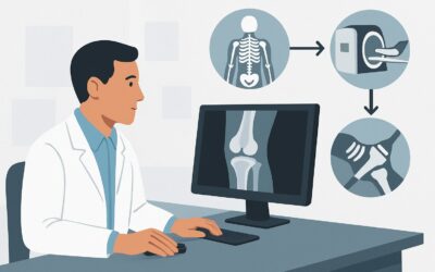
A 72-year-old male with a long history of chronic low back pain and known severe multilevel degenerative disc disease presents for a wellness visit. You note he has lost two inches in height and has developed a mild thoracic kyphosis. You suspect osteoporosis, but you know that the extensive osteophytes and sclerosis visible on his old lumbar spine radiographs will almost certainly create a falsely elevated bone mineral density (BMD) reading on a standard dual-energy X-ray absorptiometry (DXA) scan. This article addresses the specific imaging workflow for this common and challenging clinical scenario. For patients with conditions that can spuriously elevate spinal BMD, the American College of Radiology (ACR) rates DXA of the distal forearm as Usually Appropriate.
Who Fits This Clinical Scenario?
This guidance applies to a specific subset of patients: males and females aged 50 or older in whom you suspect osteoporosis, but who also have a known condition that compromises the reliability of standard BMD measurement sites. The key feature is the presence of a confounder that can falsely elevate density readings.
Inclusion Criteria:
- Age ≥ 50 years.
- Clinical suspicion for osteoporosis (e.g., due to age, risk factors, height loss, history of fragility fracture).
- Presence of advanced degenerative changes of the spine (e.g., extensive osteophytes, facet arthropathy, disc space narrowing with sclerosis), significant scoliosis, vertebral compression fractures, or extensive aortic calcification overlying the vertebrae.
Exclusion Criteria (These route to different guidelines):
- Routine Screening: This guidance is not for initial, routine osteoporosis screening in patients without the spinal confounders mentioned above. Those patients are covered in the standard screening scenario.
- Follow-up Imaging: This workflow is for initial diagnosis. Patients with established low bone density undergoing surveillance to monitor treatment response follow a different pathway.
- Younger Patients: Premenopausal females or males under 50 with risk factors for low bone mass represent a distinct clinical question and are addressed in a separate ACR variant.
What Diagnoses Are You Working Up in This Scenario?
The primary goal is to obtain an accurate, unconfounded measurement of bone mineral density to diagnose or rule out osteoporosis. The imaging choice is driven by the need to circumvent anatomical noise that would otherwise mask the true bone health status.
Osteoporosis: This is the principal diagnosis under consideration. It is a systemic skeletal disease characterized by low bone mass and microarchitectural deterioration of bone tissue, leading to an increase in fracture risk. In this scenario, the challenge is that severe degenerative joint disease (DJD) in the spine can make a patient with true osteoporosis appear to have normal or even high bone density on a standard lumbar spine DXA.
Osteopenia (Low Bone Mass): This is a precursor to osteoporosis, defined by a T-score between -1.0 and -2.5. Identifying osteopenia is clinically important as it allows for lifestyle interventions and closer monitoring to prevent progression to osteoporosis. An accurate BMD measurement is essential to correctly categorize the patient.
Secondary Causes of Low Bone Mass: While DXA measures density, it does not reveal the cause. An unexpectedly low and accurate BMD reading should prompt a workup for secondary causes, such as hyperparathyroidism, vitamin D deficiency, multiple myeloma, or glucocorticoid-induced osteoporosis. The imaging test is the first step in uncovering a potential underlying systemic condition.
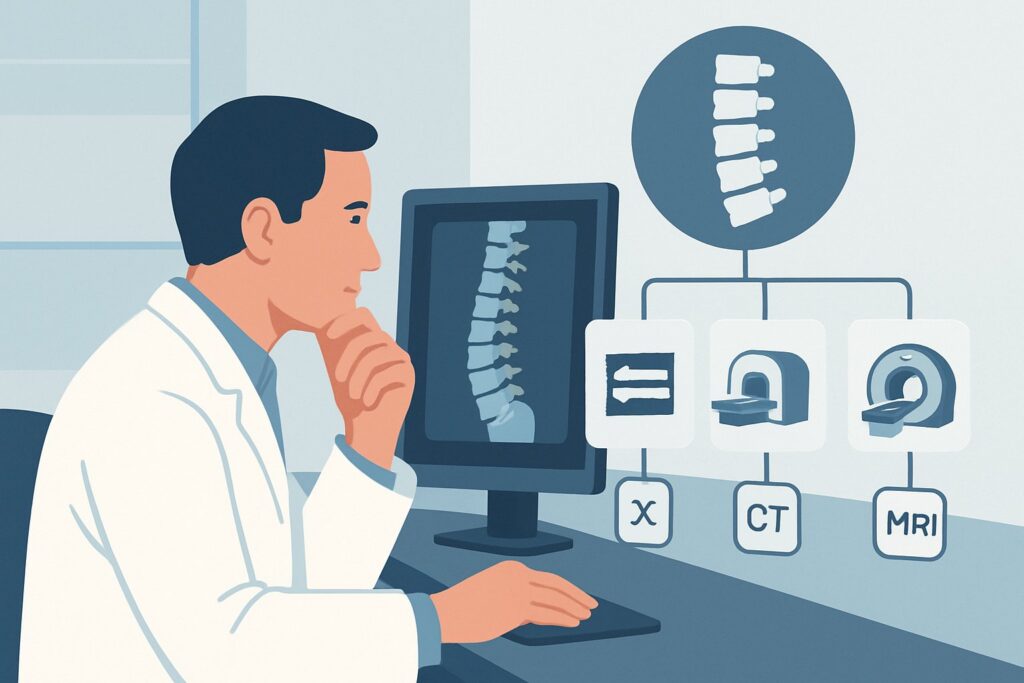
Why Is DXA of the Distal Forearm the Recommended Study for This Presentation?
In this specific clinical context, the standard approach of measuring the lumbar spine and hip is fraught with potential for error. The ACR Appropriateness Criteria recommend a strategy that mitigates these known confounders.
The core problem is that a DXA scan measures the total mineral content in the path of the X-ray beam. In a patient with severe spinal arthritis, that beam passes not only through the vertebral bone but also through osteophytes (bone spurs), sclerotic endplates, and potentially calcified aortic walls. These structures are dense and will be added to the measurement, artificially inflating the BMD value and masking any underlying bone loss. A patient could have a dangerously low T-score of -2.8 (osteoporosis) that is reported as a reassuring -0.5 (normal) due to these degenerative artifacts.
For this reason, DXA of the distal forearm is rated as Usually appropriate. The measurement is typically taken at the “1/3 radius” site, which is composed primarily of cortical bone. This location is non-weight-bearing and is not susceptible to the degenerative changes that affect the spine. It therefore serves as a more reliable “window” into the patient’s systemic skeletal health when the spine cannot be accurately assessed.
It is important to note that DXA of the lumbar spine and hip(s) is also rated Usually appropriate. In practice, a complete DXA study including all three sites (spine, hip, forearm) is often performed. However, the interpreting radiologist and ordering clinician must recognize that the lumbar spine value may be uninterpretable and should not be used for diagnosis if significant degenerative changes are present. The hip and/or forearm measurements then become the primary basis for the diagnosis.
Alternative Studies
- Quantitative Computed Tomography (QCT) of the lumbar spine and hip(s) is also Usually appropriate. QCT has the advantage of measuring true volumetric density and can isolate the trabecular bone of the vertebral body, avoiding osteophytes. However, it comes with a significantly higher radiation dose (☢☢☢ 1-10 mSv) compared to DXA (☢ <0.1 mSv) and is less widely available and standardized.
- Quantitative Ultrasound (QUS) of the calcaneus is Usually not appropriate for this indication. While it involves no radiation, QUS is considered a screening tool for fracture risk and lacks the precision and diagnostic criteria (T-scores) of DXA needed to make a formal diagnosis of osteoporosis or guide pharmaceutical therapy.
Once you’ve decided on the appropriate DXA scan, our protocol guide covers the technical details, interpretation, and reporting principles. For more information, see our in-depth guide: DXA Bone Mineral Density Scan.
What’s Next After a Distal Forearm DXA? Downstream Workflow
The results of an accurate BMD assessment will directly guide your next clinical steps. The workflow depends on whether the T-score indicates normal bone density, osteopenia, or osteoporosis.
If the result is positive for osteoporosis (T-score ≤ -2.5):
This confirms the diagnosis. The next step is to calculate the patient’s 10-year fracture risk using a tool like the Fracture Risk Assessment Tool (FRAX). This calculation, combined with the T-score, will determine the need for pharmacologic therapy (e.g., bisphosphonates, denosumab). A laboratory workup to exclude secondary causes of osteoporosis (including serum calcium, vitamin D, and parathyroid hormone levels) is also warranted before initiating treatment.
If the result indicates osteopenia (T-score between -1.0 and -2.5):
Management focuses on risk factor modification. This includes counseling on adequate calcium and vitamin D intake, weight-bearing exercise, smoking cessation, and limiting alcohol consumption. Pharmacologic therapy may be considered if the FRAX score indicates a high 10-year risk of major osteoporotic or hip fracture, even without a T-score in the osteoporotic range.
If the result is normal (T-score ≥ -1.0):
This finding provides reassurance that significant systemic bone loss is not present, despite the clinical suspicion. If the patient has symptoms like back pain, the workup should pivot to focus on the known degenerative disease as the likely cause. Repeat BMD testing can be performed at a longer interval (e.g., every 3-5 years or more), depending on the patient’s overall risk profile.
Pitfalls to Avoid (and When to Get Help)
Navigating BMD assessment in the presence of spinal artifacts requires careful attention to detail to avoid misdiagnosis.
- Pitfall 1: Accepting a “normal” spine DXA at face value. Never rely solely on a normal or even high lumbar spine T-score in a patient with known severe degenerative changes or scoliosis. Always review the hip and forearm data.
- Pitfall 2: Not providing clinical context on the order. Always specify “severe degenerative changes” or “scoliosis” as the indication. This alerts the DXA technologist and interpreting radiologist to the potential for artifact and the necessity of obtaining forearm measurements.
- Pitfall 3: Using the wrong forearm. The non-dominant forearm should be scanned. If there is a history of fracture or hardware in that arm, the dominant forearm should be used instead. This must be communicated on the order.
If results are discordant (e.g., normal forearm but very low hip BMD) or if clinical suspicion for fragility fractures remains high despite normal DXA results, consider an endocrinology or rheumatology consultation for further evaluation.
Related ACR Topics and Tools
This article focuses on a single, complex scenario. For a comprehensive overview of all clinical variants in this topic, including routine screening and follow-up, please see our parent guide. You can also use the tools below to explore adjacent scenarios, review imaging techniques, and discuss radiation dose with your patients.
- Parent Topic Hub: For breadth across all scenarios in Osteoporosis and Bone Mineral Density, see our parent guide: Osteoporosis and Bone Mineral Density: ACR Appropriateness Decoded.
- ACR Criteria Lookup: ACR Appropriateness Criteria Lookup
- Protocol Library: Imaging Protocol Library
- Dose Calculator: Radiation Dose Calculator
Frequently Asked Questions
Why can’t I just use the hip measurement from a standard DXA scan and ignore the spine?
You can, and often should, use the hip measurement (specifically the femoral neck or total hip). The hip is less prone to degenerative artifacts than the spine. However, some patients may have discordant results where one site shows osteoporosis and another does not. The World Health Organization diagnostic criteria allow for a diagnosis of osteoporosis based on the lowest T-score from the lumbar spine, femoral neck, total hip, or 1/3 radius. Therefore, obtaining an accurate reading from an unconfounded site like the forearm provides the most complete and reliable diagnostic picture.
Is the forearm T-score interpreted the same way as a spine or hip T-score for diagnosis?
Yes. For diagnostic purposes, a T-score of -2.5 or lower at the 1/3 radius site is sufficient to diagnose osteoporosis, according to the International Society for Clinical Densitometry (ISCD). However, it’s important to note that most fracture risk prediction tools, like FRAX, are primarily validated using femoral neck BMD, and the forearm is not the preferred site for monitoring the effects of therapy.
What if my patient has had a wrist fracture or has surgical hardware in their non-dominant forearm?
If the non-dominant forearm cannot be accurately measured due to prior fracture, deformity, or hardware, the technologist should scan the dominant forearm instead. If both forearms are unevaluable, you must rely on the hip measurement. If the hip is also compromised (e.g., bilateral hip replacements), a consultation for consideration of Quantitative CT (QCT) may be necessary.
Is Quantitative CT (QCT) a better option than DXA in this scenario?
QCT can be an excellent problem-solving tool because it measures true volumetric bone density and can electronically isolate the trabecular bone from cortical bone and overlying osteophytes, yielding a very accurate measurement. However, the ACR rates it as ‘Usually Appropriate,’ equivalent to DXA, not superior. This is because QCT involves a significantly higher radiation dose, is more expensive, and is less widely available than DXA. DXA of the forearm is typically the more practical and accessible first-line choice.
Does this recommendation to use the forearm apply to patients under 50 with spinal deformities?
This specific ACR variant is for patients 50 and older. For premenopausal women and men under 50, the diagnostic criteria for osteoporosis are different (requiring a Z-score ≤ -2.0 plus a secondary cause or fracture history). While the principle of avoiding spinal artifacts still applies, the clinical context and diagnostic thresholds are distinct and are covered under a separate ACR appropriateness scenario.
Reviewed by Pouyan Golshani, MD, Interventional Radiologist — May 30, 2026