How Should You Image a Palpable Lump After Mastectomy and Breast Reconstruction?
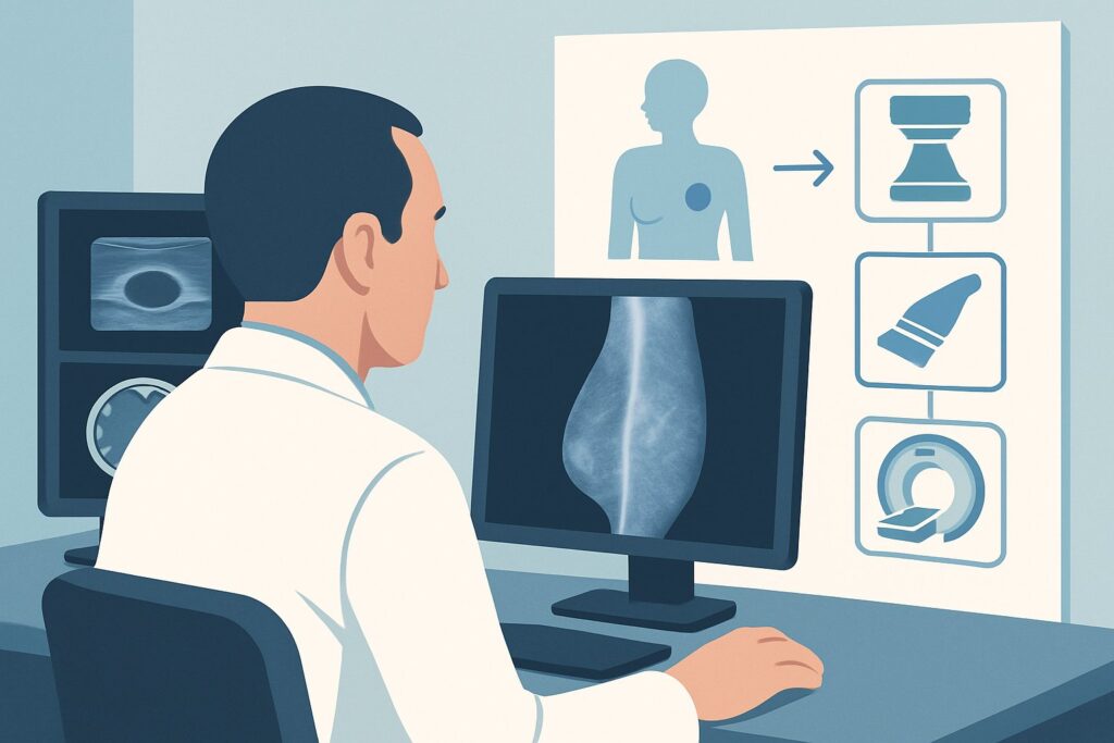
A 58-year-old female, three years post-mastectomy with a TRAM flap reconstruction for breast cancer, presents to your clinic for a follow-up. During her self-exam, she noticed a new, firm, non-tender lump near the medial aspect of her reconstructed breast. She is anxious about recurrence. You now face the critical decision of selecting the most appropriate initial imaging study to evaluate this new finding. This article provides a detailed clinical workflow for this specific scenario, guiding you through the differential diagnosis, imaging rationale, and downstream management steps. For this presentation, the American College of Radiology (ACR) finds that a breast ultrasound is Usually Appropriate as the initial imaging test.
Who Fits This Clinical Scenario?
This guidance applies specifically to a female patient who has undergone a mastectomy and subsequent breast reconstruction—either autologous (e.g., TRAM, DIEP flap) or nonautologous (implant-based)—and now presents with a new, focal symptom on the reconstructed side. The qualifying symptoms are a palpable lump or clinically significant, localized pain that prompts a diagnostic workup.
It is crucial to distinguish this symptomatic presentation from routine surveillance. This workflow does not apply to:
- Asymptomatic Screening: Patients with a history of mastectomy and reconstruction who are undergoing routine, scheduled cancer screening without any new symptoms. These scenarios, such as screening after autologous reconstruction or screening after implant reconstruction, follow different imaging pathways.
- Contralateral Breast Evaluation: This guidance is for the reconstructed side only. A new finding in the contralateral, intact breast would follow standard diagnostic mammography and ultrasound protocols.
- Systemic Symptoms: Patients presenting with diffuse pain, skin changes across the entire breast, or systemic signs concerning for metastatic disease require a different workup, which may involve systemic imaging.
The focus here is the initial diagnostic imaging step for a new, focal problem in a reconstructed breast.
What Diagnoses Are You Working Up in This Scenario?
A new lump in a reconstructed breast raises several diagnostic possibilities, ranging from benign post-surgical changes to the most feared outcome, cancer recurrence. The goal of imaging is to differentiate between these etiologies.
Cancer Recurrence: This is the primary concern. A locoregional recurrence can present as a chest wall or subcutaneous nodule. While less common than benign findings, it is the most consequential diagnosis to exclude. Recurrence can occur in the skin, subcutaneous fat, pectoralis muscle, or axillary lymph nodes, all of which are accessible to initial evaluation with ultrasound.
Fat Necrosis: This is a very common benign finding, particularly after autologous flap reconstruction. Ischemia in the transferred adipose tissue leads to necrosis, which can manifest as firm, irregular palpable masses that are often clinically indistinguishable from a malignancy. On imaging, it can have a variable appearance, from oil cysts to spiculated masses, making it a frequent mimic of cancer.
Post-Surgical Fluid Collections (Seroma/Hematoma): Fluid can accumulate at the surgical site, forming a seroma (clear fluid) or hematoma (blood). While most common in the early postoperative period, they can persist or develop later. These typically present as well-defined, palpable fluid-filled sacs.
Suture Granuloma or Scar Tissue: The body’s inflammatory reaction to suture material can create a firm nodule known as a suture granuloma. Similarly, dense scar tissue (fibrosis) at the surgical site can present as a palpable lump. Both are benign but require differentiation from a more sinister pathology.
Implant-Related Complications: In patients with nonautologous reconstruction, the lump or pain could be related to the implant itself. This includes silicone gel bleed, intracapsular or extracapsular implant rupture, or pronounced capsular contracture, which can cause focal firmness and pain.
Why Is Breast Ultrasound the Recommended Initial Study?
For a patient with a palpable lump or focal pain in a reconstructed breast, the ACR designates breast ultrasound (US) as Usually Appropriate. This recommendation is based on its high diagnostic utility, safety profile, and accessibility for this specific clinical problem.
Ultrasound excels at characterizing superficial tissues, making it ideal for evaluating the skin, subcutaneous fat, and chest wall muscle where recurrences often occur. It can reliably differentiate solid masses from simple or complex fluid collections like seromas or abscesses. For solid nodules, US can assess features concerning for malignancy, such as irregular margins, posterior acoustic shadowing, and internal vascularity. It is also the primary modality for evaluating implant integrity, capable of detecting signs of rupture. Crucially, ultrasound serves as the ideal tool for real-time guidance if a biopsy of a suspicious finding is required.
Alternative imaging modalities are rated lower for this initial workup for specific reasons:
- Diagnostic Mammography and Digital Breast Tomosynthesis (DBT): Rated as May be appropriate. While useful for evaluating breast tissue, their utility in a reconstructed breast is often limited. Post-surgical scarring, architectural distortion, and the presence of an implant can obscure underlying tissues and make interpretation difficult. Mammography is less effective for evaluating deep chest wall lesions and cannot assess implant integrity as well as US or MRI. It may serve as a useful adjunct to ultrasound but is not the preferred first step for a focal problem. These studies involve a low radiation dose (adult_rrl=☢☢ 0.1-1mSv).
- Breast MRI without and with IV Contrast: Rated as Usually not appropriate for the initial evaluation. While MRI has very high sensitivity for detecting cancer recurrence, it is not the first-line tool for a palpable lump. Post-surgical changes, such as inflammation, granulation tissue, and fat necrosis, can all enhance with contrast, leading to a high rate of false-positive findings and potentially unnecessary biopsies. Its role is primarily in problem-solving for equivocal findings on US/mammography or for high-risk screening, not as the initial diagnostic test. MRI uses no ionizing radiation (adult_rrl=O 0 mSv).
In summary, breast ultrasound provides a targeted, non-invasive (adult_rrl=O 0 mSv), and highly effective initial assessment of a focal abnormality in a reconstructed breast, directly answering the clinical question while avoiding the limitations and potential false positives of other modalities.
What’s Next After Breast Ultrasound? Downstream Workflow
The results of the breast ultrasound will guide the subsequent management steps in a clear decision tree. The key is to correlate the imaging findings with the palpable area of concern.
If the US identifies a suspicious solid mass (e.g., BI-RADS 4 or 5): The definitive next step is an ultrasound-guided core needle biopsy. This allows for histopathologic diagnosis to confirm or exclude malignancy. The patient should be referred to a breast surgeon or breast radiologist for this procedure.
If the US identifies a clearly benign finding (e.g., BI-RADS 2): When the ultrasound reveals features classic for a simple cyst, seroma, or fat necrosis that corresponds to the palpable lump, the workup is typically complete. If a seroma is large and symptomatic, ultrasound-guided aspiration can be both diagnostic and therapeutic. Otherwise, clinical reassurance and routine follow-up are appropriate.
If the US is negative or indeterminate (e.g., BI-RADS 0 or 3): This is a critical juncture. If the ultrasound shows no abnormality in the area of the palpable lump, but high clinical suspicion persists (e.g., the lump is hard, fixed, or growing), the workup should not stop. The next step is to consider one of the “May be appropriate” modalities, such as a diagnostic mammogram/DBT, to look for calcifications or other findings not visible on US. If both US and mammography are negative but clinical concern remains high, a consultation with a breast surgeon is warranted, and a contrast-enhanced breast MRI may be considered as a problem-solving tool.
Pitfalls to Avoid (and When to Get Help)
Navigating the workup of a lump in a reconstructed breast requires careful clinical-radiological correlation. Here are a few common pitfalls to avoid:
- Assuming a lump is scar tissue: Never dismiss a new, persistent palpable lump as “just scar tissue” without an imaging evaluation. While often benign, this assumption can lead to a delayed diagnosis of recurrence.
- Inadequate communication with the radiologist: When ordering the ultrasound, clearly specify the exact location of the palpable abnormality. A skin marker placed directly over the lump before imaging is best practice to ensure the radiologist evaluates the correct area.
- Stopping the workup prematurely: A negative or benign-appearing ultrasound in the face of a highly suspicious clinical exam is a red flag. The palpable finding trumps a negative imaging report.
If initial imaging is non-diagnostic and the palpable finding persists or evolves, escalate care by referring the patient to a breast surgeon for clinical evaluation and potential consideration of further imaging like MRI or an excisional biopsy.
Related ACR Topics and Tools
This article covers one specific clinical scenario. For a comprehensive overview of all related presentations and for additional resources to aid in your clinical decision-making, please refer to the following:
- For breadth across all scenarios in Imaging after Mastectomy and Breast Reconstruction, see our parent guide: Imaging after Mastectomy and Breast Reconstruction: ACR Appropriateness Decoded.
- To explore other clinical situations, use the ACR Appropriateness Criteria Lookup tool.
- For details on imaging techniques, consult the Imaging Protocol Library.
- To discuss radiation exposure with patients, the Radiation Dose Calculator can help quantify and contextualize the dose from various studies.
Frequently Asked Questions
Does the initial imaging workup change if the reconstruction is an implant versus an autologous flap?
No, the recommended initial imaging study for a new palpable lump is breast ultrasound regardless of the reconstruction type. While the differential diagnosis may shift—with implant-related issues being a concern only in nonautologous reconstructions—ultrasound remains the best first step to characterize the lump and assess surrounding tissues in both settings.
If the ultrasound is negative, can I confidently rule out cancer?
Not necessarily. While ultrasound is highly sensitive, a negative or equivocal result in the setting of a persistent and clinically suspicious palpable lump is not sufficient to rule out malignancy. This is a key scenario where clinical judgment is paramount. The next step should be a discussion with a breast specialist and consideration of further imaging, such as diagnostic mammography or breast MRI, or even a biopsy of the palpable finding.
Why is breast MRI ‘Usually not appropriate’ as the first test for a new lump?
Breast MRI is extremely sensitive but can be non-specific in the postoperative setting. Benign post-surgical changes like inflammation, scar tissue, and fat necrosis can all enhance with contrast, mimicking malignancy. Using it as a first-line test can lead to a high number of false positives, resulting in unnecessary anxiety and additional biopsies. Its primary role is as a problem-solving tool when ultrasound and mammography are inconclusive.
Does this guidance apply to pain without a distinct palpable lump?
Yes, the ACR scenario explicitly includes ‘clinically significant pain.’ Ultrasound is still the appropriate initial imaging test to investigate focal pain. It can identify underlying causes that may not be palpable, such as a small fluid collection, an inflamed suture granuloma, or early implant complications. The targeted nature of ultrasound makes it ideal for evaluating a specific area of tenderness.
Is mammography ever the first choice in this scenario?
Rarely. For a focal problem like a palpable lump, ultrasound is superior because it provides a targeted evaluation and can easily distinguish solid from cystic structures. Mammography is rated ‘May be appropriate’ and is more of a supplemental tool in this context. It might be considered if the ultrasound is negative but suspicion remains, particularly to look for suspicious microcalcifications, though these are less common in recurrences within soft tissue flaps.
Reviewed by Pouyan Golshani, MD, Interventional Radiologist — May 29, 2026