How Should You Image a Suspected Pelvic Stress Fracture During Pregnancy After Negative X-rays?
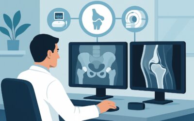
A patient in her third trimester presents to your clinic with a four-week history of worsening left-sided groin pain, exacerbated by walking. She denies any specific trauma. The pain is now significant enough to limit her daily activities. You obtain an AP radiograph of the pelvis, which shows no evidence of fracture, dislocation, or other acute osseous abnormality. Your clinical suspicion for a stress or insufficiency fracture remains high given her presentation and physiological state. The critical question is how to proceed with imaging in a way that is both diagnostically effective and safe for the fetus. This article details the American College of Radiology (ACR) Appropriateness Criteria workflow for this exact scenario. For a pregnant patient with suspected pelvic stress fracture and negative radiographs, MRI of the area of interest without IV contrast is rated Usually Appropriate.
Who Fits This Clinical Scenario for Pelvic Stress Fracture Imaging?
This clinical workflow is specifically for an adult pregnant patient with a clinical suspicion for a stress fracture involving the pelvis, hip, or sacrum, where initial radiographs have already been performed and were either negative or indeterminate. The key elements defining this scenario are the patient’s gravid state, the anatomical location of suspected injury, and the inconclusive initial imaging.
This guidance is not intended for:
- Non-pregnant adults: While the recommended imaging study may be the same, the rationale regarding radiation safety is less absolute. The decision-making process for non-pregnant adults with negative radiographs is covered in a separate ACR variant.
- Patients with a clear traumatic fracture: If there is a history of high-energy trauma and a definite fracture is seen on initial radiographs, the imaging goals shift toward surgical planning, which may involve different modalities.
- Initial imaging workup: This article addresses the next step after negative X-rays. For guidance on which study to order first for a suspected stress fracture, refer to the ACR variant on initial imaging.
The focus here is on safely and accurately diagnosing occult bony injury in a physiologically unique patient population where radiation avoidance is a primary concern.
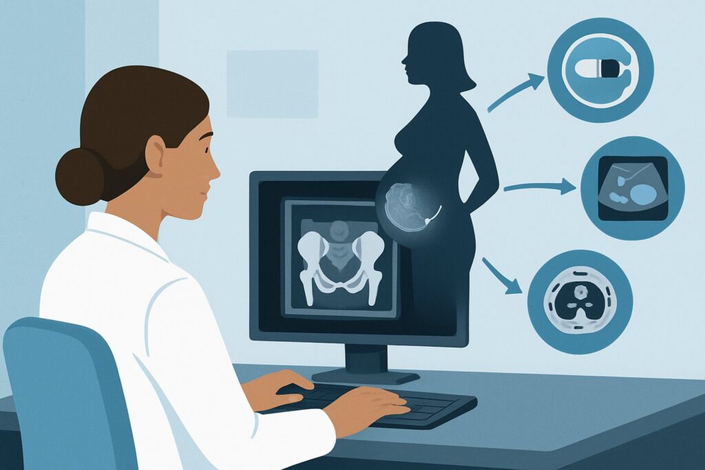
What Diagnoses Are You Working Up in a Pregnant Patient with Pelvic Pain?
When a pregnant patient presents with non-traumatic, weight-bearing pelvic or hip pain and negative radiographs, the differential diagnosis extends beyond a simple stress fracture. The choice of advanced imaging must be able to differentiate between several key possibilities.
Insufficiency Fracture: This is the primary diagnosis to confirm or exclude. Pregnancy induces a state of relative osteopenia due to fetal calcium demands and hormonal changes (e.g., increased relaxin). This makes the maternal skeleton, particularly the weight-bearing pelvic girdle and femoral heads, more susceptible to fracture from normal physiological stress. An insufficiency fracture occurs when normal stress is applied to abnormal bone.
Transient Osteoporosis of the Hip (TOH): This is a rare, self-limiting condition that typically presents in the third trimester of pregnancy or the early postpartum period. Patients experience sudden onset of hip pain that limits weight-bearing. While the etiology is unclear, the clinical and radiographic findings can mimic other serious conditions. MRI is crucial for distinguishing TOH, which shows extensive bone marrow edema without a fracture line, from other pathologies.
Osteonecrosis (Avascular Necrosis): While less common, osteonecrosis of the femoral head is a consequential diagnosis that can be associated with pregnancy. It involves the death of bone tissue due to a loss of blood supply. Early diagnosis is critical to preserving the joint. MRI is the most sensitive imaging modality for detecting early-stage osteonecrosis, often before any changes are visible on radiographs.
Musculotendinous or Ligamentous Injury: Pelvic girdle pain from soft tissue sources is extremely common in pregnancy. This can include adductor tendinopathy, gluteal tendinopathy, or sacroiliac joint ligamentous laxity. While often a clinical diagnosis, MRI can be used to rule out underlying osseous pathology and confirm a soft tissue source if the diagnosis remains uncertain.
Why Is Non-Contrast MRI the Recommended Study for a Suspected Stress Fracture in Pregnancy?
The American College of Radiology (ACR) designates MRI of the area of interest without IV contrast as Usually Appropriate for this scenario, making it the clear next step after inconclusive radiographs. The rationale is based on its superior diagnostic accuracy and unparalleled safety profile in pregnancy.
Superior Diagnostic Sensitivity and Specificity
Magnetic Resonance Imaging (MRI) is highly sensitive for detecting the earliest signs of a stress injury. It can visualize bone marrow edema, the physiological precursor to a visible fracture line, days to weeks before changes become apparent on radiographs. This allows for a definitive and timely diagnosis, which is crucial for management decisions like activity modification or protected weight-bearing to prevent fracture completion. Furthermore, MRI excels at differentiating among the key differential diagnoses, clearly distinguishing the marrow edema pattern of a stress fracture from that of transient osteoporosis of the hip or the characteristic findings of osteonecrosis.
Avoidance of Ionizing Radiation
The most compelling reason for choosing MRI in a pregnant patient is its complete lack of ionizing radiation. The radiation relative level (RRL) is zero. This eliminates any risk of radiation exposure to the fetus, a paramount consideration in all imaging decisions during pregnancy.
Rationale for Lower-Rated Alternatives
- CT Scans (with or without contrast): All variants of Computed Tomography are rated Usually not appropriate. CT uses ionizing radiation, which poses a potential risk to the developing fetus. While CT is excellent for characterizing fracture lines, the diagnostic information it provides for an occult fracture does not outweigh the radiation risk in this population when a safe and effective alternative like MRI is available.
- Bone Scan (Nuclear Scintigraphy): This study is also rated Usually not appropriate. It requires the intravenous injection of a radioactive tracer, which results in a measurable radiation dose to both the mother and the fetus (ACR RRL ☢☢☢, 1-10 mSv). It is generally contraindicated for this indication during pregnancy.
- Repeat Radiography in 10-14 days: This approach is rated Usually not appropriate. While it avoids the higher dose of CT, it suffers from low sensitivity in the early stages of a stress injury. Waiting two weeks for a potentially still-negative study delays a definitive diagnosis, prolonging the patient’s pain and risking fracture propagation.
Why Without IV Contrast?
For the evaluation of a suspected stress fracture, IV contrast is not necessary. The key findings—bone marrow edema and a potential fracture line—are best visualized on fluid-sensitive, non-contrast sequences like STIR (Short Tau Inversion Recovery). The ACR rates MRI with contrast as Usually not appropriate for this scenario, as gadolinium-based contrast agents are typically avoided during pregnancy unless the potential diagnostic benefit is critical and cannot be obtained otherwise.
What’s Next After the MRI? Downstream Workflow
The results of the non-contrast MRI will guide the subsequent clinical management and potential need for further consultation. The pathway branches based on whether the findings are positive, negative, or indeterminate.
If the MRI is positive for a stress/insufficiency fracture:
The diagnosis is confirmed. The next steps are primarily non-surgical and focus on pain management and mechanical unloading of the affected bone. This typically involves a period of protected or non-weight-bearing with crutches. Consultation with an orthopedic surgeon is recommended, especially for high-risk fractures (e.g., tension-side femoral neck fractures), to determine the optimal management strategy and follow-up plan to ensure proper healing and prevent displacement.
If the MRI is negative for fracture but shows another cause:
- Transient Osteoporosis of the Hip (TOH): Management is supportive with protected weight-bearing and analgesia. The condition is self-limiting and typically resolves within 6-12 months postpartum. Orthopedic follow-up is still advisable to monitor progress.
- Osteonecrosis: This is a more serious diagnosis requiring urgent orthopedic consultation. Management may range from core decompression to, in advanced cases, joint replacement after pregnancy.
- Soft Tissue Injury: A diagnosis of tendinopathy or ligamentous strain would prompt referral to physical therapy for conservative management.
If the MRI is negative and no alternative cause is identified:
If a high-quality MRI of the symptomatic area is entirely normal, a significant osseous or articular pathology is effectively ruled out. The focus should shift back to a clinical evaluation for other causes of pelvic girdle pain in pregnancy. Further imaging is typically not warranted unless the patient’s symptoms progress or change in character.
Pitfalls to Avoid (and When to Get Help)
Navigating this clinical scenario requires careful attention to the unique aspects of pregnancy. Here are a few common pitfalls to avoid:
- Delaying Advanced Imaging: Do not delay ordering an MRI out of an overabundance of caution. Persistent, localized, weight-bearing pain with negative radiographs warrants a definitive diagnosis to prevent a non-displaced fracture from completing.
- Ordering a Contrast-Enhanced MRI: For a stress fracture workup, gadolinium is unnecessary and should be avoided. Ensure the order explicitly states “without IV contrast.”
- Accepting an Equivocal Radiograph Report: If the initial radiograph is reported as “indeterminate” or “subtle lucency,” do not adopt a “wait and see” approach. Proceed directly to MRI to clarify the finding.
- Misattributing Pain to “Normal Pregnancy”: While musculoskeletal pain is common in pregnancy, focal, severe, and mechanically-induced pain is not normal and should be investigated thoroughly.
If the clinical picture is complex or the MRI findings are ambiguous, consultation with a musculoskeletal radiologist can provide valuable insight into the imaging findings and differential diagnosis.
Related ACR Topics and Tools
For a comprehensive overview of all clinical variants related to stress and insufficiency fractures, or to explore the technical details of imaging protocols, the following resources are available.
- For breadth across all scenarios in Stress (Fatigue/Insufficiency) Fracture, Including Sacrum, Excluding Other Vertebrae, see our parent guide: Stress (Fatigue/Insufficiency) Fracture, Including Sacrum, Excluding Other Vertebrae: ACR Appropriateness Decoded.
- ACR Appropriateness Criteria Lookup — for adjacent scenarios
- Imaging Protocol Library — for technique on the recommended study
- Radiation Dose Calculator — for cumulative dose conversations
Frequently Asked Questions
Is any amount of radiation from a pelvic radiograph safe during pregnancy?
A single pelvic radiograph delivers a very low dose of radiation, and the risk to the fetus is considered negligible when the study is clinically indicated. The decision to perform any imaging with ionizing radiation during pregnancy involves weighing this minimal risk against the benefit of making an accurate diagnosis. However, for follow-up imaging in this scenario, MRI is preferred because it involves no ionizing radiation at all.
Why is a bone scan ‘Usually not appropriate’ if it’s so sensitive for stress fractures?
While nuclear medicine bone scans are highly sensitive for detecting stress fractures, they require injecting a radioactive tracer (technetium-99m MDP). This tracer circulates throughout the mother’s body and crosses the placenta, resulting in radiation exposure to the fetus. Because a safe and equally sensitive alternative (MRI) exists, the use of a bone scan for this indication in a pregnant patient is not appropriate.
Can I order an ultrasound instead of an MRI to avoid radiation?
Ultrasound is rated ‘Usually not appropriate’ by the ACR for this indication. While it is excellent for evaluating soft tissues and can sometimes detect cortical disruptions or periosteal reactions, it has very low sensitivity for occult or intramedullary stress fractures of the pelvis, sacrum, and hip. It cannot reliably visualize bone marrow edema, which is the key early finding seen on MRI.
What if my institution has limited or no access to MRI?
In a resource-limited setting where MRI is unavailable, the decision becomes more complex. The ACR criteria are based on best practices assuming technology is available. If MRI is not an option, a discussion with a radiologist and an orthopedic specialist is crucial. Depending on the urgency and specific clinical question, options might include a limited CT scan with fetal shielding or proceeding with empiric treatment (protected weight-bearing) based on high clinical suspicion, though both are suboptimal.
Does this guidance apply to postpartum patients who are breastfeeding?
This specific guidance is for pregnant patients. For postpartum patients, even those who are breastfeeding, the primary concern of direct fetal radiation exposure is removed. While MRI remains an excellent choice, other modalities like CT or bone scan could be considered if indicated. For breastfeeding mothers, guidelines from the American College of Radiology state that it is safe to continue breastfeeding after receiving gadolinium-based contrast or nuclear medicine radiotracers without any interruption.
Reviewed by Pouyan Golshani, MD, Interventional Radiologist — May 30, 2026