How Should You Manage Liver Abscesses After ERCP? An ACR-Guided Workflow
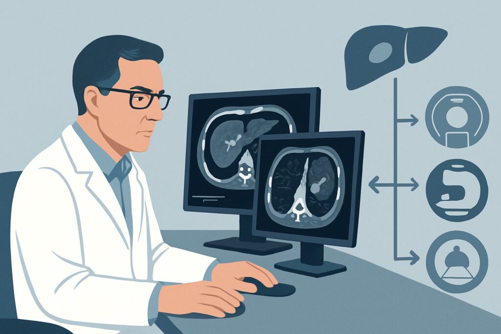
It’s late in the afternoon, and you are managing a 68-year-old patient admitted with a three-week history of worsening right upper quadrant pain, fevers, and jaundice. His history is notable for an endoscopic retrograde cholangiopancreatography (ERCP) with sphincterotomy for a suspected stone a month ago. A contrast-enhanced CT confirmed your suspicion: two well-defined liver abscesses, both measuring over 3 cm. An MRCP was negative for any residual stones or biliary obstruction. The patient is on broad-spectrum intravenous antibiotics, but his clinical picture is not improving. The next step is source control, but which approach is best? This is a common and critical decision point in interventional radiology and gastroenterology. This article details the American College of Radiology (ACR) Appropriateness Criteria for this specific scenario, explaining the rationale for the recommended management pathway. For this presentation, the ACR deems Percutaneous catheter drainage only as Usually Appropriate.
Who Fits This Clinical Scenario?
This guidance applies to a specific subset of patients with infected fluid collections. The key inclusion criteria are a recent history of biliary instrumentation (specifically ERCP with sphincterotomy), a subacute presentation (weeks, not days), and imaging findings confirming one or more liver abscesses greater than 3 cm in diameter. Crucially, follow-up imaging like Magnetic Resonance Cholangiopancreatography (MRCP) or a dedicated biliary CT has already ruled out an ongoing mechanical obstruction, such as a retained stone or stricture, as the underlying cause. The patient is typically already on appropriate antibiotic therapy but shows persistent or worsening signs of infection, such as fever, leukocytosis, and malaise.
This workflow is distinct from other clinical situations. For instance, this guidance does not apply to a patient with a suspected appendiceal abscess in the right lower quadrant, which involves a different source of infection and anatomical considerations. It also differs from the management of a sterile or infected pancreatic pseudocyst following acute pancreatitis, as the nature of the fluid collection and its relationship to the pancreatic ductal system require a different therapeutic algorithm. Finally, if the MRCP in this patient had revealed a persistent biliary obstruction, the management would shift toward relieving that obstruction, often with a combined drainage approach.
What Diagnoses Are You Working Up in This Scenario?
The primary diagnosis in this clinical context is a pyogenic liver abscess. Given the history of ERCP, the abscess is most likely iatrogenic. During ERCP, instrumentation of the biliary tree can introduce bacteria from the duodenum into the sterile biliary system. The sphincterotomy, while therapeutic for stone passage, permanently alters the protective barrier of the sphincter of Oddi, potentially allowing for ascending infection. This can lead to cholangitis and subsequent abscess formation, particularly if there is any transient biliary stasis. The 3-week delay between the procedure and worsening symptoms is classic for the gradual development of a contained, encapsulated abscess.
A less common but important consideration is a superinfected biloma. The ERCP procedure itself can cause a minor bile leak, leading to a contained collection of bile (a biloma). While initially sterile, this collection can become secondarily infected, evolving into an abscess. The imaging characteristics on CT and the clinical signs of infection are often indistinguishable from a primary pyogenic abscess.
Finally, while the negative MRCP makes it less likely, one must always consider an underlying, undiagnosed pathology that created biliary stasis and predisposed the patient to infection, such as a small, missed biliary stricture or an occult malignancy. The abscess may be the first presentation of a more subtle underlying problem that the initial ERCP was intended to investigate or treat. The fluid aspirate from drainage can sometimes yield cytologic clues if malignancy is a concern.
Why Is Percutaneous Catheter Drainage the Recommended Management for This Presentation?
For a patient with confirmed liver abscesses larger than 3 cm who is failing medical management, source control is paramount. The ACR designates Percutaneous catheter drainage only as Usually Appropriate because it provides the most effective and least invasive method for achieving definitive source control in this setting.
Antibiotics alone are often insufficient for abscesses of this size, as the encapsulated, avascular nature of the abscess cavity prevents adequate antibiotic penetration. Percutaneous drainage directly addresses this by evacuating the purulent material, decompressing the collection, and allowing the cavity to collapse. Placing a catheter, rather than performing a simple aspiration, ensures continuous drainage, prevents re-accumulation of pus, and allows for irrigation of the cavity if needed. This approach has a high technical and clinical success rate, leading to rapid defervescence and clinical improvement. Furthermore, the aspirated fluid provides a crucial sample for Gram stain and culture, enabling the transition from broad-spectrum to targeted, culture-directed antibiotic therapy.
Alternative management strategies are rated lower for specific reasons in this context:
- Needle aspiration is rated May be appropriate. While less invasive than catheter placement, it is generally less effective for abscesses larger than 3-5 cm or those containing thick, viscous fluid. It carries a higher risk of treatment failure and the need for repeat procedures due to re-accumulation.
- Surgical management is also rated May be appropriate. Open or laparoscopic surgical drainage is significantly more invasive, requires general anesthesia, and is associated with greater morbidity and longer recovery times. It is typically reserved for cases where percutaneous drainage is technically impossible due to an unsafe access route, when percutaneous attempts have failed, or in the presence of a ruptured abscess with diffuse peritonitis.
- Continued conservative management (antibiotics alone) is rated May be appropriate but is a high-risk strategy for abscesses of this size and a patient who is already clinically worsening. It is generally not recommended as a primary strategy unless the patient is too unstable for any procedure or refuses intervention.
The procedure is performed under imaging guidance, typically ultrasound (US) or computed tomography (CT), to ensure a safe and accurate access path into the abscess, avoiding major blood vessels, adjacent organs, and the pleural space. Pre-procedural assessment of coagulation parameters is essential to minimize bleeding risk.
What’s Next After Percutaneous Catheter Drainage? Downstream Workflow
The placement of a drainage catheter is the beginning, not the end, of treatment. The post-procedure workflow is critical for a successful outcome. Immediately following successful catheter placement, the patient should remain on broad-spectrum antibiotics, which will be tailored once culture and sensitivity results return in 48-72 hours.
If the patient improves: You should see a prompt clinical response, with defervescence, a decrease in white blood cell count, and improvement in right upper quadrant pain within 24-48 hours. The drain will be flushed regularly (e.g., with sterile saline) to maintain patency. It remains in place until the drainage output decreases significantly (typically to less than 10-20 mL per day), the fluid becomes serous rather than purulent, and follow-up imaging (usually CT or US) confirms resolution or near-resolution of the abscess cavity. This process can take anywhere from several days to a few weeks.
If the patient fails to improve: A lack of clinical improvement after 48-72 hours should trigger an investigation. The first step is to assess drain function with a gentle saline flush and a CT scan with contrast injected through the drain (a “drainogram” or abscessogram). This can identify drain dislodgement, catheter occlusion, or reveal previously unseen loculations within the abscess cavity that are not being adequately drained. This may necessitate repositioning the existing catheter or placing an additional one. If drainage is mechanically sound and the patient is still not improving, consider broadening antibiotic coverage for resistant organisms and re-evaluating for another source of infection.
Escalation: If the patient continues to deteriorate despite optimal percutaneous management, or if the abscess is found to be part of a more complex process (e.g., a necrotic tumor), escalation to surgical consultation for open drainage or resection is the appropriate next step.
Pitfalls to Avoid (and When to Get Help)
Several common pitfalls can complicate the management of post-ERCP liver abscesses. First, avoid delaying source control in a clinically worsening patient with a large abscess; antibiotics alone are unlikely to succeed. Second, be aware that a single drain may be insufficient for a large, complex, or multiloculated abscess; be prepared for the possibility of placing multiple catheters. Third, failing to obtain an adequate fluid sample for microbiology at the time of drainage can compromise the ability to tailor antibiotic therapy effectively. Finally, a common error is the premature removal of the drainage catheter before the abscess cavity has fully collapsed, which can lead to rapid re-accumulation and recurrence. If the patient shows signs of sepsis or hemodynamic instability despite initial drainage, or if there is evidence of abscess rupture, this constitutes a medical emergency requiring immediate escalation to an interventional radiologist and a surgical team.
Related ACR Topics and Tools
This article covers one specific variant within the broader topic of infected fluid collections. For a comprehensive overview of other clinical scenarios, from appendiceal abscesses to empyema, please consult the parent topic guide. The following GigHz tools can also support your clinical decision-making:
- For breadth across all scenarios in Radiologic Management of Infected Fluid Collections, see our parent guide: Radiologic Management of Infected Fluid Collections: ACR Appropriateness Decoded.
- ACR Appropriateness Criteria Lookup — for adjacent scenarios
- Imaging Protocol Library — for technique on the recommended study
- Radiation Dose Calculator — for cumulative dose conversations
Frequently Asked Questions
Why is percutaneous drainage preferred over just continuing antibiotics for abscesses larger than 3 cm?
For liver abscesses larger than 3 cm, antibiotics alone have a high failure rate. The abscess capsule is poorly vascularized, which prevents antibiotics from reaching the core of the infection in sufficient concentrations. Percutaneous drainage provides ‘source control’ by physically removing the pus, which rapidly reduces the bacterial load, relieves pressure, and allows the body’s immune system and antibiotics to work effectively, leading to faster clinical resolution.
What is the difference in outcome between needle aspiration and catheter drainage for this scenario?
While simple needle aspiration can be diagnostic and sometimes therapeutic for small abscesses (<3 cm), it is less effective for larger collections like those in this scenario. Catheter drainage is superior because it provides continuous evacuation of pus, preventing re-accumulation and allowing the abscess cavity to collapse over time. Needle aspiration has a higher rate of recurrence for large abscesses, often requiring repeat procedures, whereas a catheter provides more definitive, one-time management.
How do you decide between ultrasound and CT guidance for placing the drain?
The choice between ultrasound (US) and computed tomography (CT) guidance depends on the abscess’s location, visibility, and the surrounding anatomy. US is often preferred as it is real-time, avoids ionizing radiation, and is portable. However, if the abscess is deep, obscured by bowel gas or bone, or if the planned access route is complex and passes near critical structures, CT provides superior anatomical detail and a more precise path for safe catheter placement.
What if the MRCP had shown a biliary obstruction? How would that change management?
If the MRCP had revealed an underlying biliary obstruction (e.g., a retained stone or a stricture), the management strategy would change significantly. The primary goal would become relieving the obstruction to restore normal bile flow. This would typically involve a percutaneous transhepatic biliary drain (PTBD) to decompress the biliary system, in addition to draining the abscess itself. In some cases, a single catheter can be manipulated to drain both the abscess and the upstream bile duct. The ACR rates ‘Percutaneous catheter drainage with conversion to percutaneous biliary drain’ as ‘May be appropriate’ for this reason, anticipating scenarios where an obstruction is found.
How long does the drainage catheter typically stay in place?
The duration varies depending on the size of the abscess and the patient’s clinical response. The catheter is typically left in place until three criteria are met: 1) the daily drainage output is minimal (e.g., less than 10-20 mL/day), 2) the character of the fluid changes from thick pus to thin, serous fluid, and 3) follow-up imaging confirms the abscess cavity has resolved or significantly collapsed. This can take anywhere from 5-7 days to several weeks for very large or complex collections.
Reviewed by Pouyan Golshani, MD, Interventional Radiologist — May 29, 2026