How Should You Manage Obstructive Pyelonephritis in a Pregnant Patient?
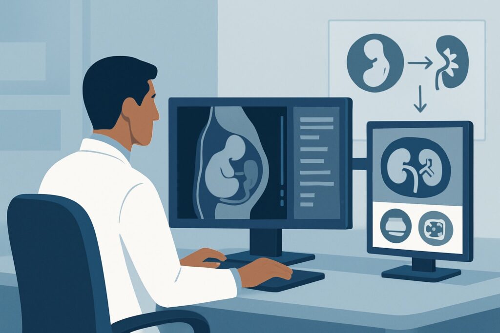
A 29-year-old patient, 26 weeks pregnant, presents to the emergency department late on a Tuesday night with a three-day history of escalating left flank pain, chills, and a fever of 38.8°C. Her white blood cell count is elevated, and a urinalysis is positive for leukocyte esterase and nitrites. A point-of-care ultrasound, performed to minimize radiation exposure, confirms new, moderate left-sided hydronephrosis. You’ve diagnosed obstructive pyelonephritis, a true obstetric and urologic emergency. The critical decision is not if to decompress the obstructed kidney, but how. This article provides a detailed clinical workflow for this specific scenario, guiding you through the differential diagnosis, procedural rationale, and downstream management based on the American College of Radiology (ACR) Appropriateness Criteria. For this presentation, both Retrograde Ureteral Stenting and Percutaneous Nephrostomy are considered ‘Usually Appropriate’ interventions.
Who Fits This Clinical Scenario of Obstructive Pyelonephritis in Pregnancy?
This guidance is specifically for a pregnant patient in the second or third trimester (20+ weeks gestation) who presents with a clinical picture of urosepsis secondary to an obstructed urinary system. The key inclusion criteria are the triad of pregnancy, evidence of infection (fever, leukocytosis, positive urinalysis), and imaging confirmation of new hydronephrosis.
The workflow detailed here is distinct from other clinical situations that may appear similar. This article does not apply to:
- Non-pregnant patients with a known obstructing stone on CT. While the need for decompression may be similar, the diagnostic and management options are broader without the constraint of avoiding fetal radiation exposure.
- Patients with chronic, non-infected hydronephrosis from malignancy. A patient with advanced cervical carcinoma and renal failure, for example, requires a different management algorithm focused on long-term drainage and oncologic care, not acute infection control.
- Patients with urinary ascites or other post-surgical complications. This scenario is specific to obstruction of the native upper urinary tract during pregnancy.
The combination of pregnancy, acute infection, and urinary obstruction creates a high-risk situation requiring urgent intervention tailored to protect both the mother and the fetus.
What Diagnoses Are You Working Up in a Pregnant Patient with Hydronephrosis and Fever?
When a pregnant patient presents with these signs and symptoms, you are evaluating a narrow but critical differential diagnosis. The primary goal of intervention is to address the urgent problem—infected, obstructed urine—regardless of the precise underlying cause.
Obstructive Pyelonephritis Secondary to Physiologic Hydronephrosis: This is the most common underlying condition. During pregnancy, elevated progesterone levels cause smooth muscle relaxation in the ureters, leading to decreased peristalsis and urinary stasis. Compounding this is the mechanical compression of the ureters, particularly the right ureter, by the enlarging gravid uterus. This “physiologic” hydronephrosis is present in up to 90% of pregnancies by the third trimester but becomes pathologic when the static urine becomes infected, leading to pyelonephritis.
Obstructive Pyelonephritis Secondary to Urolithiasis: While physiologic changes are the most frequent cause, a true obstructing stone must be considered. Pregnancy does not protect against kidney stones, and their diagnosis is complicated by the necessary avoidance of CT. Ultrasound is the first-line imaging modality but has limited sensitivity for detecting ureteral stones. The clinical management of decompression, however, is the same in the acute setting.
Pyonephrosis: This represents a urologic emergency where the obstructed renal collecting system is filled with purulent material. The clinical presentation is identical to severe obstructive pyelonephritis. It is not a separate diagnosis so much as the severe end of the same disease spectrum. The distinction is academic in the acute phase, as the management—urgent decompression—is identical and life-saving.
Why Are Retrograde Ureteral Stenting and PCN the Recommended Interventions?
The ACR panel rates both Retrograde Ureteral Stenting and Percutaneous Nephrostomy (PCN) as ‘Usually Appropriate’ for this clinical scenario. The primary goal is to emergently decompress the infected collecting system while minimizing risk to the fetus, particularly from ionizing radiation and anesthesia. The choice between the two often depends on local expertise, resource availability, and a multidisciplinary discussion with the patient, obstetrics, urology, and interventional radiology.
Retrograde Ureteral Stenting is typically performed by a urologist in an operating room setting. Under cystoscopic guidance, a thin, flexible stent is passed up the ureter from the bladder, bypassing the obstruction.
- Advantages: It provides internal drainage, avoiding an external tube and bag, which improves patient comfort and mobility and reduces the risk of accidental dislodgement.
- Considerations: This procedure often requires spinal or general anesthesia, which carries its own set of considerations and risks in pregnancy. It also involves fluoroscopy (X-ray) to confirm placement, though exposure can be significantly minimized with lead shielding over the abdomen and adherence to low-dose protocols (ALARA – As Low As Reasonably Achievable).
Percutaneous Nephrostomy (PCN) is performed by an interventional radiologist. Under local anesthesia, a small tube is placed directly through the skin of the flank into the kidney’s collecting system.
- Advantages: The primary advantage is the avoidance of general or spinal anesthesia. The procedure is guided almost exclusively by ultrasound, virtually eliminating fetal radiation exposure.
- Considerations: The patient is left with an external drainage tube and collection bag, which can be cumbersome, require regular care, and is at risk for being dislodged.
Why Other Options Are Rated Lower
Alternative management strategies are deemed ‘Usually not appropriate’ because they fail to adequately address the immediate danger.
- Medical management without decompression: This is ‘Usually not appropriate’ because systemic antibiotics cannot effectively penetrate an obstructed, infected collecting system. Delaying decompression in the face of obstructive pyelonephritis risks progression to sepsis, septic shock, and adverse maternal-fetal outcomes.
- Percutaneous antegrade ureteral stenting: This hybrid procedure, also ‘Usually not appropriate’ in this setting, involves first performing a PCN and then attempting to pass a stent down the ureter. It is more complex, offers no clear benefit over the primary options, and potentially increases procedural time and radiation exposure.
What’s Next After Urinary Decompression? Downstream Workflow
Successful decompression is marked by clinical improvement—defervescence, normalization of white blood cell count, and resolution of flank pain—typically within 24 to 48 hours, alongside continued appropriate antibiotic therapy. The management plan, however, extends through the remainder of the pregnancy and into the postpartum period.
If the patient improves: The drainage catheter (either the internal ureteral stent or the external nephrostomy tube) is left in place. These tubes often require exchange every 6-12 weeks to prevent encrustation and occlusion, a decision made in consultation with urology or interventional radiology. The definitive management of any underlying cause, such as a kidney stone, is almost always deferred until after delivery.
If the patient fails to improve: Persistent fever or pain after 48 hours should trigger an investigation for inadequate drainage.
- With a ureteral stent: A renal ultrasound should be performed to assess for any decrease in hydronephrosis and to confirm stent position. If drainage is suboptimal, the stent may be occluded or malpositioned, requiring cystoscopic revision or replacement.
- With a PCN tube: The external tube should be checked for kinks or blockages. A nephrostogram (injecting a small amount of contrast into the tube under minimal fluoroscopy) can confirm patency and position. The tube may need to be repositioned or exchanged for a larger one.
Close collaboration between the primary obstetrics team, the procedural service (urology or IR), and infectious disease specialists is paramount for navigating the remainder of the pregnancy safely.
Pitfalls to Avoid in Pregnant Patients with Obstructive Pyelonephritis
Managing this high-stakes scenario requires vigilance to avoid common and potentially serious errors.
- Pitfall 1: Delaying Decompression. Do not treat this as uncomplicated pyelonephritis. Once obstruction is confirmed on ultrasound in a septic pregnant patient, the clock is ticking. Every hour of delay increases the risk of maternal sepsis and preterm labor.
- Pitfall 2: Ordering a CT Scan. The diagnosis can and should be made with clinical findings and ultrasound. CT should be avoided due to fetal radiation exposure. If complex anatomy must be evaluated, MRI without gadolinium is the preferred cross-sectional imaging modality in pregnancy.
- Pitfall 3: Inadequate Communication. The decision between a stent and a PCN tube should be a shared one, involving the patient and a multidisciplinary team. Discuss the pros and cons of each (anesthesia vs. external tube) to align the plan with the patient’s priorities and the institution’s capabilities.
When to Escalate: If the patient develops signs of hemodynamic instability, altered mental status, or lactic acidosis, escalate immediately to a higher level of care (ICU/critical care) in parallel with arranging for emergent decompression.
Related ACR Topics and Tools
This article covers one specific variant within the broader topic of managing urinary tract obstruction. For a comprehensive overview of other clinical scenarios and for tools to assist in your daily practice, please refer to the following GigHz resources:
- For breadth across all scenarios in Radiologic Management of Urinary Tract Obstruction, see our parent guide: Radiologic Management of Urinary Tract Obstruction: ACR Appropriateness Decoded.
- Imaging Appropriateness Selector — for adjacent scenarios
- Imaging Protocol Library — for technique on the recommended study
- Radiation Dose Calculator — for cumulative dose conversations
Frequently Asked Questions
Is right-sided hydronephrosis more common than left-sided in pregnancy?
Yes. Physiologic hydronephrosis of pregnancy more commonly affects the right kidney. This is thought to be due to a combination of dextrorotation of the uterus and the course of the right ovarian vein, which crosses over the right ureter and can contribute to its compression.
Why is medical management alone considered ‘Usually not appropriate’?
In an obstructed urinary system, urine flow is blocked. Systemically administered antibiotics have difficulty reaching the site of infection in sufficient concentrations to be effective. This can lead to uncontrolled infection, abscess formation (pyonephrosis), and sepsis. Decompressing the system to restore urine flow is essential for both source control and allowing antibiotics to work.
If we choose a retrograde ureteral stent, how much radiation is the fetus exposed to?
The radiation dose from a properly performed fluoroscopically-guided stent placement is very low. By using techniques like lead shielding over the uterus, limiting the fluoroscopy time (‘beam-on’ time), and using low-dose settings, the fetal exposure can be kept well below the threshold associated with deterministic effects. The risk of untreated maternal sepsis far outweighs the minimal radiation risk from the procedure.
Can an MRI be used to diagnose the cause of obstruction in this scenario?
Yes, if ultrasound is inconclusive and a definitive diagnosis of the obstructing cause is needed to guide management, a non-contrast MRI of the abdomen and pelvis is the preferred next imaging step. It avoids ionizing radiation and can clearly visualize the urinary tract to identify stones, strictures, or other causes of obstruction.
What happens to the stent or nephrostomy tube after the patient delivers?
The drainage tube is typically removed or managed definitively in the postpartum period, usually 4-6 weeks after delivery. If the cause was a kidney stone, the patient can then undergo standard procedures (like CT imaging and lithotripsy or ureteroscopy) to treat the stone without concern for fetal radiation exposure.
Reviewed by Pouyan Golshani, MD, Interventional Radiologist — May 26, 2026