Imaging Acute Ankle Trauma When Ottawa Rules Are Inapplicable: An ACR Workflow
A 68-year-old patient with diabetic peripheral neuropathy presents to the urgent care clinic after twisting his ankle stepping off a curb. He has moderate swelling and ecchymosis over the lateral malleolus, but his report of tenderness is unreliable due to diminished sensation in his feet. You know the Ottawa Ankle Rules are not validated in this population, so you cannot use them to clinically clear his ankle. What is the appropriate initial imaging study to order to rule out a fracture? This article provides a detailed workflow for this specific clinical scenario, where exclusionary criteria prevent the use of standard clinical decision rules. For this presentation, the American College of Radiology (ACR) Appropriateness Criteria rate `Radiography ankle` as Usually Appropriate.
Who Fits This Clinical Scenario?
This guidance applies to a specific subset of patients: adults or children aged five years or older who have sustained acute trauma to the ankle, but for whom standard clinical decision rules are not reliable. The key feature of this scenario is the presence of an exclusionary criterion that invalidates the use of the Ottawa Ankle Rules (OAR). These criteria include conditions that impair a patient’s ability to feel or localize pain, such as:
- Peripheral neuropathy (eg, from diabetes mellitus)
- Other neurologic disorders affecting sensation
- Altered mental status or intoxication
- Significant distracting injuries
This workflow is distinct from the more common presentation of an acute ankle injury in a neurologically intact patient where the OAR can be applied to determine the need for imaging. It also differs from scenarios involving persistent ankle pain for more than one week or follow-up imaging after initial radiographs have already been obtained. This guide focuses exclusively on the initial imaging decision when the physical exam is an unreliable guide.
What Diagnoses Are You Working Up in This Scenario?
When a reliable physical examination is not possible, imaging becomes the primary tool for diagnosis. The main objective is to identify or exclude significant osseous and ligamentous injuries that require specific management. The differential diagnosis is broad, but the imaging choice is tailored to efficiently assess the most critical possibilities.
Ankle Fracture
This is the most important diagnosis to exclude. Fractures can involve the lateral, medial, or posterior malleoli. More complex patterns, such as bimalleolar or trimalleolar fractures, result in an unstable ankle joint requiring orthopedic consultation and often surgical fixation. Because the patient cannot reliably bear weight or localize pain, a fracture cannot be ruled out on clinical grounds alone.
Foot Fractures (Talar or Calcaneal)
The mechanism of injury for an ankle sprain can also transmit force to the bones of the hindfoot and midfoot. Fractures of the talus (eg, lateral process) or the anterior process of the calcaneus can mimic the signs of a lateral ankle sprain. These are often subtle on radiographs but are important to identify.
Significant Ligamentous Injury
While radiographs are not the primary modality for visualizing ligaments, they can reveal secondary signs of severe ligamentous disruption. A syndesmotic injury (high ankle sprain), for example, may present with widening of the tibiofibular clear space or the medial clear space on a mortise view, indicating instability of the ankle joint.
Soft-Tissue Injury Without Fracture
A simple ankle sprain remains a common possibility. However, in this clinical scenario, it is a diagnosis of exclusion. Imaging is required to confidently rule out a fracture before initiating treatment for a sprain, such as early mobilization and physical therapy.
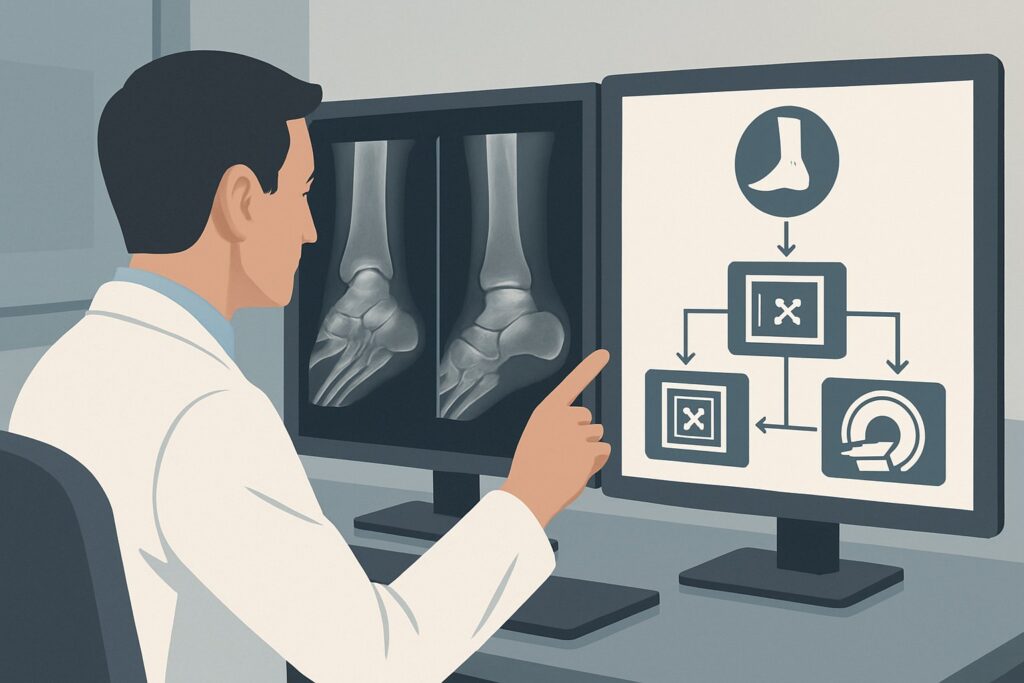
Why Is Ankle Radiography the Recommended Initial Study?
The ACR designates a standard three-view `Radiography ankle` series as Usually Appropriate for this presentation. This recommendation is based on the test’s high diagnostic yield for the primary clinical question—the presence of a fracture—balanced against its low cost, wide availability, and minimal radiation exposure.
In a patient whose sensory deficits make the physical exam unreliable, imaging is not optional; it is a diagnostic necessity. Radiography is highly sensitive and specific for detecting the vast majority of clinically significant ankle and hindfoot fractures. A standard series, including anteroposterior (AP), lateral, and mortise views, provides a comprehensive assessment of the osseous structures and the alignment of the ankle mortise.
Alternative imaging modalities are rated lower for this initial workup for several key reasons:
- CT ankle without IV contrast is rated as May be appropriate. While CT offers superior detail for detecting subtle or complex fracture patterns, it is not the recommended first-line study. It imparts a higher radiation dose than radiography and is best reserved as a problem-solving tool if radiographs are negative but clinical suspicion for an occult fracture remains high, or for preoperative planning once a fracture is identified.
- MRI ankle without IV contrast is rated Usually not appropriate for the initial evaluation of acute trauma in this context. MRI excels at evaluating soft tissues like ligaments, tendons, and cartilage. However, it is more costly, less readily available, and not as efficient as radiography for the primary goal of identifying an acute fracture. Its role is typically in the subacute setting for assessing ligamentous integrity or looking for bone marrow edema if radiographs are negative and symptoms persist.
From a safety perspective, the radiation dose from an ankle radiograph series is minimal (adult relative radiation level ☢ <0.1 mSv; pediatric ☢ <0.03 mSv), which is a fraction of the average annual background radiation. This makes it a very safe initial step for both adult and pediatric patients.
What’s Next After Ankle Radiography? Downstream Workflow
The results of the initial ankle radiographs will dictate the subsequent clinical pathway. The decision tree branches based on whether the images are positive, negative, or indeterminate for a significant injury.
If Radiographs Are Positive for Fracture:
The next step depends on the fracture pattern and stability. For nondisplaced, stable fractures (eg, an isolated lateral malleolus fracture), management may involve immobilization with a cast or walking boot and non-weight-bearing status. For displaced or unstable fractures (eg, bimalleolar fracture or any fracture with mortise widening), an urgent orthopedic consultation is necessary for consideration of surgical fixation. In some complex cases, the orthopedist may order a preoperative CT scan to better delineate the fracture anatomy.
If Radiographs Are Negative for Fracture:
A negative radiograph series effectively rules out a significant osseous injury in most cases. The patient can be presumptively diagnosed with an ankle sprain or soft-tissue contusion. Management typically includes the RICE protocol (Rest, Ice, Compression, Elevation), a short period of protected weight-bearing in a brace or boot, and subsequent physical therapy. If pain and mechanical symptoms persist despite conservative management, the patient may fit the criteria for a different ACR scenario, such as acute trauma with persistent pain, where advanced imaging like MRI may become appropriate.
If Radiographs Are Indeterminate or Suspicious:
Occasionally, radiographs may be equivocal. There might be a high clinical suspicion for a fracture (eg, based on mechanism and severe swelling) despite negative X-rays, or a subtle finding that is difficult to characterize. In this situation, a `CT ankle without IV contrast` (May be appropriate) is the logical next step to clarify the presence and extent of an occult osseous injury.
Pitfalls to Avoid (and When to Get Help)
In this specific scenario, where clinical assessment is limited, several pitfalls can lead to diagnostic errors. First, avoid the temptation to forgo imaging based on a seemingly benign mechanism of injury; the patient’s inability to report pain reliably means you must maintain a high index of suspicion for fracture. Second, ensure a complete three-view radiographic series is obtained, as some fractures are only visible on the mortise or lateral view. Third, do not forget to scrutinize the entire image for associated injuries, such as a base of the fifth metatarsal fracture or a talar process fracture. If radiographs are negative but the patient has significant swelling, inability to bear weight, and a high-risk mechanism, escalate care by considering a CT or obtaining an orthopedic consultation.
Related ACR Topics and Tools
Navigating imaging guidelines requires access to reliable, up-to-date resources. For a comprehensive overview of all clinical variants related to this topic, please consult our parent guide. For other tools to assist in ordering and interpreting imaging, see the resources below.
- For breadth across all scenarios in Acute Trauma to the Ankle, see our parent guide: Acute Trauma to the Ankle: ACR Appropriateness Decoded.
- To explore other clinical presentations, use the Imaging Appropriateness Selector.
- For details on imaging techniques, visit the Imaging Protocol Library.
- To discuss radiation exposure with patients, use the Radiation Dose Calculator.
Frequently Asked Questions
Why can’t I just use the Ottawa Ankle Rules on a patient with diabetic neuropathy?
The Ottawa Ankle Rules (OAR) rely on the patient’s ability to feel and localize pain over specific bony landmarks and their ability to bear weight. In patients with peripheral neuropathy, sensation is impaired, making their response to palpation unreliable. Therefore, the OAR lose their high sensitivity and are not validated for use in this population, necessitating imaging to rule out a fracture.
If I’m already concerned about a subtle fracture, why not order a CT scan first?
While CT is more sensitive for subtle fractures, ankle radiography is the recommended initial study because it is highly effective at identifying the vast majority of clinically significant fractures with much lower radiation dose and cost. The ACR recommends a stepwise approach: start with radiography, and if the results are negative but clinical suspicion remains very high, then proceed to CT. This strategy optimizes diagnostic yield while minimizing radiation exposure.
Does this guidance apply to a patient who is intoxicated?
Yes. Intoxication is considered an exclusionary criterion that makes the Ottawa Ankle Rules unreliable, similar to neuropathy. An intoxicated patient may not be able to provide a reliable history or accurately report pain. Therefore, this workflow, which recommends initial ankle radiography, would apply.
What if the radiographs are negative but I suspect a syndesmotic (high ankle) sprain?
If initial radiographs are negative for fracture but you have a strong clinical suspicion for a syndesmotic injury (based on mechanism and tenderness over the anterior inferior tibiofibular ligament), the patient enters a different clinical pathway. While radiographs can sometimes show subtle mortise widening, MRI is the definitive imaging modality for evaluating ligamentous injuries. This would be considered after the initial fracture workup is complete and symptoms persist.
Is an ultrasound useful in this acute trauma scenario?
According to the ACR, ultrasound of the ankle is rated ‘Usually not appropriate’ for the initial evaluation of acute ankle trauma when a fracture is suspected. While ultrasound is excellent for evaluating some soft tissue structures like tendons, it is not a reliable tool for excluding osseous fractures. Radiography remains the standard of care for the initial assessment.
Reviewed by Pouyan Golshani, MD, Interventional Radiologist — May 26, 2026