Imaging Suspected Discitis or Epidural Abscess: An ACR-Guided Workflow for High-Risk Patients
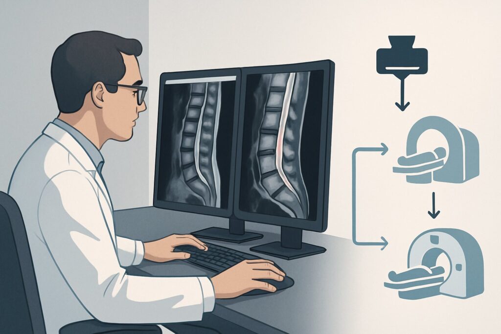
A 58-year-old man with a history of type 2 diabetes and intravenous drug use presents to the emergency department with three weeks of worsening, deep lumbar back pain and a new low-grade fever. He denies any specific trauma. His C-reactive protein (CRP) is significantly elevated, raising your suspicion for a spinal infection. You know that early and accurate diagnosis is critical to prevent devastating neurologic complications, but which imaging study is the right first step to confirm or rule out discitis-osteomyelitis or a spinal epidural abscess? This article provides a detailed workflow for this specific clinical scenario, guiding you through the differential, study rationale, and downstream decisions. According to the American College of Radiology (ACR) Appropriateness Criteria, the most definitive initial study, MRI of the spine area of interest without and with IV contrast, is rated Usually appropriate.
Who Fits This Clinical Scenario?
This guidance applies to patients presenting with new or worsening back or neck pain where a spinal infection is a primary concern. The key feature of this scenario is the presence of one or more “red flag” risk factors that significantly increase the pre-test probability of infection.
Inclusion criteria for this workflow:
- Symptoms: New or worsening axial spine pain (neck or back), with or without fever.
- Risk Factors: At least one of the following is present: diabetes mellitus, history of intravenous (IV) drug use, active cancer, human immunodeficiency virus (HIV), or end-stage renal disease requiring dialysis.
- Lab Findings: Abnormal inflammatory markers (such as elevated erythrocyte sedimentation rate [ESR] or C-reactive protein [CRP]) are often present and strengthen the indication for imaging.
This workflow is for the initial imaging workup. It is crucial to distinguish this presentation from closely related but distinct clinical situations that follow different diagnostic pathways. This guidance does not apply if the patient has:
- New, definitive neurologic deficits or cauda equina syndrome: This represents a surgical emergency. While MRI is still the study of choice, the urgency and management protocol are escalated.
- A recent spine intervention: Post-operative changes can mimic infection, requiring a different interpretive lens and sometimes alternative imaging modalities.
- An overlying decubitus ulcer or open wound: This scenario points to direct contiguous spread of infection, which can alter the imaging approach.
What Diagnoses Are You Working Up in This Scenario?
When a patient with risk factors presents with severe back pain and elevated inflammatory markers, your differential diagnosis must prioritize conditions that carry high morbidity. The choice of imaging is driven by its ability to differentiate among these possibilities.
Vertebral Discitis and Osteomyelitis This is the most common form of spinal infection, involving the intervertebral disc and the adjacent vertebral body endplates. It typically arises from hematogenous spread of bacteria, with Staphylococcus aureus being the most frequent pathogen. Patients with diabetes or a history of IV drug use are at particularly high risk. The infection causes inflammation and eventual destruction of bone and disc material, leading to severe, unremitting pain.
Spinal Epidural Abscess (SEA) This is a neurosurgical emergency. An SEA is a collection of pus in the space between the dura mater and the vertebral column. It can arise as a complication of discitis-osteomyelitis or from direct hematogenous seeding. As the abscess expands, it can compress the spinal cord or nerve roots, leading to progressive neurologic deficits, paralysis, and death if not treated promptly. Early detection is paramount.
Psoas or Paraspinal Abscess Infection can extend from the vertebral bodies and discs into the surrounding soft tissues, most commonly the psoas muscles. A psoas abscess can present with back pain, flank pain, fever, and a limp. It often coexists with vertebral osteomyelitis and requires specific drainage procedures in addition to antibiotic therapy.
Malignancy Mimicking Infection In a patient with a known history of cancer, metastatic disease to the spine is a key consideration. Both metastases and infection can cause lytic bone destruction, soft tissue masses, and intense pain. Similarly, primary bone tumors or multiple myeloma can present this way. Imaging characteristics, particularly on MRI with contrast, are crucial for distinguishing between tumor and infection.
Why Is MRI of the Spine Without and With IV Contrast the Recommended Study?
For a patient with risk factors for spine infection, MRI is the cornerstone of diagnosis due to its unparalleled soft tissue resolution and high sensitivity for detecting inflammation in bone marrow, discs, and neural elements.
The ACR rates MRI spine area of interest without and with IV contrast as Usually appropriate. This is the definitive, first-line examination. An MRI without contrast is also rated Usually appropriate and is highly sensitive for detecting the bone marrow edema of osteomyelitis and disc space fluid of discitis. However, the addition of gadolinium-based IV contrast is critical for fully characterizing the extent of disease, especially for the most feared complication. Contrast enhancement allows for the clear delineation of phlegmon (inflammatory tissue) versus a rim-enhancing, fluid-filled epidural abscess, a distinction that directly impacts the need for emergent surgical decompression. It also helps define the extent of any associated paraspinal abscesses and non-viable tissue.
Why are other studies rated lower for this initial workup?
- CT of the spine with IV contrast is rated May be appropriate. While superior to radiographs for assessing bony destruction and readily available, CT has significantly lower sensitivity for early osteomyelitis, which manifests as marrow edema long before cortical bone is destroyed. It is particularly poor at visualizing the spinal cord and differentiating an epidural phlegmon from a frank abscess. CT is primarily reserved for patients with absolute contraindications to MRI or as an adjunct for pre-operative planning or CT-guided biopsy.
- Radiography (X-rays) is also rated May be appropriate. It is often the very first imaging performed for back pain but is notoriously insensitive for early spine infection. Findings such as disc space narrowing and endplate erosion are late-stage signs, often appearing 2-8 weeks after the onset of infection. A normal radiograph does not rule out a serious underlying spinal infection in a high-risk patient.
Because MRI uses no ionizing radiation (0 mSv), it is the safest option from a radiation exposure standpoint, a key consideration if follow-up imaging is needed. When ordering, be sure to specify the spinal region that corresponds to the patient’s point of maximal tenderness to ensure the correct area is imaged.
Once you’ve decided on an MRI, our protocol guide covers the technical details for acquisition and interpretation: MRI Lumbar Spine Without Contrast.
What’s Next After MRI? Downstream Workflow
The MRI result is a critical branch point that dictates immediate management, consultation, and further diagnostic steps.
- If the MRI is positive for discitis-osteomyelitis or epidural abscess: This is an actionable, urgent finding. The next steps include immediate consultation with both an infectious disease specialist for antibiotic guidance and a spine surgeon (neurosurgery or orthopedic spine) for consideration of surgical debridement or decompression. A CT-guided biopsy of the infected disc or bone is often required to obtain tissue for culture and guide targeted antibiotic therapy, especially if blood cultures are negative. The patient requires admission for intravenous antibiotics and close neurologic monitoring.
- If the MRI is negative for infection: In a high-risk patient with persistent symptoms and elevated inflammatory markers, a negative MRI is reassuring but may not be the end of the workup. Re-evaluate the patient for non-infectious causes of severe back pain, such as an acute vertebral compression fracture, rapidly progressive malignancy, or a severe inflammatory spondyloarthropathy. If clinical suspicion for infection remains exceptionally high despite a negative initial MRI, consultation with a spine or infectious disease specialist is warranted. In rare cases of very early infection, repeat MRI in 3-5 days may be considered.
- If the MRI is indeterminate: Sometimes, findings can be ambiguous, showing phlegmonous changes without a drainable fluid collection or non-specific marrow edema. In these cases, management typically involves admission for empiric IV antibiotics and serial neurologic exams. A follow-up MRI may be performed to assess for progression to a frank abscess.
Pitfalls to Avoid (and When to Get Help)
Navigating a suspected spine infection requires vigilance to avoid common diagnostic and management errors.
- Imaging the wrong spinal level: A thorough physical exam to localize the point of maximal tenderness is essential. Imaging the lumbar spine when the pathology is in the thoracic region is a critical miss.
- Accepting a “normal” radiograph: In a high-risk patient, a negative X-ray provides false reassurance. Maintain a high index of suspicion and proceed to MRI if clinical concern persists.
- Delaying the MRI: A spinal epidural abscess is a neurologic emergency. Unnecessary delays in imaging can lead to irreversible spinal cord injury.
- Over-relying on a normal WBC count: The white blood cell count can be normal in up to 50% of patients with spine infections. ESR and CRP are far more sensitive inflammatory markers for this condition.
If your patient develops any new or worsening neurologic symptoms—such as leg weakness, saddle anesthesia, or bowel/bladder dysfunction—escalate immediately to the on-call spine surgery service. This signals potential spinal cord compression and may require emergent surgical intervention.
Related ACR Topics and Tools
This article covers one specific scenario in depth. For a broader view of all clinical variants or to explore the tools used in this workflow, see the resources below.
- For breadth across all scenarios in Suspected Spine Infection, see our parent guide: Suspected Spine Infection: ACR Appropriateness Decoded.
- ACR Appropriateness Criteria Lookup: Look up ratings for thousands of clinical scenarios.
- Imaging Protocol Library: Review detailed acquisition parameters for recommended studies.
- Radiation Dose Calculator: Discuss cumulative radiation exposure with patients when considering CT or nuclear medicine studies.
Frequently Asked Questions
Why is MRI so much better than CT for suspected spine infection?
MRI is superior because of its excellent soft tissue contrast. It can detect inflammation in the bone marrow (osteomyelitis) and disc space (discitis) days to weeks before bony changes become visible on CT or X-rays. Crucially, MRI can clearly visualize the spinal cord and nerves, allowing it to identify and characterize a spinal epidural abscess, which is often missed or underestimated by CT.
Is IV contrast always necessary for an MRI in this scenario?
While an MRI without contrast is also rated ‘Usually appropriate’ and is very sensitive for bone and disc infection, the addition of IV gadolinium contrast is strongly recommended. Contrast is essential for distinguishing between a phlegmon (an area of inflammation) and a true, rim-enhancing fluid collection (an abscess). This distinction is critical, as a frank abscess often requires urgent surgical drainage, whereas a phlegmon may be managed with antibiotics alone.
What imaging should I order if my patient has a pacemaker and cannot get an MRI?
If MRI is absolutely contraindicated, the next best test is a CT of the spine with IV contrast, which the ACR rates as ‘May be appropriate’. While less sensitive than MRI, it can identify significant bony destruction, malalignment, and large soft tissue abscesses. In complex or equivocal cases after CT, a nuclear medicine study like a tagged white blood cell scan may be considered in consultation with radiology.
How quickly do I need to get the imaging for a suspected spine infection?
The imaging should be performed urgently. A spinal epidural abscess can cause rapid and irreversible neurologic damage. If there are any neurologic signs or symptoms (weakness, sensory changes, bowel/bladder issues), the MRI should be considered emergent. For a stable patient with no neurologic deficits but high clinical suspicion, the MRI should still be obtained as soon as reasonably possible, typically within hours in an emergency setting.
What if the initial MRI is negative but my clinical suspicion for spine infection is still very high?
This situation requires careful clinical judgment. A negative MRI is highly reassuring, but in very early infection, findings can be subtle or absent. First, reconsider alternative diagnoses (malignancy, inflammatory conditions). If inflammatory markers are very high and the patient’s pain is severe and unremitting, management may include admission for observation, empiric antibiotics, and a repeat MRI in 3-5 days. Consultation with an infectious disease or spine specialist is highly recommended in this scenario.
Reviewed by Pouyan Golshani, MD, Interventional Radiologist — June 27, 2026