MRI vs. CT for Complicated Acute Rhinosinusitis: An ACR-Guided Workflow
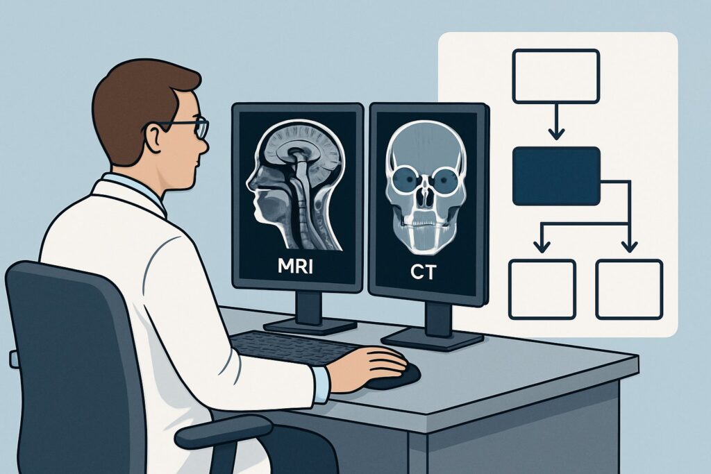
A 34-year-old patient presents to the emergency department with a four-day history of worsening facial pain and purulent nasal discharge, diagnosed as acute rhinosinusitis by their primary care physician. Tonight, however, their symptoms have escalated. They report new, painful swelling around the right eye, double vision, and pain with eye movement. You are concerned about the spread of infection beyond the sinuses. The critical clinical question is no longer if the patient has sinusitis, but whether they have developed a sight- or life-threatening orbital or intracranial complication. This is a time-sensitive decision, and choosing the right initial imaging study is paramount. According to the American College of Radiology (ACR) Appropriateness Criteria, for a patient with suspected orbital or intracranial complications of acute rhinosinusitis, MRI head without and with IV contrast is rated Usually appropriate.
Who Fits This Clinical Scenario?
This guidance applies specifically to patients with a clinical diagnosis of acute rhinosinusitis (symptoms for less than four weeks) who now exhibit “red flag” signs or symptoms suggestive of extrasinus extension. These alarm bells indicate the infection may have breached the bony confines of the paranasal sinuses and invaded adjacent critical structures.
Inclusion criteria for this workflow include one or more of the following findings in the context of acute sinusitis:
- Orbital Signs: Proptosis (bulging eye), ophthalmoplegia (paralysis or weakness of eye muscles), pain with eye movements, chemosis (swelling of the conjunctiva), or decreased visual acuity.
- Intracranial Signs: Severe, persistent headache (especially if localized or different from typical sinus pressure), altered mental status, seizures, focal neurologic deficits, or signs of meningeal irritation.
- “Pott’s Puffy Tumor”: A tender, boggy swelling over the frontal bone, indicating a subperiosteal abscess secondary to frontal sinusitis.
Conversely, this workflow is not for patients with uncomplicated acute rhinosinusitis, who typically require no imaging. It also differs from the workup for recurrent or chronic sinusitis, where imaging is used to evaluate anatomy for potential surgery, or for suspected invasive fungal sinusitis in an immunocompromised host, which presents a distinct and aggressive clinical picture.
What Diagnoses Are You Working Up in This Scenario?
When ordering imaging for complicated acute rhinosinusitis, you are investigating a spectrum of dangerous conditions that require prompt and specific management. The differential diagnosis guides the choice of imaging modality, as the primary goal is to identify purulent collections that may require surgical drainage and to assess for vascular or central nervous system involvement.
Orbital Cellulitis and Abscess: This is the most common complication. It’s crucial to distinguish preseptal (periorbital) cellulitis, an infection of the eyelid anterior to the orbital septum, from the more dangerous postseptal (orbital) cellulitis. Postseptal infection can progress to a subperiosteal or intra-orbital abscess—a collection of pus that displaces the orbital contents, compresses the optic nerve, and requires urgent surgical intervention to prevent permanent vision loss.
Cavernous Sinus Thrombosis: A rare but often fatal complication, this involves the formation of a septic thrombus in the cavernous sinus, typically from the spread of infection from the sphenoid or ethmoid sinuses. Patients may present with bilateral orbital signs, cranial nerve palsies (III, IV, V1, V2, VI), and severe headache. Imaging must be able to visualize the dural venous sinuses and their contents.
Intracranial Abscess or Empyema: Infection can spread directly through bone or via valveless veins into the intracranial space, leading to an epidural abscess, subdural empyema, or an intraparenchymal brain abscess. These conditions present with severe headache, fever, focal neurologic deficits, or seizures and represent a neurosurgical emergency.
Meningitis: Inflammation of the meninges can also result from the direct extension of sinusitis. While lumbar puncture is diagnostic, imaging is performed first to rule out a mass lesion or abscess that could cause herniation during the procedure. Imaging may show leptomeningeal enhancement.
Why Is MRI Head Without and With IV Contrast the Recommended Study?
The ACR rates MRI head without and with IV contrast as Usually appropriate because of its superior soft-tissue contrast, which is essential for distinguishing between the key diagnoses in this scenario. While other studies can be useful, MRI provides the most definitive information to guide urgent management.
The power of MRI lies in its ability to differentiate a drainable, ring-enhancing abscess from a phlegmon (a region of severe inflammation without a liquid pus collection). This distinction is critical, as phlegmon is typically managed with IV antibiotics alone, while an abscess requires surgical drainage. MRI, particularly with diffusion-weighted imaging (DWI), can clearly identify the restricted diffusion characteristic of viscous pus within an abscess cavity. Furthermore, it excels at detecting early signs of intracranial spread, such as dural enhancement, cerebritis, or thrombosis within the dural venous sinuses, which may be subtle or invisible on CT.
How do alternative studies compare for this specific scenario?
- CT maxillofacial with IV contrast: This study is also rated Usually appropriate. Its primary advantages are speed and superior visualization of bony anatomy, making it excellent for identifying the sinus of origin and any bony erosion that may be a pathway for infection. It is a strong alternative if MRI is contraindicated or unavailable. However, its ability to differentiate phlegmon from a small abscess is inferior to MRI, and it is less sensitive for early intracranial complications like meningitis or cavernous sinus thrombosis.
- CT head without IV contrast: This study is rated Usually not appropriate and represents a significant diagnostic pitfall. While it can show sinus opacification and perhaps gross orbital swelling, it cannot reliably identify an enhancing abscess rim, dural inflammation, or venous sinus thrombosis. Ordering a non-contrast study in this setting fails to answer the critical clinical questions and will almost always necessitate a follow-up contrast-enhanced study, delaying diagnosis and treatment.
From a safety perspective, MRI avoids ionizing radiation (0 mSv), a particularly important consideration in pediatric patients. In contrast, a CT maxillofacial with IV contrast delivers a dose of 0.1-1 mSv (☢☢) in adults, and a CT head with IV contrast delivers 1-10 mSv (☢☢☢). The use of IV gadolinium contrast with MRI is essential for this indication and is generally safe in patients with adequate renal function.
What’s Next After MRI? Downstream Workflow
The results of the MRI will dictate the immediate next steps in management, often involving urgent subspecialty consultation. The workflow branches based on the key findings.
If the MRI is positive for a subperiosteal or orbital abscess: This is a surgical emergency. An immediate consultation with both Otolaryngology (ENT) and Ophthalmology is required. The patient will need admission for high-dose IV antibiotics and prompt surgical drainage to decompress the orbit and prevent optic nerve damage.
If the MRI shows phlegmon/cellulitis without a drainable abscess: Management is typically medical. The patient should be admitted for IV antibiotics and close clinical monitoring of their vision and neurologic status. If the patient fails to improve or worsens on antibiotics, a repeat scan may be warranted to assess for interval development of an abscess.
If the MRI demonstrates cavernous sinus thrombosis or an intracranial abscess/empyema: This requires an emergent multidisciplinary response. Consult Neurosurgery and/or Neurology immediately. Treatment involves broad-spectrum IV antibiotics that cross the blood-brain barrier, and potentially anticoagulation for thrombosis or surgical evacuation for an intracranial collection.
If the MRI is negative: A negative MRI is highly reassuring and largely rules out a complicated, space-occupying infectious process. If significant clinical signs persist, reconsider the differential diagnosis. The patient may be managed with antibiotics for severe but uncomplicated sinusitis. If neurologic symptoms are the primary driver and the MRI is negative, the workup may shift towards other etiologies, potentially including a lumbar puncture if meningitis is still a concern and there is no contraindication.
Pitfalls to Avoid (and When to Get Help)
In this high-stakes clinical scenario, several common errors can delay diagnosis and worsen outcomes. Awareness of these pitfalls is key to effective management.
- Ordering a Non-Contrast Study: The single biggest pitfall is ordering a CT or MRI without intravenous contrast. The key pathologies—abscesses, dural enhancement, vascular complications—are defined by their enhancement patterns. A non-contrast study is inadequate.
- Delaying the Scan: Complications of sinusitis can progress rapidly, with vision loss or neurologic injury occurring within hours. Once suspected, imaging should be obtained emergently.
- Choosing CT Over MRI Without a Specific Reason: While contrast-enhanced CT is a valid option, MRI provides superior diagnostic information for the most critical questions. Unless MRI is contraindicated (e.g., incompatible implant, patient instability) or immediately unavailable, it is the preferred first-line study.
- Underestimating Clinical Signs: Do not dismiss subtle proptosis or mild pain with eye movements. These can be the earliest signs of a developing orbital abscess. Maintain a high index of suspicion.
If you see any red flag signs of orbital or intracranial involvement, escalate care immediately. This is not a condition to be managed in an outpatient setting and requires urgent imaging and subspecialty consultation.
Related ACR Topics and Tools
Navigating imaging decisions requires access to reliable, evidence-based resources. For a comprehensive overview of all clinical variants related to sinus imaging, see our parent guide. For tools to help with ordering and interpretation, see the resources below.
- For breadth across all scenarios in Sinonasal Disease, see our parent guide: Sinonasal Disease: ACR Appropriateness Decoded.
- To look up other clinical scenarios, use the Imaging Appropriateness Selector.
- For detailed technical parameters of the recommended study, explore the Imaging Protocol Library.
- To discuss radiation exposure with patients, especially when CT is considered, use the Radiation Dose Calculator.
Frequently Asked Questions
Why is MRI preferred over CT when CT with contrast is also rated ‘Usually Appropriate’?
MRI is generally preferred due to its superior soft-tissue contrast, which allows for a more confident distinction between a phlegmon (inflammation) and a drainable abscess. It is also more sensitive for detecting early intracranial complications like dural enhancement or venous sinus thrombosis. CT is a fast and excellent alternative, especially if MRI is unavailable or contraindicated.
Is a non-contrast MRI or CT ever sufficient in this scenario?
Almost never. Intravenous contrast is critical to identify the ring-enhancing wall of an abscess and to assess for dural or leptomeningeal inflammation. According to the ACR, a non-contrast CT head is ‘Usually not appropriate,’ and a non-contrast MRI head is only ‘May be appropriate,’ as it would miss key findings. Always order these studies ‘without and with IV contrast’ in this setting.
What should I order if my patient has a contraindication to MRI, like a pacemaker?
If MRI is contraindicated, the best alternative is ‘CT maxillofacial with IV contrast.’ This study is also rated ‘Usually appropriate’ by the ACR. It provides excellent detail of the sinuses and bony anatomy and, with contrast, can identify most abscesses, though it is less sensitive than MRI for early intracranial changes.
Does this imaging guidance apply to children?
Yes, the principles of diagnosis are the same in children, who can also develop these serious complications. The preference for MRI is even stronger in the pediatric population due to the complete avoidance of ionizing radiation. If CT must be performed, pediatric-specific low-dose protocols are essential.
What specific clinical signs should trigger this imaging pathway instead of just treating for uncomplicated sinusitis?
Any ‘red flag’ sign suggesting the infection has spread beyond the sinuses should trigger this emergent imaging pathway. Key signs include proptosis (bulging eye), pain with eye movements, decreased vision, double vision, severe localized headache, altered mental status, or swelling over the forehead (Pott’s puffy tumor).
Reviewed by Pouyan Golshani, MD, Interventional Radiologist — May 26, 2026