What Surveillance Imaging Is Best for an Adult After an Ischemic Stroke? An ACR-Guided Workflow
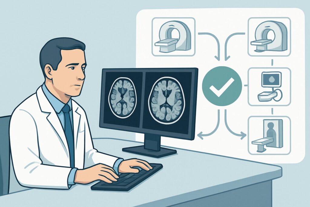
A 68-year-old male with a history of a left middle cerebral artery ischemic stroke one year ago presents for his annual primary care visit. He is neurologically stable, compliant with his secondary prevention medications, and has no new complaints. You are considering whether routine surveillance imaging is warranted to assess for changes and, if so, which study provides the most value with the least risk. This decision requires balancing the desire to monitor for silent events against the costs and potential harms of repeated imaging.
This article provides a detailed clinical workflow for surveillance imaging in an adult with a prior ischemic infarct, guided by the American College of Radiology (ACR) Appropriateness Criteria. For this specific scenario, a CT head without IV contrast is rated Usually appropriate.
Who Fits This Clinical Scenario for Stroke Surveillance Imaging?
This guidance applies to a specific patient population: adults with a known, previously documented ischemic infarct who are now being evaluated for surveillance purposes. The key inclusion criteria are:
- Adult patient.
- Confirmed history of a prior ischemic stroke. The initial event has already been diagnosed and imaged.
- Asymptomatic or neurologically stable. The patient has no new, worsening, or fluctuating focal neurologic deficits. The imaging is being performed for routine follow-up, not to evaluate an acute event.
It is critical to distinguish this scenario from similar but distinct clinical presentations that require a different imaging approach. This workflow does not apply if the patient presents with:
- New or worsening focal neurologic deficits: This presentation suggests an acute ischemic stroke or transient ischemic attack (TIA) and requires an urgent workup. This falls under the ACR scenarios for initial imaging of a suspected acute stroke.
- Resolved symptoms of a TIA: A patient with transient symptoms that have fully resolved needs an initial workup to identify the source of emboli or stenosis, which is a different clinical question than long-term surveillance.
- History of intraparenchymal hemorrhage: Follow-up imaging for a hemorrhagic stroke has a different set of goals, primarily assessing for hematoma evolution, and follows a separate ACR guideline.
What Diagnoses Are You Working Up in This Scenario?
Surveillance imaging after an ischemic stroke is not typically performed to find a new, emergent diagnosis but rather to monitor the brain parenchyma for changes that might influence long-term management. The primary goals are to assess for the evolution of the known infarct and to detect clinically silent events.
Evolution of the Prior Infarct: The most common and expected finding is the chronic stage of the known infarct. This includes established encephalomalacia (loss of brain tissue), gliosis (scarring), and ex-vacuo dilatation of the adjacent ventricle. Confirming these stable, expected changes provides a baseline and reassurance that no unexpected pathology has developed in that area.
New Silent Infarct: A key objective is to identify new areas of ischemia that occurred without producing overt clinical symptoms. The presence of new silent infarcts is a powerful marker of ongoing, poorly controlled cerebrovascular disease and may prompt an intensification of secondary prevention strategies, such as stricter blood pressure control, lipid management, or re-evaluation of antiplatelet or anticoagulant therapy.
Chronic Hemorrhagic Transformation or Microhemorrhages: Patients on antiplatelet or anticoagulant therapy are at risk for bleeding. While a large, symptomatic hemorrhage would present acutely, surveillance imaging can detect evidence of chronic, small-scale bleeding, such as hemosiderin deposition from prior petechial hemorrhagic transformation or cerebral microbleeds. This finding can be particularly relevant when considering the risks and benefits of ongoing antithrombotic therapy.
Exclusion of Alternative Pathologies: Less commonly, a patient’s subtle cognitive decline or non-specific symptoms might be attributed to their prior stroke. Surveillance imaging can help exclude other slowly progressive conditions that can mimic or co-exist with chronic cerebrovascular disease, such as a low-grade tumor, normal pressure hydrocephalus, or a chronic subdural hematoma.
Why Is CT Head Without IV Contrast the Recommended Study for This Presentation?
The ACR designates a CT head without IV contrast as Usually appropriate for routine surveillance imaging in an adult with a prior ischemic infarct. This recommendation is based on its ability to adequately answer the primary clinical questions in a rapid, accessible, and cost-effective manner.
A non-contrast CT is highly effective at visualizing the chronic sequelae of a prior infarct, which appears as a well-demarcated area of low attenuation (darkness) consistent with encephalomalacia and gliosis. It is also sufficiently sensitive to detect most new, subacute-to-chronic ischemic events, which would also appear as areas of low attenuation. Crucially, it remains the fastest and most reliable modality for excluding acute or chronic hemorrhage, which appears hyperdense (bright).
The radiation dose for a non-contrast head CT is relatively low (adult relative radiation level ☢☢☢, corresponding to 1-10 mSv), a reasonable trade-off for the diagnostic information gained in periodic surveillance.
Why Are Other Studies Rated Lower for Routine Surveillance?
While more advanced imaging may seem appealing, it is often not necessary for this specific indication and may not change management. The ACR rates other common studies as follows:
- MRI head without IV contrast: Rated May be appropriate (Disagreement). While MRI, particularly with sequences like Diffusion-Weighted Imaging (DWI) and Susceptibility-Weighted Imaging (SWI), is more sensitive than CT for detecting small, acute/subacute silent infarcts and cerebral microbleeds, there is disagreement among the expert panel about its routine use. The lack of clear evidence that these more sensitive findings consistently lead to changes in management that improve outcomes, combined with MRI’s higher cost, longer scan time, and limited accessibility, tempers the recommendation for its use in every asymptomatic surveillance case.
- CTA or MRA of the head and neck: Rated May be appropriate. These are vascular imaging studies designed to evaluate the arteries, not the brain parenchyma. They are not the correct initial choice for parenchymal surveillance. However, they become appropriate if the clinical question shifts to assessing the progression of a known stenosis (e.g., carotid or intracranial atherosclerosis) that was the presumed cause of the initial stroke. In that context, they are used to answer a different question, not for routine parenchymal follow-up.
For this scenario, the non-contrast CT provides the best balance of diagnostic utility and practicality. Once you’ve decided on this study, our protocol guide covers the technical parameters and interpretation principles in detail: CT Brain Without Contrast.
What’s Next After CT Head Without Contrast? Downstream Workflow
The results of the surveillance CT will guide your next steps in managing the patient’s secondary stroke prevention. The clinical workflow typically branches based on one of three outcomes.
If the study shows stable, expected chronic changes: This is the most common and reassuring outcome. The findings confirm the known history without demonstrating new or unexpected pathology.
- Next Step: Continue current medical management and secondary prevention strategies. Reassure the patient. No further routine imaging is indicated until a change in clinical status occurs.
If the study shows a new, previously unknown infarct (a silent infarct): This finding indicates that the patient’s cerebrovascular disease is active despite current therapy.
- Next Step: This should trigger a comprehensive re-evaluation of the patient’s risk factors and secondary prevention regimen. This includes assessing medication adherence, optimizing blood pressure and lipid control (e.g., higher intensity statin), and potentially reconsidering the antiplatelet or anticoagulant strategy.
If the study is indeterminate or shows unexpected findings: The CT may reveal subtle findings that are unclear, or it might show evidence of chronic microhemorrhages.
- Next Step: This is the primary situation where escalating to a more advanced modality is warranted. An MRI head without IV contrast, which is rated May be appropriate, would be the logical next step to better characterize the indeterminate finding or to quantify the burden of cerebral microbleeds using susceptibility-weighted sequences.
Pitfalls to Avoid (and When to Get Help)
When ordering surveillance imaging for a post-stroke patient, several common pitfalls can lead to suboptimal care or unnecessary testing. Be mindful of the following:
- Ordering surveillance imaging too frequently: There is no established high-level evidence to guide the optimal frequency of surveillance. Avoid annual or routine imaging without a specific clinical question or concern, as it can lead to incidental findings and patient anxiety without clear benefit.
- Using the wrong study for the question: Do not order a CTA or MRA if your goal is to assess the brain parenchyma. Conversely, do not rely on a non-contrast CT to evaluate for progression of arterial stenosis. Match the modality to the clinical question.
- Ignoring contraindications to advanced imaging: If you consider escalating to MRI, always screen for contraindications such as incompatible pacemakers, aneurysm clips, or other metallic implants.
- Misinterpreting age-related changes: Differentiating chronic small vessel ischemic changes from a new silent infarct on CT can be challenging. A low threshold for radiologist consultation or comparison with prior studies is essential.
If the surveillance CT shows a new infarct in a patient who is already on maximal medical therapy, consider a consultation with a stroke neurologist to discuss advanced secondary prevention options.
Related ACR Topics and Tools
This article focuses on a single clinical scenario. For a comprehensive overview of imaging recommendations across all stroke-related presentations, from TIA to acute stroke to hemorrhage, please see our parent guide. You can also use the tools below to explore other scenarios, protocols, and radiation safety topics.
- For breadth across all scenarios in Cerebrovascular Diseases-Stroke and Stroke-Related Conditions, see our parent guide: Cerebrovascular Diseases-Stroke and Stroke-Related Conditions: ACR Appropriateness Decoded.
- ACR Appropriateness Criteria Lookup — for adjacent scenarios
- Imaging Protocol Library — for technique on the recommended study
- Radiation Dose Calculator — for cumulative dose conversations
Frequently Asked Questions
How often should surveillance imaging be performed after an ischemic stroke?
There is no consensus guideline on the optimal frequency. Imaging should be driven by a specific clinical question rather than a fixed schedule. Many clinicians consider a baseline follow-up scan 6-12 months post-stroke, with further imaging only if there is a clinical change or a need to reassess risk before modifying therapy.
If my patient has new symptoms, is a non-contrast CT still the right first step?
No. If a patient with a prior stroke develops new or worsening neurologic symptoms, this should be treated as a suspected acute stroke. The imaging workup is more urgent and often involves a combination of non-contrast CT, CT angiography (CTA), and possibly CT perfusion to assess for a new ischemic event and treatment eligibility.
Why is MRI only ‘May be appropriate’ if it’s more sensitive for silent strokes?
The ACR panel’s ‘Disagreement’ on this rating reflects the clinical uncertainty. While MRI is technically more sensitive, it’s not clear that detecting additional, subtle silent infarcts or microbleeds in an otherwise stable, asymptomatic patient consistently leads to management changes that improve long-term outcomes. Therefore, the higher cost and lower availability of MRI make the more practical non-contrast CT the ‘Usually appropriate’ choice for routine surveillance.
Does this guidance apply to patients with a history of hemorrhagic stroke?
No, this guidance is specific to ischemic stroke. Follow-up imaging for intraparenchymal hemorrhage follows a different ACR Appropriateness Criteria variant, as the clinical questions (e.g., hematoma stability, detection of underlying lesions like an AVM or tumor) are different.
Should I order a CT with IV contrast for surveillance?
Generally, no. A CT head with IV contrast is rated ‘Usually not appropriate’ for this scenario. Contrast is used to assess for blood-brain barrier breakdown (e.g., in an acute/subacute infarct, tumor, or infection) and is not necessary for routine surveillance of a chronic, stable infarct. It adds risk (contrast reaction, nephrotoxicity) and radiation dose without providing relevant information for this specific clinical question.
Reviewed by Pouyan Golshani, MD, Interventional Radiologist — May 30, 2026