What Is the Best Imaging for Suspected Spine Infection from a Decubitus Ulcer?
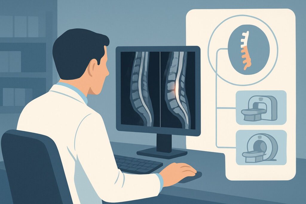
An 82-year-old patient from a skilled nursing facility presents with a non-healing sacral decubitus ulcer, new-onset localized back pain, fever, and elevated inflammatory markers. You are concerned about direct extension of the infection to the underlying spine, a common and dangerous complication. The critical clinical question is whether there is underlying discitis-osteomyelitis or a spinal epidural abscess. Deciding on the right initial imaging study is crucial for timely diagnosis and management, as a delay can lead to severe neurologic consequences. According to the American College of Radiology (ACR) Appropriateness Criteria, for this specific presentation, an MRI of the spine area of interest without and with IV contrast is rated Usually Appropriate. This article provides a detailed workflow for this exact clinical scenario.
Who Fits This Clinical Scenario?
This guidance is specifically for patients where a spine infection is suspected due to a contiguous, overlying source. The key inclusion criterion is the presence of a decubitus ulcer, pressure sore, or other wound directly overlying the spine. This creates a direct pathway for bacteria to invade deeper structures, making the clinical suspicion for discitis-osteomyelitis or an epidural abscess particularly high.
This workflow is distinct from other presentations of suspected spine infection. It does not apply to patients who present with:
- New or worsening back pain without an overlying wound: This scenario suggests a hematogenous (blood-borne) source of infection rather than direct extension and follows a different diagnostic pathway.
- Recent spinal intervention or surgery: Post-procedural infections have a unique set of imaging considerations, as post-operative changes can mimic or obscure signs of infection.
- Acute, severe neurologic deficits or cauda equina syndrome: While infection may be the cause, this presentation is a neurologic emergency that may require a different pace and sequence of imaging and consultation.
Correctly identifying your patient’s scenario is the first step to ensuring the most diagnostically useful and appropriate imaging is ordered.
What Diagnoses Are You Working Up in This Scenario?
When a patient has a wound overlying the spine, your imaging order is intended to confirm or exclude several specific, high-morbidity diagnoses that can arise from direct microbial invasion.
Vertebral Discitis-Osteomyelitis
This is often the primary concern. It refers to infection of the intervertebral disc and the adjacent vertebral bodies. In this scenario, bacteria from the skin ulcer can track directly through the posterior soft tissues to the bone and disc space. Early diagnosis is key to preventing vertebral collapse, deformity, and further spread.
Spinal Epidural Abscess (SEA)
This is the most feared complication and a true neurologic emergency. An SEA is a collection of pus in the space between the dura mater and the vertebral column. As the abscess expands, it can compress the spinal cord or nerve roots, leading to rapidly progressive neurologic deficits, paralysis, and even death. The presence of an overlying wound significantly increases the risk of an SEA.
Paraspinal or Psoas Abscess
The infection may not be confined to the spinal column itself but can spread into the adjacent soft tissues, forming abscesses in the paraspinal muscles or, if lower in the lumbar spine, the psoas muscle. These collections can be a persistent source of sepsis and may require separate drainage procedures.
Phlegmon or Deep Soft Tissue Infection
In some cases, the infection may be limited to a severe cellulitis or phlegmon (an unencapsulated inflammatory mass) in the deep soft tissues without frank abscess or bone involvement. Distinguishing a drainable abscess from a non-drainable phlegmon is a critical role of advanced imaging.
Why Is MRI Without and With IV Contrast the Recommended Study?
The ACR rates MRI spine area of interest without and with IV contrast as Usually Appropriate because it is uniquely capable of evaluating all the potential pathologies in this scenario with high sensitivity and specificity. Its superior soft-tissue contrast is essential for detecting the early signs of infection that other modalities miss.
The rationale for MRI’s superiority includes:
- Detecting Early Osteomyelitis: MRI can detect abnormal signal in the bone marrow from edema and inflammation days to weeks before bony destruction becomes visible on radiographs or CT. T2-weighted and STIR sequences are particularly sensitive for this.
- Defining Abscesses: The combination of pre- and post-contrast T1-weighted sequences is the gold standard for identifying and characterizing fluid collections like an epidural or paraspinal abscess. The classic finding is a fluid collection with a peripherally enhancing rim after gadolinium administration.
- Assessing Neural Element Compression: MRI provides direct visualization of the spinal cord, cauda equina, and nerve roots, allowing for precise assessment of any compression or inflammation caused by an abscess or phlegmon.
Why are other studies rated lower for this scenario?
- Radiography spine area of interest is rated May be appropriate. While easy to obtain, plain films are notoriously insensitive for early spine infection. Findings like disc space narrowing and endplate erosion are late-stage signs, and a negative radiograph cannot rule out a clinically significant infection.
- CT spine area of interest with IV contrast is also rated May be appropriate. CT is excellent for assessing bony destruction but is far less sensitive than MRI for detecting early marrow changes, epidural phlegmon, or small abscesses. It is primarily a second-line option when MRI is contraindicated or unavailable.
From a safety perspective, MRI is the preferred modality as it involves no ionizing radiation (0 mSv). The use of intravenous gadolinium contrast is critical for characterizing abscesses and inflammation, but requires appropriate screening for renal dysfunction. The “without and with” contrast protocol is essential; the pre-contrast images provide a baseline to accurately assess enhancement patterns. Once you’ve decided on this study, our protocol guide covers the technical details. For technique, reading principles, and sequence specifics, see our guide: MRI Lumbar Spine Without Contrast.
What’s Next After MRI? Downstream Workflow
The results of the spine MRI will directly guide your next steps in management, which often involves a multidisciplinary team including infectious disease, spine surgery, and interventional radiology.
- Positive for Epidural Abscess: This is a surgical emergency. The immediate next step is an urgent consultation with a spine surgeon for consideration of surgical decompression and washout. Delays can result in permanent neurologic injury. Concurrently, an infectious disease consult should be placed to guide antibiotic therapy based on blood cultures and, eventually, operative cultures.
- Positive for Discitis-Osteomyelitis (without large abscess): Management is typically non-operative initially. The next step is to obtain tissue for culture to guide long-term antibiotic therapy. This is usually done via CT-guided biopsy by an interventional radiologist. An infectious disease consultation is essential.
- Negative for Spine Infection: If the MRI is negative for osteomyelitis and abscess, the focus returns to managing the superficial wound and cellulitis. The source of the patient’s systemic symptoms (fever, elevated inflammatory markers) must be re-evaluated. However, a negative high-quality MRI provides strong evidence against a deep spinal infection, allowing you to de-escalate concern for this specific pathology.
- Indeterminate Findings: Occasionally, MRI findings can be equivocal, such as in a patient with extensive degenerative changes or post-operative hardware. In these cases, a nuclear medicine study like a Gallium scan (May be appropriate) could be considered to provide complementary functional information about inflammation, though this is less common now with modern MRI techniques.
Pitfalls to Avoid (and When to Get Help)
Navigating this clinical scenario requires vigilance to avoid common diagnostic and management errors.
- Pitfall 1: Relying on a negative radiograph. Never rule out a spine infection based on normal plain films. They are highly insensitive in the early stages of the disease.
- Pitfall 2: Ordering MRI without contrast. While a non-contrast MRI can show marrow edema, it is inadequate for diagnosing an epidural abscess. IV contrast is mandatory if an abscess is on the differential.
- Pitfall 3: Delaying imaging in the setting of neurologic symptoms. If a patient with a spinal wound develops new weakness, sensory changes, or bowel/bladder dysfunction, do not wait. This is a red flag for cord compression, and imaging should be obtained emergently.
If you see any neurologic red flags or the MRI confirms a spinal epidural abscess, escalate immediately with an urgent call to your on-call spine surgery service.
Related ACR Topics and Tools
This article covers one specific variant of suspected spine infection. For a comprehensive overview of all related clinical scenarios, from post-operative concerns to patients with no clear infectious source, please consult our parent topic hub. You can also use the tools below to explore other ACR guidelines, imaging protocols, and radiation dose information.
- For breadth across all scenarios in Suspected Spine Infection, see our parent guide: Suspected Spine Infection: ACR Appropriateness Decoded.
- ACR Appropriateness Criteria Lookup — for adjacent scenarios
- Imaging Protocol Library — for technique on the recommended study
- Radiation Dose Calculator — for cumulative dose conversations
Frequently Asked Questions
Why is MRI with and without contrast ‘Usually Appropriate’ while MRI with contrast only is ‘Usually Not Appropriate’?
The pre-contrast (without contrast) sequences, particularly T1-weighted images, are essential to establish a baseline. They help differentiate tissues that are naturally bright on T1 (like fat or subacute hemorrhage) from areas of true pathologic enhancement after contrast is given. Without the pre-contrast comparison, it can be difficult to confidently diagnose an abscess or inflamed tissue, making the study less accurate.
What if my patient has a pacemaker or other contraindication to MRI?
If MRI is absolutely contraindicated, the next best test is ‘CT spine area of interest with IV contrast,’ which is rated ‘May be appropriate.’ While less sensitive than MRI, it can still identify significant bony destruction and larger fluid collections or abscesses. A nuclear medicine study, such as a 3-phase bone scan, may also be considered to look for osseous inflammatory changes, but it lacks the anatomic detail of CT or MRI.
Does the location of the decubitus ulcer (e.g., sacral vs. thoracic) change the imaging recommendation?
No, the recommendation for MRI without and with IV contrast remains the same regardless of the spinal level. However, the ‘area of interest’ must be specified correctly on the order. You should order the MRI for the specific spinal segment directly underlying the wound (e.g., ‘MRI Lumbar Spine’ for a sacral ulcer, ‘MRI Thoracic Spine’ for a mid-back wound).
My patient’s inflammatory markers (ESR/CRP) are normal. Can I rule out a spine infection?
While highly sensitive, normal inflammatory markers do not definitively rule out a spine infection, especially in immunocompromised or elderly patients who may not mount a robust systemic response. If clinical suspicion remains high due to the overlying wound and localized pain, imaging with MRI is still warranted.
Should I wait for blood cultures to come back before ordering the MRI?
No, you should not delay imaging. If there is clinical concern for a serious diagnosis like an epidural abscess, the MRI should be performed as soon as possible. Blood cultures should be drawn before starting antibiotics, but imaging should proceed concurrently. A positive blood culture can help tailor antibiotic therapy later, but it doesn’t replace the need for anatomic evaluation with MRI.
Reviewed by Pouyan Golshani, MD, Interventional Radiologist — May 29, 2026