New VCF on Radiograph with Malignancy History: Should You Order MRI or CT?
It’s a busy afternoon in clinic when you review the lumbar spine radiographs for your 72-year-old patient with a history of breast cancer, who presents with new, severe, focal back pain. The report confirms your suspicion: a new L2 vertebral compression fracture (VCF). The critical question is no longer if there is a fracture, but why. Is this an unfortunate but benign osteoporotic fracture, or is it a pathologic fracture from metastatic disease? Choosing the next imaging study is crucial for guiding therapy, from simple pain management to urgent oncology and radiation oncology consultation. This article provides a step-by-step workflow for this specific scenario, based on the American College of Radiology (ACR) Appropriateness Criteria, which rate MRI of the spine without and with IV contrast as Usually Appropriate.
Who Fits This Clinical Scenario?
This guidance is specifically for patients who meet all the following criteria: a new, symptomatic vertebral compression fracture has been identified on initial radiographs, and the patient has a known history of malignancy. The presence of symptoms, such as focal pain, is a key differentiator, as it suggests an acute or subacute process that requires prompt characterization.
This workflow does not apply to several similar-sounding but distinct clinical situations. If your patient has a new symptomatic VCF but no known history of malignancy, the workup prioritizes benign causes, and the imaging rationale shifts. Similarly, this guidance is not intended for patients with an asymptomatic VCF found incidentally, even with a cancer history, as the urgency and differential diagnosis are different. Finally, patients with new back pain who have a previously treated VCF or a history of vertebral augmentation represent a separate scenario where post-treatment changes can complicate the imaging interpretation, requiring a distinct approach.
What Diagnoses Are You Working Up in This Scenario?
In a patient with a known malignancy, a new symptomatic VCF raises immediate concern for a pathologic fracture. The primary goal of advanced imaging is to differentiate this from other potential causes. The differential diagnosis drives the choice of imaging modality.
Pathologic Fracture from Metastasis: This is the primary concern and the reason for the specific imaging pathway. Cancers that commonly metastasize to bone include breast, lung, prostate, kidney, and thyroid. A metastatic lesion weakens the vertebral body, leading to its collapse under physiologic load. Identifying a malignant fracture is critical as it impacts cancer staging and often requires urgent oncologic or radiation therapy, particularly if there is concern for spinal cord compression.
Benign Osteoporotic Compression Fracture: Patients with cancer are often older and may have treatment-related or age-related osteoporosis, making them susceptible to benign VCFs. While common, this is a diagnosis of exclusion in this clinical context. Imaging must confidently rule out underlying malignancy before a fracture can be attributed to osteoporosis alone.
Multiple Myeloma or Lymphoma: While technically malignancies, these may represent a new or primary diagnosis rather than a metastasis from a known solid tumor. These hematologic malignancies can present with lytic bone lesions and pathologic fractures, and advanced imaging is key to their detection.
Infection (Osteomyelitis/Diskitis): Though less common, spinal infection can weaken a vertebral body and cause collapse. Patients with cancer can be immunocompromised from their disease or its treatment, increasing their risk. Imaging features of infection, such as disc space involvement and adjacent soft tissue abscess, are important to identify as the management is entirely different.
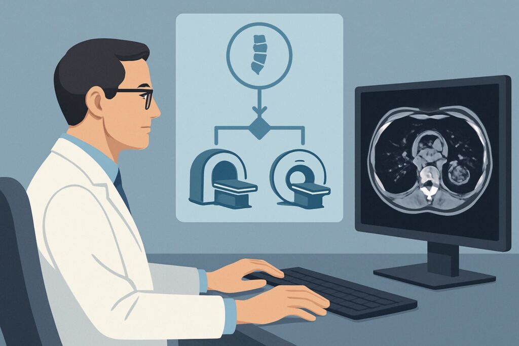
Why Is MRI of the Spine Without and With IV Contrast the Recommended Study?
The ACR panel rates ‘MRI spine area of interest without and with IV contrast’ as Usually Appropriate because of its superior ability to evaluate bone marrow and soft tissues, which is essential for distinguishing a malignant fracture from a benign one. MRI provides detailed information that other modalities cannot, directly addressing the key questions in this scenario.
The strength of MRI lies in its sensitivity for detecting marrow-replacing lesions. On T1-weighted images, normal fatty marrow is bright; tumor infiltration replaces this fat and appears dark. Post-contrast sequences are critical for assessing enhancement patterns. Malignant lesions typically demonstrate avid enhancement, helping to differentiate them from the edema of a benign fracture. Furthermore, MRI is unparalleled in evaluating the posterior elements of the vertebra, the epidural space, and the spinal cord itself, identifying any signs of neural compression that constitute a neurologic emergency.
How do alternative studies compare for this specific presentation?
- CT spine area of interest without IV contrast is also rated Usually Appropriate. CT provides excellent bone detail and can clearly define the fracture anatomy. However, it is less sensitive than MRI for detecting bone marrow infiltration and cannot directly visualize the spinal cord. It is a valuable alternative when MRI is contraindicated or unavailable.
- A whole-body bone scan (scintigraphy) is rated May be appropriate. While it can detect areas of increased bone turnover throughout the skeleton, it is notoriously non-specific. It cannot reliably distinguish between a fracture, metastasis, infection, or degenerative change. Its primary role is in systemic staging, not in characterizing a specific, known fracture.
From a safety perspective, MRI is the preferred choice as it involves no ionizing radiation (0 mSv). This is a significant advantage in patients who may have undergone multiple prior imaging studies as part of their cancer care. Once you’ve decided on the study, our protocol guide can help with the technical details. While the top recommendation includes contrast to evaluate for malignancy, the fundamental sequences are covered in our MRI Lumbar Spine Without Contrast protocol guide.
What’s Next After MRI? Downstream Workflow
The results of the contrast-enhanced MRI will guide your next steps, branching into distinct clinical pathways. The goal is to move from diagnosis to appropriate management swiftly and confidently.
If the MRI confirms a pathologic fracture: Findings suggestive of malignancy include abnormal marrow signal replacing normal fat, convex posterior border of the vertebral body, pedicle involvement, and an associated soft tissue mass. The presence of epidural extension or cord compression is an emergency. The next step is an urgent consultation with Medical Oncology and Radiation Oncology to discuss systemic therapy, radiation, and potential surgical intervention for spinal stabilization.
If the MRI suggests a benign osteoporotic fracture: Features favoring a benign cause include a preserved normal fatty marrow signal, a fluid sign (intravertebral cleft), and lack of a discrete soft tissue mass or posterior element involvement. In this case, the patient can be managed conservatively with pain control, bracing, and treatment for osteoporosis. The focus shifts away from the oncologic workup for this specific finding.
If the MRI is indeterminate or equivocal: Sometimes, the imaging features overlap, and a definitive distinction cannot be made. In this situation, the next step is often an ‘Image-guided biopsy spine area of interest,’ which the ACR rates as May be appropriate. A biopsy provides a tissue diagnosis to definitively guide treatment. Alternatively, ‘FDG-PET/CT’ (May be appropriate) may be considered to assess the metabolic activity of the lesion and evaluate for other sites of disease, which can help clarify the diagnosis.
Pitfalls to Avoid (and When to Get Help)
Navigating this scenario requires careful attention to detail to avoid common missteps. First, the most significant pitfall is anchoring on a diagnosis of a benign osteoporotic fracture without a definitive imaging workup, potentially delaying the diagnosis of metastatic disease. In a patient with a known cancer history, a new VCF should be considered malignant until proven otherwise. Second, failing to order IV contrast with the MRI can severely limit its diagnostic utility, as enhancement patterns are key to differentiating tumor from edema. Third, underappreciating subtle signs of spinal cord or nerve root compression on imaging or in the clinical exam can lead to catastrophic neurologic decline; any new neurologic symptom warrants immediate escalation and imaging.
Related ACR Topics and Tools
This article focuses on one specific clinical question. For a comprehensive overview of all related scenarios, from asymptomatic fractures to post-treatment evaluation, please consult our parent guide. Additional GigHz tools can help you apply these criteria in your daily practice.
- For breadth across all scenarios in Management of Vertebral Compression Fractures, see our parent guide: Management of Vertebral Compression Fractures: ACR Appropriateness Decoded.
- To look up other clinical scenarios, use the ACR Appropriateness Criteria Lookup tool.
- For detailed procedural techniques, explore the Imaging Protocol Library.
- To discuss cumulative radiation exposure with patients, reference the Radiation Dose Calculator.
Frequently Asked Questions
Why not just order a CT scan? It’s faster and more available.
While a non-contrast CT of the spine is also rated ‘Usually Appropriate’ by the ACR and is excellent for evaluating bone anatomy, it is significantly less sensitive than MRI for assessing bone marrow infiltration and the spinal cord. In a patient with a history of malignancy, the primary question is whether the fracture is pathologic, which is a question of marrow and soft tissue. MRI with and without contrast is superior for answering this specific question.
What if my patient has a pacemaker or other contraindication to MRI?
If MRI is contraindicated, ‘CT spine area of interest without IV contrast’ is the best alternative and is also rated ‘Usually Appropriate’. While less sensitive for marrow disease, it can reveal lytic or blastic changes suggestive of metastasis. If the CT is inconclusive, a ‘Bone scan whole body’ or ‘FDG-PET/CT’ (both rated ‘May be appropriate’) could be considered for further evaluation, followed by a CT-guided biopsy if a tissue diagnosis is needed.
Is a bone scan a reasonable first choice after the radiograph?
A bone scan is rated ‘May be appropriate’ but is generally not the preferred next step for characterizing a known fracture. Its limitation is a lack of specificity; it shows areas of increased bone turnover but cannot reliably differentiate a malignant process from a benign fracture, infection, or degenerative change. Its main utility is in screening the entire skeleton for other potential lesions, not in defining the nature of the known VCF.
Does every VCF in a patient with a cancer history require a biopsy?
No. A biopsy is typically reserved for cases where advanced imaging, like a contrast-enhanced MRI, is indeterminate or equivocal. If the MRI shows classic features of a benign osteoporotic fracture or definitive features of a pathologic fracture consistent with the patient’s known primary cancer, a biopsy may not be necessary to guide the immediate next steps in management.
What specific MRI findings strongly suggest a malignant fracture?
Key MRI features that point towards a malignant VCF include: involvement of the posterior elements (like the pedicles), a convex posterior border of the vertebral body (bulging into the spinal canal), the presence of an associated epidural or paraspinal soft tissue mass, and diffuse, abnormal T1-hypointense marrow signal that avidly enhances with contrast. In contrast, benign fractures more often spare the posterior elements and may show a fluid-filled cleft or a band-like pattern of edema with preserved fatty marrow.
Reviewed by Pouyan Golshani, MD, Interventional Radiologist — May 30, 2026