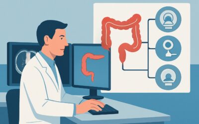
Which Imaging Study Should You Order for an Unstable Lower GI Bleed?
It’s 3 AM, and your patient in the intensive care unit is crashing. They have a known history of diverticulosis, and after a large-volume, bright red bowel movement, their blood pressure is dropping and their heart rate is climbing. They are now receiving their sixth unit of packed red blood cells in the last 18 hours. The gastroenterology and interventional radiology teams are on the phone, and everyone is looking to you for the next step. What is the right imaging study to order for a patient with a massive, active lower gastrointestinal (GI) bleed who is hemodynamically unstable? This article provides a clinical workflow for this specific, high-stakes scenario. According to the American College of Radiology (ACR) Appropriateness Criteria, the definitive next step is a study rated as ‘Usually Appropriate’: CTA abdomen and pelvis without and with IV contrast.
Who Fits This Clinical Scenario?
This guidance is specifically for patients with a suspected lower GI bleed who are critically ill. The key inclusion criteria are:
* **Hemodynamic Instability:** This includes hypotension (e.g., systolic blood pressure < 90 mmHg), persistent tachycardia, signs of shock, or requiring vasopressor support. * **Massive Transfusion Requirement:** The patient has required more than 5 units of packed red blood cells within a 24-hour period to maintain hemodynamic stability. * **Evidence of Active Bleeding:** There is ongoing hematochezia or clinical suspicion of a brisk, active hemorrhage.This workflow does **not** apply to patients who are hemodynamically stable, even if they have observed hematochezia. Those patients may be better served by a different diagnostic pathway, often starting with colonoscopy. Similarly, this guidance is distinct from the workup for a patient with an obscure, intermittent, or slow GI bleed, where the diagnostic urgency and imaging choices differ significantly. This article is for the acute, life-threatening hemorrhage where time is critical and the primary goal is to localize the bleeding source for immediate intervention.
What Diagnoses Are You Working Up in This Scenario?
In a massive lower GI bleed, the imaging study is intended to rapidly identify the source from a specific list of potential causes. The differential diagnosis in this high-acuity setting is focused on pathologies known to cause brisk, arterial hemorrhage.
**Diverticular Hemorrhage** is the most common cause of major lower GI bleeding. The hemorrhage occurs when a penetrating artery at the neck or dome of a diverticulum erodes. This bleeding is typically arterial, abrupt, and can be massive. CTA is highly effective at identifying the specific diverticulum with active contrast extravasation.
**Angiodysplasia** (also known as arteriovenous malformation or AVM) is another primary consideration, particularly in older adults. These are dilated, tortuous submucosal vessels that are prone to bleeding. While often associated with slower, chronic bleeding, they can also be the source of a massive, acute hemorrhage, most commonly in the right colon.
**Ischemic Colitis**, while often causing self-limited bleeding, can occasionally present with significant hematochezia and instability, especially in patients with underlying vascular disease or low-flow states. CTA can not only identify a potential bleeding site but also show ancillary findings like bowel wall thickening, edema, or lack of enhancement characteristic of ischemia.
**Aortoenteric Fistula** is a rare but catastrophic cause of massive GI bleeding that must be considered in any patient with a prior history of aortic surgery or aneurysm repair. It represents a direct communication between the aorta and the bowel lumen. This is a surgical emergency, and CTA is the primary and most effective imaging modality for making this critical diagnosis.
Why Is CTA Abdomen and Pelvis Without and With IV Contrast the Recommended Study?
For a hemodynamically unstable patient with a massive lower GI bleed, the ACR rates CTA abdomen and pelvis without and with IV contrast as ‘Usually Appropriate’. The rationale is rooted in the need for speed, accuracy, and actionable information to guide immediate, life-saving intervention.
The primary advantage of multiphase CTA is its ability to rapidly and precisely localize the site of active bleeding. The protocol involves a non-contrast phase to identify baseline hyperdense material (like surgical clips or intramural hematoma), followed by arterial and portal venous phases. Active bleeding appears as a “blush” of high-density contrast extravasating into the bowel lumen, which increases in size and intensity on delayed phases. This localization is critical for guiding the next step, which is often transcatheter arteriography.
Let’s compare this to the alternatives:
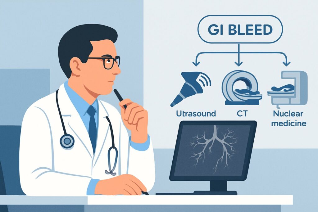
* **Tagged Red Blood Cell (RBC) Scan:** This study is rated ‘Usually not appropriate’ in this scenario. While sensitive for detecting slower bleeds, it takes too long to perform (often over an hour) in a patient who is hemodynamically unstable. Furthermore, its spatial localization is often imprecise, providing a general region of bleeding rather than an exact vessel, which is less helpful for subsequent intervention.
* **Diagnostic/Therapeutic Colonoscopy:** Rated as ‘May be appropriate (Disagreement)’, colonoscopy is extremely challenging in this setting. The patient’s instability makes sedation risky, and the colon is typically filled with blood, making visualization nearly impossible without a full bowel preparation—a process that is not feasible in an emergency.
The radiation dose for CTA is a relevant consideration (ACR Relative Radiation Level ☢☢☢☢, or 10-30 mSv), but in the context of a life-threatening hemorrhage, the diagnostic benefit of quickly finding the bleeding source far outweighs the risk. The speed and anatomic detail provided by CTA make it the superior choice for directing therapy and improving outcomes.
Once you’ve decided on CTA abdomen and pelvis without and with IV contrast, our protocol guide covers the technique, contrast, and reading principles: CT Abdomen/Pelvis Without Contrast (Renal Stone).
What’s Next After CTA Abdomen and Pelvis Without and With IV Contrast? Downstream Workflow
The results of the CTA will dictate the immediate next steps in patient management, typically in close coordination with interventional radiology and gastroenterology.
* **If the CTA is positive (shows active extravasation):** This is a clear indication for intervention. The patient should be immediately prepared for transcatheter arteriography. The CTA serves as a roadmap for the interventional radiologist, who will selectively catheterize the bleeding artery (e.g., a branch of the superior or inferior mesenteric artery) and perform embolization to stop the hemorrhage. This is also rated ‘Usually appropriate’ by the ACR as the direct therapeutic follow-up.
* **If the CTA is negative (no active extravasation):** A negative CTA in a patient who was clinically bleeding suggests the hemorrhage may have temporarily stopped or is too slow to be detected by the scan (typically <0.5 mL/min). The next step depends on the patient's clinical course. If they remain unstable or have another episode of bleeding, a repeat CTA may be considered. If they stabilize, they may become a candidate for colonoscopy once medically optimized and the colon is prepped, which can identify non-bleeding stigmata of a recent hemorrhage or other lesions like tumors or ulcers.* **If the CTA is indeterminate or shows a non-bleeding cause:** The scan may reveal an alternative diagnosis for the patient's condition, such as severe ischemic colitis, a tumor, or an aortoenteric fistula. Each of these findings triggers a different downstream pathway—consultation with general or vascular surgery, for instance.
Pitfalls to Avoid (and When to Get Help)
In this high-pressure scenario, several common pitfalls can compromise the diagnostic utility of the imaging and delay care.
* **Delaying the Scan:** Time is critical. Do not wait for the patient to be perfectly stable before ordering the CTA; the purpose of the scan is to find the cause of the instability to allow for definitive treatment.
* **Inadequate IV Access:** A high-quality multiphase CTA requires a good-bore (e.g., 18-gauge) intravenous line and a high rate of contrast injection. Ensure adequate IV access is established before the patient goes to the scanner.
* **Ordering the Wrong Protocol:** Simply ordering a “CT Abdomen/Pelvis with contrast” may result in a standard single-phase scan. You must specifically request a “GI Bleed Protocol” or “Multiphase CTA” to ensure non-contrast, arterial, and venous phases are acquired.
* **Lack of Communication:** Before the scan, call the radiologist to provide clinical context. After the scan, if it is positive, immediately contact the on-call interventional radiologist to mobilize the team for embolization.
If the CTA is positive for a life-threatening cause like an aortoenteric fistula, escalate immediately to vascular surgery while also coordinating with interventional radiology.
Related ACR Topics and Tools
This article focuses on a single, critical decision point. For a comprehensive overview of imaging for all related clinical presentations, further resources are available.
* For breadth across all scenarios in Radiologic Management of Lower Gastrointestinal Tract Bleeding, see our parent guide: Radiologic Management of Lower Gastrointestinal Tract Bleeding: ACR Appropriateness Decoded.
* The GigHz platform offers several tools to support evidence-based imaging decisions:
- ACR Appropriateness Criteria Lookup — for adjacent scenarios
- Imaging Protocol Library — for technique on the recommended study
- Radiation Dose Calculator — for cumulative dose conversations
Frequently Asked Questions
Why not go straight to interventional radiology for an angiogram instead of getting a CTA first?
While transcatheter arteriography is the primary treatment, it is more invasive and time-consuming as a purely diagnostic tool. A ‘blind’ angiogram without a known target can involve extensive catheter work and contrast load. CTA is much faster at confirming active bleeding and provides a precise roadmap, making the subsequent angiogram quicker, safer, and more targeted, which is critical in an unstable patient.
What if the patient has renal failure? Can we still perform a CTA with contrast?
In a patient with a life-threatening hemorrhage, the risk of not performing the CTA and failing to localize the bleed is far greater than the risk of contrast-induced nephropathy. This is a classic risk-versus-benefit decision. The ACR and most institutional guidelines support the use of IV contrast in this emergency setting, often with pre- and post-procedure hydration if clinically feasible.
How sensitive is CTA for detecting a lower GI bleed?
The sensitivity of multiphase CTA is highly dependent on the rate of bleeding at the exact moment of the scan. It can reliably detect bleeding rates as low as 0.3 to 0.5 mL/minute. If the bleeding is intermittent and has temporarily stopped during the scan, the CTA will be negative. This is why a negative scan doesn’t completely rule out a source, but a positive scan is highly specific and actionable.
Is there any role for MRA in this acute, unstable setting?
No. The ACR rates MRA of the abdomen and pelvis as ‘Usually not appropriate’ for this scenario. MRA is significantly slower to acquire than CTA, is more susceptible to motion artifact (a major issue in an unstable patient), and is less readily available on an emergency basis in most hospitals. CTA remains the superior modality for speed and diagnostic utility in acute GI bleeding.
What if the CTA suggests an upper GI source of bleeding instead?
While the clinical presentation might suggest a lower GI source, massive hematochezia can sometimes be caused by a very brisk upper GI bleed (e.g., a duodenal ulcer eroding into an artery). CTA of the abdomen and pelvis covers the upper abdomen and can often identify these sources. If an upper GI source is found, the downstream workflow shifts immediately towards consultation with gastroenterology for an emergent upper endoscopy (EGD) and/or interventional radiology for embolization.
Reviewed by Pouyan Golshani, MD, Interventional Radiologist — May 21, 2026