Which Imaging Study Is Best for Active Lower GI Bleeding in a Stable Patient?
It’s 10 p.m. in the emergency department, and you are evaluating a 68-year-old male with multiple episodes of bright red blood per rectum that started this evening. His vital signs are stable, with a blood pressure of 125/80 mm Hg and a heart rate of 88 beats per minute. He is alert and reports no significant abdominal pain. After initial resuscitation with intravenous fluids, his hemoglobin is stable. The immediate clinical question is how to localize the source of this active lower gastrointestinal (GI) bleeding to guide further management. This article provides a detailed workflow for this specific scenario, grounded in the American College of Radiology (ACR) Appropriateness Criteria. For a hemodynamically stable patient with active lower GI bleeding, the ACR rates Computed Tomography Angiography (CTA) of the abdomen and pelvis without and with IV contrast as *Usually Appropriate*.
Who Fits This Clinical Scenario?
This guidance applies to a specific and common clinical presentation: a patient with observed, active lower gastrointestinal bleeding who is hemodynamically stable. The key inclusion criteria are:
- Active Bleeding: The patient is currently passing blood, observed as either hematochezia (bright red blood) or melena (dark, tarry stools). This implies an ongoing process, not a resolved or intermittent one.
- Hemodynamic Stability: The patient does not exhibit signs of shock, such as significant hypotension, tachycardia, or altered mental status. They are not requiring vasopressors or massive transfusion protocols to maintain their blood pressure.
It is critical to distinguish this scenario from similar but distinct presentations that require a different diagnostic and management pathway. This workflow does not apply if:
- The patient is hemodynamically unstable. A patient with active bleeding and signs of shock requires a more aggressive, emergent approach, often involving direct consultation with interventional radiology or surgery, bypassing diagnostic imaging for immediate intervention.
- The bleeding is obscure or intermittent. A patient with recurrent, non-localized bleeding who is not actively bleeding at the time of evaluation falls into a different ACR variant, where studies like capsule endoscopy or tagged red blood cell scans may have a more prominent role.
- The bleeding site is already known. If a recent colonoscopy identified and attempted to treat a bleeding source, but bleeding recurs, the workflow shifts toward repeat therapeutic intervention rather than initial localization.
What Diagnoses Are You Working Up in This Scenario?
When ordering imaging for active lower GI bleeding, the primary goal is to pinpoint the location and, if possible, the cause of hemorrhage. The differential diagnosis in this patient population is broad, but imaging is tailored to identify the most common and clinically significant sources.
Diverticular Hemorrhage: This is the most common cause of major lower GI bleeding in adults, accounting for a substantial portion of cases. Bleeding occurs when a small artery (vasa recta) penetrating the colon wall at the site of a diverticulum ruptures. The bleeding is typically arterial, can be brisk, and is often painless. CTA is highly effective at identifying the specific diverticulum with active contrast extravasation.
Angiodysplasia (Arteriovenous Malformation): These are small, abnormal blood vessels in the wall of the GI tract, most commonly in the cecum and ascending colon. They are a frequent cause of bleeding, particularly in older adults. While the bleeding can be less brisk than diverticular hemorrhage, CTA can often detect these lesions by identifying an early-draining vein or a tuft of abnormal vessels.
Ischemic Colitis: While more commonly associated with abdominal pain and bloody diarrhea, segmental ischemia of the colon can present as hematochezia. CTA can reveal characteristic findings like bowel wall thickening, mucosal enhancement changes, and potential vascular occlusion, suggesting ischemia as the underlying etiology.
Post-procedural Bleeding: A common scenario is delayed bleeding after a procedure like a polypectomy. This can occur hours to weeks after the initial intervention. CTA can localize extravasation from the polypectomy site, guiding subsequent endoscopic or interventional treatment.
Less common but critical considerations include neoplasms (which may erode into a vessel) and inflammatory bowel disease, though these are less likely to present with acute, brisk, painless hemorrhage without other preceding symptoms.
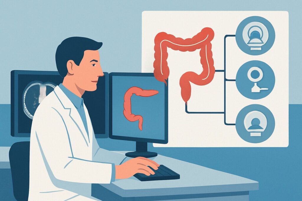
Why Is CTA of the Abdomen and Pelvis the Recommended Study for This Presentation?
For a stable patient with active lower GI bleeding, CTA of the abdomen and pelvis without and with IV contrast is rated *Usually Appropriate* by the ACR because it offers a rapid, accurate, and widely available method to localize the source of bleeding. Its primary advantage is the ability to detect active arterial extravasation into the bowel lumen, which appears as a high-density focus of contrast on arterial phase images that pools and changes shape on later phases.
The rationale for its high rating includes:
- High Diagnostic Accuracy: CTA has a high sensitivity for detecting active bleeding rates as low as 0.3 to 0.5 mL/minute. This allows for precise localization of the bleeding site to a specific colonic segment, which is invaluable for planning subsequent therapy, whether it be catheter-based embolization or targeted surgery.
- Speed and Availability: CT scanners are available 24/7 in most hospitals. A complete multiphase CTA can be acquired in minutes, providing a rapid diagnosis that is crucial even in a stable patient, as their condition can change quickly.
- Anatomic Roadmap: Beyond just finding the bleed, CTA provides a detailed vascular map of the mesenteric arteries. This is essential for interventional radiologists who may need to perform subsequent transcatheter arteriography and embolization.
In contrast, other modalities are rated differently for this specific scenario. While diagnostic/therapeutic colonoscopy is also *Usually Appropriate*, it may be difficult to perform in a patient with active, brisk bleeding due to poor visualization from blood in the colon. Technetium-99m-labeled red blood cell (RBC) scan is also *Usually Appropriate* and is more sensitive for slower, intermittent bleeding, but it provides less precise anatomic localization and takes significantly longer to perform. Transcatheter arteriography is rated *May be appropriate* as a primary diagnostic step but is more often used as a therapeutic intervention after the bleeding site has been localized by CTA.
The recommended CTA study carries a radiation dose of ☢☢☢☢ (10-30 mSv). This is a significant but justified exposure given the high clinical stakes of localizing an active bleed. The protocol requires both non-contrast and multiple post-contrast phases to confidently identify extravasation. Once you’ve decided on CTA abdomen and pelvis without and with IV contrast, our protocol guide covers the technique, contrast, and reading principles: CT Abdomen/Pelvis Without Contrast (Renal Stone).
What’s Next After CTA? Downstream Workflow
The results of the CTA will directly guide the next steps in management. The post-imaging workflow is a critical decision tree for the clinical team.
If the CTA is positive (shows active contrast extravasation): This is a clear indication for intervention. The patient should be immediately referred to Interventional Radiology for transcatheter arteriography and embolization. The CTA serves as a roadmap, allowing the interventional radiologist to selectively catheterize the bleeding vessel and deploy embolic agents (e.g., coils, particles) to achieve hemostasis. This is a highly effective, minimally invasive treatment.
If the CTA is negative (no active extravasation seen): A negative CTA in a patient with clinically active bleeding suggests the bleeding rate was below the scanner’s detection threshold at the moment of imaging, or the bleeding has temporarily stopped. In this case, the next step is often colonoscopy. With the colon now clearer, the endoscopist may be able to identify the source (e.g., a diverticulum with a visible vessel, angiodysplasia) and provide endoscopic therapy like clipping or cautery. If colonoscopy is also non-diagnostic, the patient may fit the criteria for the “obscure recurrent bleeding” scenario, prompting consideration of other studies like a tagged RBC scan or capsule endoscopy.
If the CTA is indeterminate or shows a non-bleeding cause: The scan may reveal an alternative diagnosis, such as a tumor, severe colitis, or an aortoenteric fistula. In these cases, the downstream pathway is dictated by the specific finding and will involve consultation with the appropriate service (e.g., surgery, gastroenterology) for management of that condition.
Pitfalls to Avoid (and When to Get Help)
Several common pitfalls can compromise the diagnostic utility of imaging in this scenario. Be mindful of the following:
- Inadequate Bowel Preparation: While a full prep is not feasible, retained high-density material (e.g., oral contrast from a prior study, metallic objects) can mimic or obscure contrast extravasation. The non-contrast phase of the CTA is essential to identify these mimics.
- Incorrect CT Protocol: Ordering a standard “CT abdomen/pelvis with contrast” is insufficient. You must specifically order a multiphase “CTA for GI Bleed” protocol, which includes non-contrast, arterial, and portal venous phases to properly characterize potential bleeding.
- Delaying the Scan: Bleeding can be intermittent. The highest yield for CTA is when the patient is actively bleeding. Unnecessary delays between the clinical observation of bleeding and the scan can lead to a false-negative result.
If the patient’s hemodynamic status deteriorates at any point during the workup, escalate immediately. This involves activating your institution’s massive transfusion protocol and obtaining emergent consultation with Interventional Radiology and/or General Surgery for definitive intervention.
Related ACR Topics and Tools
For a comprehensive overview of all clinical variants and imaging modalities related to lower gastrointestinal bleeding, please refer to our parent topic guide. It provides a broader context for the specific workflow detailed in this article. Additionally, several tools can assist in applying these criteria and understanding the recommended studies.
- For breadth across all scenarios in Radiologic Management of Lower Gastrointestinal Tract Bleeding, see our parent guide: Radiologic Management of Lower Gastrointestinal Tract Bleeding: ACR Appropriateness Decoded.
- ACR Appropriateness Criteria Lookup — for adjacent scenarios
- Imaging Protocol Library — for technique on the recommended study
- Radiation Dose Calculator — for cumulative dose conversations
Frequently Asked Questions
Why not go straight to colonoscopy in a stable patient with active lower GI bleeding?
While colonoscopy is rated ‘Usually Appropriate’ and can be both diagnostic and therapeutic, performing it during active, brisk bleeding can be very challenging. The colon may be filled with blood, obscuring visualization and making it difficult to identify the source. CTA can rapidly localize the bleeding segment, allowing for a more targeted and often more successful subsequent intervention, whether it be angiography or a focused colonoscopy.
What if the patient has renal insufficiency? Can I still order a CTA with contrast?
This is a risk-benefit decision. In a patient with active, significant bleeding, the risk of not localizing the source often outweighs the risk of contrast-induced nephropathy (CIN). Most modern guidelines support the use of iodinated contrast when clinically indicated, even in patients with chronic kidney disease, provided they are not on dialysis. Pre- and post-procedure hydration can help mitigate the risk. Always discuss with the radiology team if you have concerns.
How does a tagged red blood cell (RBC) scan compare to CTA in this scenario?
A tagged RBC scan is also ‘Usually Appropriate’ and is more sensitive for detecting slower or intermittent bleeding (as low as 0.1 mL/min). However, it takes much longer to perform (often several hours) and provides poor anatomic localization, often only regionalizing the bleed to a quadrant of the abdomen. For a stable patient with clinically active bleeding, CTA is generally preferred for its speed and precise anatomic detail, which better guides immediate intervention.
Is there a role for MRA in working up active lower GI bleeding?
No, Magnetic Resonance Angiography (MRA) is rated ‘Usually not appropriate’ for this scenario. MRA is slower to acquire, more susceptible to motion artifact from bowel peristalsis, and less sensitive for detecting active contrast extravasation compared to CTA. It does not offer a diagnostic advantage in this acute setting.
What if the CTA is negative but I’m still convinced the patient is bleeding?
A negative CTA means there was no active extravasation at a rate of ~0.3-0.5 mL/min or higher during the scan. If clinical signs of bleeding persist (e.g., ongoing hematochezia, dropping hemoglobin), the bleeding may be intermittent or slower than the CTA’s detection threshold. The next step is typically colonoscopy. If that is also negative, a tagged RBC scan may be considered to look for slower, intermittent bleeding over a longer period.
Reviewed by Pouyan Golshani, MD, Interventional Radiologist — May 21, 2026