Post-Cholecystectomy Bile Leak: Why Is Endoscopic Stenting the ACR-Recommended First Step?
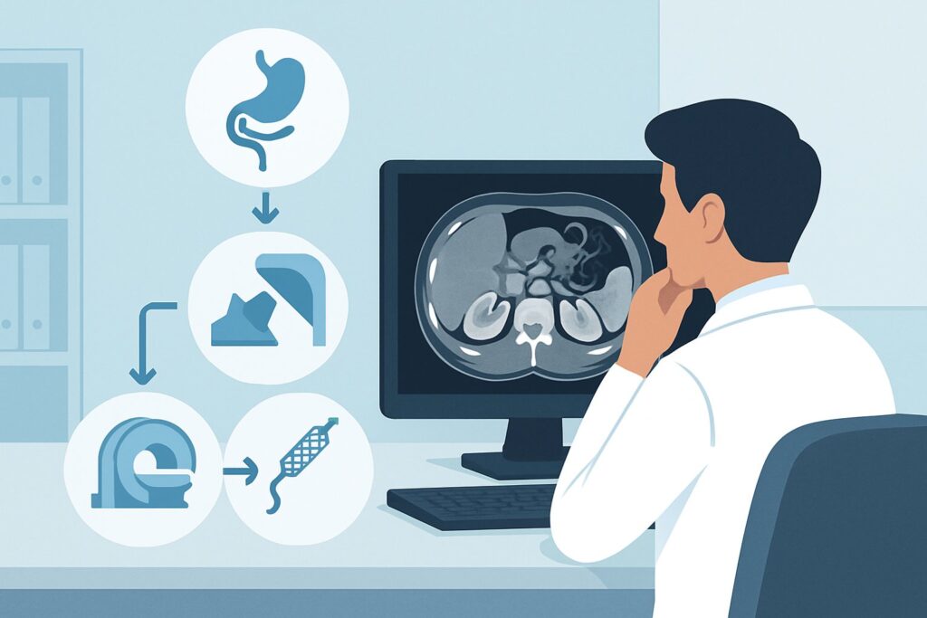
A 54-year-old male is on postoperative day four following a laparoscopic cholecystectomy for symptomatic cholelithiasis. He now presents to the emergency department with worsening right upper quadrant abdominal pain, low-grade fever, and nausea. His labs are notable for a rising total bilirubin and alkaline phosphatase. A CT scan of the abdomen confirms a 6 cm subhepatic fluid collection and shows mild dilation of the common bile duct. You suspect a bile leak with a component of biliary obstruction. The next step is therapeutic, not just diagnostic, but which intervention is the most appropriate initial choice?
This article provides a detailed clinical workflow for the initial therapeutic procedure in a patient with a suspected bile leak and dilated bile ducts after laparoscopic cholecystectomy, based on the American College of Radiology (ACR) Appropriateness Criteria. For this specific scenario, the ACR rates an Endoscopic internal biliary catheter (removable plastic stent) as Usually Appropriate.
Who Fits This Clinical Scenario?
This guidance is intended for a specific and common postoperative complication. The key inclusion criteria for this workflow are:
- A recent history of laparoscopic cholecystectomy.
- Clinical and/or imaging findings suggestive of a bile leak (e.g., a new subhepatic or perihepatic fluid collection, or bilious fluid from a surgical drain).
- Imaging evidence of biliary ductal dilation, suggesting a component of obstruction or impaired bile flow.
It is critical to distinguish this presentation from other causes of biliary obstruction, which follow different management pathways. This article does not apply to patients with:
- Suspected malignant obstruction: If a patient with a history of pancreatic or biliary cancer presents with dilated ducts, the workup and management priorities are different. This scenario is covered in the ACR variant for malignant common bile duct obstruction.
- Primary sclerosing cholangitis: Patients with this underlying condition have a distinct pattern of stricturing and require a different diagnostic and therapeutic approach.
- Simple choledocholithiasis without a leak: If the primary suspicion is a retained common bile duct stone without evidence of a bile leak, the therapeutic urgency and procedural goals differ.
Correctly identifying your patient’s scenario is the first step to selecting the most effective and least invasive therapeutic procedure.
What Diagnoses Are You Working Up in This Scenario?
When a patient presents with a bile leak and dilated ducts after cholecystectomy, you are managing a direct iatrogenic injury. The differential diagnosis is less about the underlying disease and more about the specific nature and location of the injury to the biliary tree, which will guide the therapeutic intervention.
Cystic Duct Stump Leak: This is the most common type of post-cholecystectomy bile leak. It occurs when the surgical clip or ligature on the cystic duct stump becomes dislodged or fails, allowing bile to leak from the junction with the common bile duct. It is often associated with distal obstruction (e.g., a retained stone or papillary edema) which increases pressure in the biliary system and forces bile out of the path of least resistance at the stump.
Injury to a Duct of Luschka: These are small, accessory bile ducts that drain directly from the liver into the gallbladder or common bile duct. They are an anatomic variant and can be easily transected during gallbladder dissection from the liver bed. A leak from a duct of Luschka is another common cause of postoperative biloma.
Common Bile Duct (CBD) Injury: This is a more severe and consequential complication. The injury can range from a small laceration or thermal injury from electrocautery to a complete transection or ligation of the duct. A significant CBD injury often causes both a leak and, critically, distal obstruction, which explains the finding of dilated upstream ducts. Identifying this specific injury is crucial as it may ultimately require surgical repair.
Why Is an Endoscopic Internal Biliary Catheter the Recommended Initial Procedure?
The ACR panel designates an Endoscopic internal biliary catheter (removable plastic stent) as a Usually Appropriate initial therapeutic procedure. This approach, performed via Endoscopic Retrograde Cholangiopancreatography (ERCP), is favored because it directly addresses the underlying pathophysiology in a minimally invasive manner.
The primary goal is to create a low-pressure pathway for bile to flow from the liver directly into the duodenum. By placing a plastic stent across the Sphincter of Oddi, the procedure reduces the pressure gradient that drives bile out of the leak site. This allows the leak to heal spontaneously over several weeks. During the ERCP, a cholangiogram is performed, which precisely maps the biliary anatomy and confirms the location and extent of the leak. This diagnostic information is invaluable for planning further management.
Another procedure rated as Usually Appropriate is a Percutaneous internal/external biliary catheter. This involves placing a drainage catheter through the skin and liver into the biliary tree (Percutaneous Transhepatic Cholangiography, or PTC). While also effective at decompressing the biliary system, it is often considered a second-line approach for several reasons. It requires a transhepatic puncture, which carries risks of bleeding and pneumothorax. It also typically involves an external drainage bag, which can be cumbersome for the patient and carries a risk of catheter dislodgement. Endoscopic stenting provides internal drainage, which is more physiologic and generally better tolerated.
Conversely, more aggressive or less effective options are rated lower:
- Surgery: Rated as May be appropriate, open or laparoscopic surgical repair is significantly more invasive and is typically reserved for cases where endoscopic or percutaneous management fails, or for severe injuries like complete ductal transection that are not amenable to non-surgical repair.
- Permanent biliary metallic stent: This is rated Usually not appropriate. Because a post-surgical bile leak is a benign and temporary condition, a permanent metal stent is unnecessary. These stents can be difficult or impossible to remove and are associated with long-term complications like sludge formation and obstruction, making them unsuitable for benign disease.
What’s Next After an Endoscopic Biliary Stent? Downstream Workflow
The placement of an endoscopic stent is the beginning, not the end, of the management pathway. The next steps depend on the procedural findings and the patient’s clinical response.
If the Stent is Placed Successfully and the Leak is Minor: The patient should show rapid clinical improvement, with resolution of pain and fever and a downward trend in liver function tests. The associated fluid collection (biloma) is often managed concurrently with a separate percutaneous drain if it is large, symptomatic, or shows signs of infection. The biliary stent is typically left in place for 4 to 8 weeks to allow the leak to heal completely. A repeat ERCP is then performed to remove the stent and perform a cholangiogram to confirm resolution of the leak.
If Endoscopic Access Fails: In a small number of cases, the endoscopist may be unable to cannulate the bile duct, particularly in patients with altered anatomy (e.g., prior gastric bypass). In this situation, the next logical step is to pursue the other Usually Appropriate option: a percutaneous internal/external biliary catheter placed by interventional radiology.
If a Complex Injury is Found: If the initial ERCP reveals a complete transection of the common bile duct or a complex stricture with a large gap, stenting alone will not be sufficient. While a stent or percutaneous drain can serve as a crucial temporizing measure to control sepsis and stabilize the patient, these findings typically necessitate surgical consultation for a definitive repair, such as a hepaticojejunostomy. This aligns with surgery’s rating as May be appropriate in select cases.
Pitfalls to Avoid (and When to Get Help)
Navigating a post-cholecystectomy bile leak requires timely and coordinated care. Here are a few common pitfalls to avoid:
- Delaying intervention: A contained biloma can evolve into biliary peritonitis or an abscess if not addressed promptly. Early consultation with gastroenterology (for ERCP) and/or interventional radiology (for PTC or biloma drainage) is key.
- Forgetting to drain the biloma: Placing a biliary stent addresses the “faucet” but doesn’t remove the “puddle.” A large, symptomatic, or infected biloma requires separate percutaneous drainage for source control.
- Choosing a permanent solution for a temporary problem: Avoid the use of permanent metallic or covered stents for benign post-surgical leaks. These are designed for malignant obstructions and can cause significant long-term problems.
If the patient presents with signs of sepsis, hemodynamic instability, or diffuse peritonitis, this is a clinical emergency. Escalate immediately for multidisciplinary evaluation by GI, IR, and surgery to facilitate urgent biliary decompression and source control.
Related ACR Topics and Tools
This article covers one specific scenario in depth. For a broader view of all clinical variants, or to explore the tools used to make these evidence-based decisions, please see the following resources:
- For breadth across all scenarios in Radiologic Management of Biliary Obstruction, see our parent guide: Radiologic Management of Biliary Obstruction: ACR Appropriateness Decoded.
- To search for other clinical presentations, use the Imaging Appropriateness Selector.
- To review procedural details for various imaging studies, explore the Imaging Protocol Library.
- To discuss cumulative radiation exposure with patients, the Radiation Dose Calculator can be a helpful tool.
Frequently Asked Questions
Why is a removable plastic stent used instead of a permanent metal one for a bile leak?
A post-surgical bile leak is a benign and temporary condition. A removable plastic stent is sufficient to bridge the healing process and can be easily taken out after 4-8 weeks. A permanent metal stent is designed for malignant (cancerous) blockages, is difficult or impossible to remove, and can lead to long-term complications like blockage from sludge or tissue overgrowth, making it inappropriate for this benign scenario.
What is the main difference between an endoscopic and a percutaneous approach for this problem?
The endoscopic approach (ERCP) involves passing a scope through the mouth, stomach, and into the small intestine to access the bile duct opening. The percutaneous approach (PTC) involves passing a needle and catheter through the skin and liver to access the bile ducts from above. The endoscopic route is generally preferred first as it avoids a skin puncture and an external drain, providing a more physiologic internal drainage pathway.
Does the biloma (fluid collection) from the leak always need a separate drain?
Not always, but often. If the biloma is small and the patient is asymptomatic, stenting the bile duct to stop the leak may be enough to allow the body to reabsorb the fluid. However, if the collection is large, causing pain, or showing signs of infection, a separate percutaneous drain placed by interventional radiology is typically required for effective source control.
How long does the biliary stent typically stay in place?
For a standard post-cholecystectomy bile leak, a plastic biliary stent is typically left in place for 4 to 8 weeks. This duration is usually sufficient to allow the site of the leak to heal completely. A follow-up procedure (typically another ERCP) is then performed to remove the stent and confirm via cholangiogram that the leak has resolved.
What happens if the endoscopic approach (ERCP) fails to place a stent?
If ERCP is unsuccessful due to technical difficulty or unusual patient anatomy, the next step is typically to consult interventional radiology for a percutaneous transhepatic cholangiography (PTC) with placement of an internal/external biliary drainage catheter. This provides an alternative route to decompress the biliary system and control the leak.
Reviewed by Pouyan Golshani, MD, Interventional Radiologist — May 26, 2026