Should You Order an MRI for a Child with Precocious Puberty? An ACR-Guided Workflow

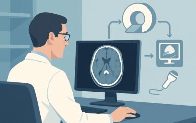
A 7-year-old girl presents for evaluation of breast development that began six months ago. Her bone age is advanced, and laboratory testing confirms elevated gonadotropins, consistent with central precocious puberty. As the managing clinician, you must now determine the appropriate initial imaging to investigate for a central nervous system cause. The decision involves balancing diagnostic yield with the unique considerations of pediatric imaging, including the avoidance of ionizing radiation. This workflow article outlines the evidence-based approach for this specific scenario. According to the American College of Radiology (ACR) Appropriateness Criteria, an MRI of the sella without and with IV contrast is Usually appropriate as the initial imaging study.
Who Fits This Clinical Scenario for Precocious Puberty Imaging?
This guidance applies to a specific pediatric population: girls younger than 8 years of age and boys younger than 9 years of age who present with signs of puberty and have a confirmed diagnosis of central precocious puberty (CPP), also known as gonadotropin-dependent precocious puberty. The key inclusion criterion is biochemical evidence that the hypothalamic-pituitary-gonadal (HPG) axis has been prematurely activated.
This workflow is not intended for:
- Peripheral Precocious Puberty: This gonadotropin-independent form is caused by autonomous production of sex steroids from the gonads or adrenal glands (e.g., from an ovarian cyst or adrenal tumor). These cases require a different imaging workup, typically starting with pelvic or adrenal ultrasound.
- Benign Pubertal Variants: Children with isolated premature thelarche (breast development without other signs of puberty) or premature adrenarche (early development of pubic hair) often do not require neuroimaging, as these are typically non-progressive, benign conditions.
- Adults with Pituitary Dysfunction: Patients presenting with adult-onset hypopituitarism, hyperfunctioning pituitary adenomas (like Cushing disease or acromegaly), or diabetes insipidus follow distinct imaging pathways. This guidance is strictly for the initial workup of CPP in children.
What Diagnoses Are You Working Up with Brain Imaging in Precocious Puberty?
While most cases of central precocious puberty in girls are idiopathic, imaging is crucial to rule out an underlying structural lesion in the brain. The differential diagnosis for a central cause of precocious puberty drives the choice of imaging modality.
The primary concern is often a hypothalamic hamartoma. This is a benign, non-neoplastic, congenital malformation of the hypothalamus. These lesions contain ectopic GnRH-secreting neurons that function outside of normal central inhibition, leading to the premature activation of puberty. They are a classic, albeit uncommon, cause of CPP, particularly in very young children (under 3 years old) and boys.
Less commonly, other central nervous system (CNS) tumors can trigger precocious puberty by disrupting normal hypothalamic inhibitory pathways. These include optic pathway gliomas (especially in the context of neurofibromatosis type 1), astrocytomas, craniopharyngiomas, and germinomas. Identifying these lesions is critical, as they require urgent neuro-oncologic management.
Finally, imaging helps identify structural changes resulting from a prior CNS insult. Precocious puberty can be a delayed consequence of brain injury from trauma, CNS infection (meningitis or encephalitis), hydrocephalus, or cranial radiation therapy. In the absence of any identifiable lesion, the diagnosis becomes idiopathic central precocious puberty, which is a diagnosis of exclusion and the most common finding, particularly in girls over the age of 6.
Why Is MRI of the Sella the Recommended Initial Study for Precocious Puberty?
The ACR rates MRI sella without and with IV contrast as Usually appropriate because it provides the highest diagnostic yield for the key pathologies in the differential diagnosis while avoiding ionizing radiation.
The superior soft-tissue contrast of Magnetic Resonance Imaging (MRI) is essential for visualizing the small, complex structures of the hypothalamus and pituitary gland. It can clearly delineate a hypothalamic hamartoma, which typically appears as a non-enhancing mass isointense to gray matter, and differentiate it from the optic chiasm and pituitary stalk. This level of detail is unmatched by other modalities.
The use of intravenous contrast is critical for a comprehensive evaluation. While a classic hypothalamic hamartoma does not enhance, many of the other neoplastic and inflammatory causes on the differential—such as gliomas, germinomas, or post-infectious changes—often demonstrate avid enhancement. A protocol including both pre- and post-contrast sequences provides a complete assessment, allowing the radiologist to confidently exclude these more aggressive pathologies. An `MRI sella without IV contrast` is also rated Usually appropriate, but the addition of contrast significantly increases diagnostic confidence.
Alternative studies are rated lower for clear reasons:
- CT sella without and with IV contrast: This is rated Usually not appropriate. CT has significantly lower soft-tissue resolution compared to MRI, making it difficult or impossible to detect small, non-calcified, non-enhancing lesions like a hypothalamic hamartoma. Furthermore, CT exposes the child’s developing brain to ionizing radiation (pediatric dose ☢☢☢ 0.3-3 mSv), a critical consideration when a non-radiation alternative (MRI) offers superior diagnostic information.
- Radiography of the sella: This is also rated Usually not appropriate. This is an outdated technique that provides no useful information about the hypothalamic or pituitary soft tissues. It can only visualize the bony sella turcica and is insensitive for detecting the underlying causes of CPP.
What Is the Downstream Workflow After a Pediatric Brain MRI for Precocious Puberty?
The results of the MRI will dictate the subsequent clinical pathway, which almost always involves a multidisciplinary team.
- If the MRI is positive for a hypothalamic hamartoma: The patient should be referred to a pediatric endocrinologist for medical management, typically with a gonadotropin-releasing hormone (GnRH) agonist to suppress puberty. A referral to pediatric neurosurgery is also warranted for evaluation, as surgical intervention may be considered, especially if the child also has associated gelastic (laughing) seizures.
- If the MRI is positive for another CNS tumor (e.g., glioma, craniopharyngioma): This finding requires urgent referral to a pediatric neuro-oncology team, which includes neurosurgeons, oncologists, and endocrinologists. The immediate next steps will involve further staging, biopsy, and development of a comprehensive treatment plan.
- If the MRI is negative (normal study): This is the most frequent outcome, especially for girls. A negative MRI confirms the diagnosis of idiopathic central precocious puberty. The patient should be managed by a pediatric endocrinologist. The primary treatment is typically a GnRH agonist to halt pubertal progression, preserve adult height potential, and address any psychosocial concerns related to early development.
Pitfalls to Avoid (and When to Get Help)
When managing the initial imaging workup for a child with precocious puberty, several pitfalls can compromise diagnostic accuracy and patient care.
First, avoid ordering a CT scan as the initial study. The combination of ionizing radiation exposure and poor soft-tissue resolution for key pathologies makes it an inappropriate choice when MRI is available. Second, ensure the MRI order is specific: “MRI sella/pituitary without and with IV contrast, pediatric precocious puberty protocol.” A general “MRI brain” may not use the thin slices and specific sequences needed to properly evaluate the hypothalamic-pituitary region. Finally, do not delay imaging in very young children (e.g., under 3) or in boys, as the likelihood of an underlying organic cause is significantly higher in these groups.
If the clinical picture is complex, such as a child with CPP and other neurologic symptoms like seizures or visual changes, an urgent consultation with pediatric neurology or neurosurgery is warranted before or concurrent with imaging.
Related ACR Topics and Tools
This article focuses on a single, specific clinical scenario. For a comprehensive overview of all related scenarios and their recommended imaging pathways, please consult the parent topic article. The tools below can assist in navigating adjacent criteria, understanding imaging protocols, and discussing radiation safety with families.
- For breadth across all scenarios in Neuroendocrine Imaging, see our parent guide: Neuroendocrine Imaging: ACR Appropriateness Decoded.
- Imaging Appropriateness Selector — for adjacent scenarios
- Imaging Protocol Library — for technique on the recommended study
- Radiation Dose Calculator — for cumulative dose conversations
Frequently Asked Questions
Is a brain MRI necessary for every girl under 8 with signs of puberty?
Not necessarily. Imaging is indicated for confirmed central precocious puberty (CPP), where lab tests show premature activation of the HPG axis. For girls with isolated premature thelarche or adrenarche, which are benign variants, neuroimaging is not typically required. The decision should be guided by a thorough clinical and biochemical evaluation, often in consultation with a pediatric endocrinologist.
Why is the risk of an underlying brain tumor higher in boys with precocious puberty?
While the exact reason is not fully understood, clinical data consistently show that boys with central precocious puberty have a much higher incidence of an identifiable CNS lesion compared to girls. For girls, especially those between 6 and 8 years old, the cause is idiopathic in over 90% of cases. For boys, the rate of underlying pathology is significantly higher, making neuroimaging a mandatory part of their workup.
Does a hypothalamic hamartoma always cause precocious puberty?
No. Hypothalamic hamartomas are rare, and not all of them are hormonally active. Some are discovered incidentally on imaging for other reasons (like headaches or seizures) and do not cause any endocrine dysfunction. The classic presentation, however, is central precocious puberty, often accompanied by gelastic (laughing) seizures.
What if my patient cannot have an MRI due to a contraindication like a non-compatible implanted device?
This is a rare situation in young children but can occur. In such cases, a high-resolution CT of the sella with and without IV contrast would be the next best option, despite its limitations. This decision should be made in close consultation with a pediatric radiologist and endocrinologist to weigh the risks of radiation and lower diagnostic yield against the clinical need to rule out a CNS lesion.
If the initial MRI is negative, is any follow-up imaging needed?
For a technically adequate, high-quality MRI that is interpreted as normal in a child with idiopathic CPP, routine follow-up imaging is generally not recommended. Repeat imaging would only be considered if the child develops new or progressive neurologic symptoms, such as headaches, visual changes, or seizures, which would suggest a new or previously missed pathology.
Reviewed by Pouyan Golshani, MD, Interventional Radiologist — June 27, 2026