What Is the Initial Imaging for Suspected Motor Neuron Disease? An ACR-Guided Workflow

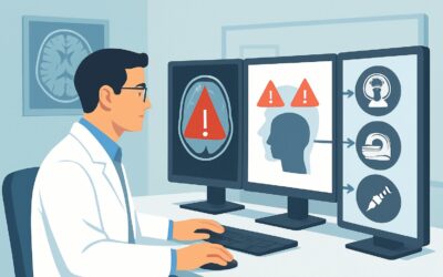
A 62-year-old patient presents to your clinic with nine months of progressive, asymmetric weakness in his left hand, now accompanied by visible muscle twitching in his arm and shoulder. He also reports difficulty with speech articulation. On examination, you note fasciculations, atrophy, and weakness in the left upper extremity, along with a brisk jaw jerk and hyperreflexia in the lower extremities. There is no sensory loss. You suspect a motor neuron disease (MND). The critical next step is to initiate a diagnostic workup, but which imaging study provides the most value while efficiently excluding crucial mimics? This article details the American College of Radiology (ACR) guided workflow for this specific scenario, starting with the top-rated initial study: MRI head without IV contrast, which the ACR rates as Usually appropriate.
Who Fits This Clinical Scenario?
This guidance applies to adult patients presenting with a subacute or chronic history of progressive motor weakness suggestive of motor neuron disease. The classic clinical picture involves a combination of upper motor neuron (UMN) signs (e.g., spasticity, hyperreflexia, Babinski sign) and lower motor neuron (LMN) signs (e.g., weakness, atrophy, fasciculations). Crucially, sensory examination is typically normal, and symptoms cannot be explained by a single nerve root or peripheral nerve distribution.
This workflow is specifically for the initial imaging workup where MND is a primary consideration. It is important to distinguish this presentation from clinically distinct scenarios that require different imaging pathways:
- Parkinsonian Syndromes: Patients whose primary symptoms are bradykinesia, rigidity, tremor, and postural instability. While motor symptoms are present, the constellation of signs points toward a basal ganglia disorder, not a primary UMN/LMN pathology.
- Chorea: Patients presenting with involuntary, irregular, and non-rhythmic movements. This presentation suggests a workup for conditions like Huntington disease.
- Rapidly Progressive Dementia: Patients whose dominant feature is a swift cognitive decline, which may be accompanied by motor signs. This presentation raises concern for etiologies like Creutzfeldt-Jakob disease, which has a different imaging signature.
This article focuses squarely on the patient whose signs and symptoms point toward a primary degeneration of motor neurons, such as in amyotrophic lateral sclerosis (ALS).
What Diagnoses Are You Working Up in This Scenario?
The primary goal of initial imaging in suspected MND is not to “see” the disease itself, but to meticulously exclude other conditions that can mimic its presentation. A “normal” imaging study is often a key piece of evidence that supports the clinical diagnosis of MND. The differential diagnosis includes several important mimics.
Cervical Spondylotic Myelopathy: This is one of the most critical mimics to exclude. Structural compression of the cervical spinal cord from degenerative disc disease, osteophytes, or ligamentous hypertrophy can produce a combination of UMN signs below the level of compression and LMN signs at the level of compression, closely mirroring MND.
Brainstem or Foramen Magnum Tumors: A slow-growing mass, such as a meningioma or glioma, located at the craniocervical junction or within the brainstem can compress motor pathways and cranial nerve nuclei. This can cause progressive weakness, dysarthria, and dysphagia, mimicking bulbar-onset ALS.
Multiple Sclerosis (MS): While MS typically presents with sensory and visual symptoms, a primary progressive or motor-predominant variant can sometimes manifest with progressive spastic weakness. Demyelinating plaques in the corticospinal tracts of the brain or spinal cord are the underlying cause.
Stroke Mimics: A strategically located lacunar infarct in the brainstem or internal capsule, or a chronic watershed infarct, can sometimes result in motor deficits that evolve over time. While the onset is typically acute, the presentation can occasionally be misidentified as a progressive disorder.
Amyotrophic Lateral Sclerosis (ALS): This is the archetypal MND. While imaging is primarily for excluding mimics, certain advanced MRI techniques may show subtle findings in established ALS, such as T2/FLAIR hyperintensity along the corticospinal tracts or hypointensity of the motor cortex (“motor band sign” on SWI). However, these findings are often absent in early disease and are not required for diagnosis.
Why Is MRI Head Without IV Contrast the Recommended Initial Study?
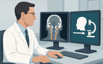
The ACR designates MRI head without IV contrast as Usually appropriate for the initial evaluation of suspected motor neuron disease. This recommendation is based on its superior ability to visualize brain parenchyma and the brainstem to rule out the structural mimics that constitute the primary differential diagnosis.
The rationale for this choice is multi-faceted:
- High Soft-Tissue Resolution: MRI provides unparalleled detail of the brain’s gray and white matter, the brainstem, and the cerebellum. It can readily identify tumors, demyelinating plaques from MS, evidence of prior strokes, and other structural lesions that CT might miss.
- Exclusion of Key Mimics: The primary purpose of the scan is to find an alternative diagnosis. A high-quality brain MRI can confidently exclude a brainstem glioma or a foramen magnum meningioma, which are critical “can’t-miss” diagnoses.
- Safety Profile: This study involves no ionizing radiation (Adult RRL=O 0 mSv). As contrast is not typically required for the initial exclusionary scan, the risks associated with gadolinium-based contrast agents are also avoided.
Why are alternative studies rated lower for this initial workup?
Understanding why other common neuroimaging studies are not the first choice is key to efficient and effective diagnosis. The ACR provides specific ratings that clarify their roles:
- CT head without IV contrast is rated as May be appropriate. While fast and widely available, its sensitivity for the subtle parenchymal abnormalities seen in MS or small brainstem lesions is significantly lower than MRI. It also involves ionizing radiation (Adult RRL=☢☢☢ 1-10 mSv). It serves as an alternative when MRI is contraindicated or immediately unavailable.
- MRI spine without and with IV contrast is also rated as May be appropriate. A cervical spine MRI is essential for ruling out spondylotic myelopathy. However, the initial ACR guidance prioritizes a head MRI unless the clinical signs point strongly to a spinal cord origin (e.g., a clear sensory level, severe neck pain, or Lhermitte’s sign). Often, the cervical spine MRI is the logical next step after a negative head MRI.
- FDG-PET/CT brain is rated Usually not appropriate. This is a functional imaging study that assesses metabolic activity and is not used for the initial structural evaluation in this context. It involves substantial radiation (Adult RRL=☢☢☢ 1-10 mSv) and provides no significant diagnostic advantage over MRI for this indication.
What’s Next After MRI Head Without IV Contrast? Downstream Workflow
The result of the initial head MRI is a critical branch point in the diagnostic algorithm. The next steps depend entirely on whether a structural mimic is identified.
If the head MRI is negative (i.e., normal or shows only non-specific findings):
A normal head MRI is the most common outcome in patients who truly have MND. This negative result is diagnostically valuable because it excludes major mimics within the brain and brainstem. The workup should then proceed based on the clinical picture:
- The next essential step is typically electrodiagnostic studies (EMG and nerve conduction studies). These tests are crucial for confirming the presence of widespread active and chronic denervation with reinnervation, which is the electrophysiologic hallmark of MND.
- If clinical suspicion for a cervical myelopathy remains despite the normal head MRI (e.g., based on neck pain or specific reflex patterns), an MRI of the cervical spine is the appropriate next imaging study.
If the head MRI is positive for a mimic:
The diagnostic pathway pivots completely. For example:
- If a foramen magnum meningioma is found, the patient should be referred to Neurosurgery for evaluation and management. The working diagnosis is no longer MND.
- If imaging shows demyelinating plaques consistent with Multiple Sclerosis, the patient should be referred to a Neurologist specializing in MS for further workup, including spinal cord imaging and cerebrospinal fluid analysis.
If the head MRI is indeterminate:
Occasionally, findings may be ambiguous. In such cases, adding IV contrast (MRI head without and with IV contrast, rated May be appropriate) can help characterize a potential lesion, such as determining if it is an active demyelinating plaque or a tumor. Consultation with a neurologist and/or neuroradiologist is recommended.
Pitfalls to Avoid (and When to Get Help)
Navigating the workup for suspected MND requires careful clinical-radiologic correlation. Several common pitfalls can delay diagnosis or lead to misinterpretation.
- The “Normal Means Nothing” Fallacy: Do not interpret a normal head MRI as unhelpful. In the context of suspected MND, a normal study that excludes mimics is a powerful piece of evidence that strengthens the clinical diagnosis.
- Stopping at the Brain: If a patient has any signs or symptoms suggesting a spinal cord issue (e.g., cape-like sensory loss, bowel/bladder changes, a clear sensory level), do not delay ordering an MRI of the spine. The head and spine should be considered as two parts of a whole neuraxis evaluation.
- Over-interpreting Incidentalomas: Be cautious about attributing a patient’s profound motor symptoms to non-specific age-related findings, such as mild white matter ischemic changes or minimal degenerative disc disease. The imaging findings must be sufficient to explain the full clinical picture.
If the clinical picture is atypical, rapidly progressing, or if imaging results are complex, early consultation with a neuromuscular specialist is essential for guiding further diagnostic steps and management.
Related ACR Topics and Tools
Staying current with evidence-based imaging guidelines is crucial for providing high-quality care. For a broader overview of related neurodegenerative conditions, see the parent topic hub article. For specific questions about other scenarios or imaging techniques, the following GigHz resources are available:
- For breadth across all scenarios in Movement Disorders and Neurodegenerative Diseases, see our parent guide: Movement Disorders and Neurodegenerative Diseases: ACR Appropriateness Decoded.
- To explore other clinical presentations, use the ACR Appropriateness Criteria Lookup.
- For technical details on how specific studies are performed, visit the Imaging Protocol Library.
- To discuss radiation exposure with patients, consult the Radiation Dose Calculator.
Frequently Asked Questions
Why not start with an MRI of the spine for suspected motor neuron disease?
While an MRI of the cervical spine is crucial for ruling out spondylotic myelopathy, the ACR recommends starting with a head MRI unless clinical signs specifically point to the spinal cord. This is because many mimics, such as brainstem tumors or multiple sclerosis plaques, are intracranial. A head MRI provides a high-yield initial screen for these conditions. The spine MRI is often the second step if the head MRI is negative.
Is intravenous contrast necessary for the initial head MRI?
No, for the initial evaluation, an MRI of the head *without* IV contrast is rated ‘Usually appropriate’ and is sufficient. The primary goal is to assess for structural abnormalities, which are well-visualized on non-contrast sequences. Contrast is rated ‘May be appropriate’ and is typically reserved for cases where the non-contrast scan reveals an indeterminate lesion that requires further characterization.
What specific MRI findings can suggest ALS, even if the main goal is to exclude mimics?
While the primary role of MRI is exclusionary, in some patients with established ALS, subtle findings may be present. These include T2/FLAIR hyperintensity along the corticospinal tracts in the posterior limb of the internal capsule, cerebral peduncles, or precentral gyrus. Another sign described is atrophy of the motor cortex. These findings are not sensitive or specific enough for initial diagnosis but can be corroborative in the right clinical context.
If the head MRI is normal, does the patient still need an EMG?
Yes, absolutely. A normal head MRI is an expected finding in motor neuron disease. The diagnosis of MND relies on clinical findings supported by electrodiagnostic evidence of widespread denervation. An EMG/NCS is a mandatory next step after a normal MRI to confirm the diagnosis and determine the extent of lower motor neuron involvement.
When should I consider a CT scan instead of an MRI?
A CT head without contrast is rated ‘May be appropriate’ and should be considered a second-line option. It is primarily used when MRI is unavailable, contraindicated (e.g., due to an incompatible implanted device or severe claustrophobia), or in an emergency setting to rule out acute hemorrhage or a large mass. However, its sensitivity for key mimics like demyelinating disease or small brainstem lesions is significantly lower than MRI.
Reviewed by Pouyan Golshani, MD, Interventional Radiologist — May 29, 2026