What Is the Next Imaging Study for Known Acute Subarachnoid Hemorrhage on CT?
It’s 2 a.m. in the emergency department, and the non-contrast head CT for your 52-year-old patient with a sudden, severe headache is back. The report confirms your suspicion: acute subarachnoid hemorrhage (SAH), with hyperdense blood filling the basal cisterns. The diagnosis is made, but the critical next question is clinical priority number one: what is the source of the bleed? Identifying an underlying aneurysm or vascular malformation is paramount for guiding neurosurgical or endovascular intervention and preventing a devastating re-bleed. This article details the evidence-based imaging workflow for this precise scenario, explaining why the American College of Radiology (ACR) rates Arteriography cervicocerebral as Usually appropriate to definitively characterize the cause.
Who Fits This Clinical Scenario for SAH Workup?
This guidance applies specifically to patients who have a confirmed diagnosis of acute subarachnoid hemorrhage on a non-contrast computed tomography (CT) scan. The key inclusion criterion is the definitive presence of hyperdense material (blood) within the subarachnoid spaces, such as the sulci, fissures, or basal cisterns. The clinical context is typically a patient presenting with a “thunderclap” headache, often described as the “worst headache of life,” though other neurologic deficits may be present.
It is crucial to distinguish this scenario from similar but distinct clinical presentations that follow different diagnostic pathways:
- Suspected SAH with a negative CT: If clinical suspicion for SAH is high but the initial non-contrast head CT is negative, the next step is typically a lumbar puncture to test for xanthochromia, not immediate vascular imaging.
- Surveillance of a known, treated aneurysm: Patients with a previously coiled or clipped aneurysm require routine follow-up imaging, but this is a surveillance scenario, not an acute workup.
- Screening for an unruptured aneurysm: Asymptomatic individuals with a strong family history or genetic predisposition for cerebral aneurysms may undergo screening, which follows a separate set of recommendations.
- Monitoring for vasospasm: This workup is for identifying the initial source of the bleed. Evaluating for delayed cerebral ischemia from vasospasm, which typically occurs 3-14 days after the initial hemorrhage, is a different clinical question.
What Diagnoses Are You Working Up in This Scenario?
Once SAH is confirmed on CT, the immediate goal of subsequent imaging is to identify the source of the hemorrhage to facilitate definitive treatment. The differential diagnosis is focused on vascular lesions prone to rupture.
The most common and life-threatening cause is a ruptured saccular (berry) aneurysm. These aneurysms account for approximately 85% of non-traumatic subarachnoid hemorrhages. They typically form at arterial branch points within the circle of Willis, such as the junction of the anterior communicating artery or the posterior communicating artery. Identifying the precise location, size, and neck morphology of the aneurysm is essential for planning treatment.
Less common but critical to identify are other vascular malformations. An arteriovenous malformation (AVM) is a tangle of abnormal vessels directly connecting arteries and veins without an intervening capillary bed, creating a high-flow, high-pressure shunt that is prone to rupture. A dural arteriovenous fistula (dAVF) is an abnormal connection within the dura mater. Both require detailed vascular imaging to characterize their feeding arteries, draining veins, and nidus structure.
In some cases, particularly when blood is isolated to the cisterns around the midbrain, the diagnosis may be a perimesencephalic nonaneurysmal SAH. This is a diagnosis of exclusion, made after comprehensive vascular imaging is negative for a bleeding source. It typically has a much better prognosis than aneurysmal SAH. Other rare causes, such as ruptured mycotic aneurysms, vasculitis, or arterial dissections, must also be considered.
Why Is Vascular Imaging the Essential Next Step After a CT Shows Subarachnoid Hemorrhage?
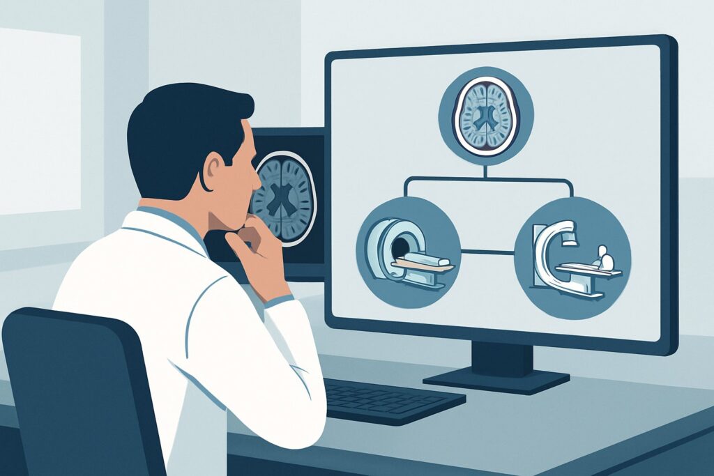
With a diagnosis of SAH established by non-contrast CT, the ACR guidelines designate both CTA head with IV contrast and Arteriography cervicocerebral as Usually appropriate. The choice between them often depends on institutional preference, available expertise, and the need for potential immediate intervention.
Arteriography, also known as digital subtraction angiography (DSA), is widely considered the gold standard for detecting the source of a subarachnoid hemorrhage. Its primary advantage is its superior spatial and temporal resolution, which allows for the detection of very small aneurysms (under 3 mm) that can be missed by non-invasive modalities. DSA provides dynamic, real-time information about blood flow, which is invaluable for characterizing the complex architecture of AVMs and dural fistulas. A key benefit of DSA is its dual diagnostic and therapeutic capability; if an aneurysm is identified, endovascular treatment like coiling can often be performed in the same session, saving critical time.
CTA of the head with IV contrast is also rated Usually appropriate and is the first-line vascular imaging study at many institutions. It is fast, non-invasive, and has high sensitivity and specificity for detecting aneurysms, especially those larger than 3 mm. Modern multidetector CT scanners can generate high-quality 3D reconstructions that are extremely useful for pre-procedural planning.
Alternatives are rated lower for specific reasons in this acute setting:
- MRA head without IV contrast is rated May be appropriate. It avoids both ionizing radiation (Adult RRL: O 0 mSv) and iodinated contrast, making it a viable option for patients with severe contrast allergies or significant renal impairment. However, its spatial resolution is generally lower than CTA and DSA, potentially missing smaller aneurysms.
- MRI head without IV contrast is rated Usually not appropriate for the initial search for a bleeding source. While MRI is excellent for evaluating brain parenchyma, it lacks the vascular detail and speed of CTA or DSA needed in this emergent clinical scenario.
Both CTA and Arteriography involve ionizing radiation (Adult RRL: ☢☢☢ 1-10 mSv). The risk of a missed or delayed diagnosis of a ruptured aneurysm, which carries a high risk of re-bleeding and death, far outweighs the radiation risk in this context.
What’s Next After Vascular Imaging? Downstream Workflow
The results of your vascular study will dictate the immediate next steps in patient management, which almost always involves urgent consultation with neurosurgery and/or neurointerventional radiology.
If the study is positive for a ruptured aneurysm: The patient requires immediate intervention to secure the aneurysm and prevent re-hemorrhage. The treatment options are surgical clipping (a neurosurgical procedure) or endovascular coiling/flow diversion (a neurointerventional procedure). The choice depends on the aneurysm’s size, location, and morphology, as well as the patient’s clinical condition and institutional expertise.
If the initial study (e.g., CTA) is negative: A negative CTA in the setting of diffuse SAH is a high-stakes situation. Because small aneurysms can be missed on CTA, the standard of care is often to proceed with Arteriography (DSA) for a more definitive evaluation. A “negative CTA, positive DSA” is a well-documented occurrence.
If all vascular imaging (including DSA) is negative: If a comprehensive DSA is negative for a bleeding source, the patient is categorized as having angiogram-negative SAH. A repeat DSA may be considered in 7-14 days, as vasospasm or thrombosis could initially obscure a small aneurysm. If the bleeding pattern was consistent with perimesencephalic SAH, and the DSA is negative, the prognosis is generally favorable, and management becomes supportive.
Pitfalls to Avoid (and When to Get Help)
Navigating the workup for acute SAH requires precision and speed. Common pitfalls to avoid include:
- Delaying vascular imaging: The highest risk of aneurysm re-bleeding is within the first 24 hours. Vascular imaging to identify the source should be obtained emergently once SAH is confirmed.
- Stopping at a negative CTA: In patients with a classic aneurysmal bleeding pattern on non-contrast CT (e.g., blood in the anterior interhemispheric fissure suggesting an ACOM aneurysm), a negative CTA should be viewed with high suspicion and prompt a discussion about proceeding to DSA.
- Misinterpreting the bleeding pattern: The distribution of blood on the initial CT can provide valuable clues to the location of the ruptured aneurysm. Forgetting to correlate the imaging findings can make the search for the source less efficient.
- Inadequate imaging protocol: Ensure the CTA protocol is optimized for aneurysm detection, including thin slices and appropriate timing of the contrast bolus to achieve dense arterial opacification.
If there is any uncertainty regarding the imaging findings or the appropriate next step, immediate consultation with neuroradiology, neurosurgery, and/or a neurointerventional specialist is critical.
Related ACR Topics and Tools
This article covers a single, critical scenario. For a comprehensive overview of imaging for all related conditions, from aneurysm screening to vasospasm evaluation, please consult the parent topic guide.
- For breadth across all scenarios in Cerebrovascular Diseases-Aneurysm, Vascular Malformation, and Subarachnoid Hemorrhage, see our parent guide: Cerebrovascular Diseases-Aneurysm, Vascular Malformation, and Subarachnoid Hemorrhage: ACR Appropriateness Decoded.
- To explore adjacent clinical scenarios, use the ACR Appropriateness Criteria Lookup.
- For detailed procedural techniques, see the Imaging Protocol Library.
- To discuss radiation exposure with patients, use the Radiation Dose Calculator.
Frequently Asked Questions
Why are both CTA and Arteriography (DSA) rated ‘Usually appropriate’?
Both are excellent modalities for detecting the source of a subarachnoid hemorrhage. CTA is non-invasive, fast, and widely available, making it a common first choice. Arteriography (DSA) is more invasive but offers higher spatial resolution for detecting very small aneurysms and allows for immediate endovascular treatment. The choice often depends on institutional protocols and the specific clinical situation.
If my initial CTA is negative, is the workup complete?
Not necessarily. If the pattern of hemorrhage on the non-contrast CT is highly suggestive of an aneurysm, a negative CTA should prompt consideration of a follow-up Arteriography (DSA). DSA is more sensitive for small aneurysms that can be missed on CTA. This decision should be made in consultation with neuroradiology and neurosurgery.
Is MRA a good alternative to avoid radiation and contrast?
MRA without contrast is rated ‘May be appropriate’ by the ACR. It is a reasonable option for patients in whom iodinated contrast is strictly contraindicated (e.g., severe allergy, advanced renal failure). However, it is generally less sensitive than CTA or DSA for small aneurysms, so it is not a first-line choice in patients who can tolerate contrast and radiation.
What if the patient has traumatic SAH instead of spontaneous SAH?
This workflow is for spontaneous, non-traumatic subarachnoid hemorrhage, where the primary concern is a ruptured aneurysm. Traumatic SAH is managed differently; while vascular injury (e.g., dissection) can occur, the immediate focus is on managing intracranial pressure and other traumatic injuries. CTA may still be performed to rule out a vascular injury if the mechanism is concerning.
How quickly does this vascular imaging need to be done?
Emergently. The risk of a catastrophic re-bleed from an unsecured ruptured aneurysm is highest in the first 24 hours. Once SAH is diagnosed on CT, the workup to find and treat the source should proceed without delay.
Reviewed by Pouyan Golshani, MD, Interventional Radiologist — May 29, 2026