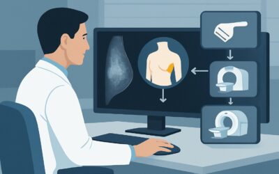
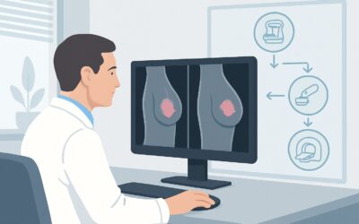
Should You Order an Ultrasound for Clinically Evident Male Gynecomastia?

A 52-year-old male presents to your clinic concerned about bilateral breast enlargement and tenderness that has developed over the past six months. His physical examination reveals soft, non-tender, concentric subareolar tissue without a discrete palpable mass, skin changes, or axillary adenopathy. Your initial thought might be to order a breast ultrasound to confirm the diagnosis and rule out anything more serious. But is that the correct next step according to evidence-based guidelines?
This article provides a detailed workflow for the specific clinical scenario of a male patient of any age with symptoms and physical exam findings consistent with gynecomastia or pseudogynecomastia. For this presentation, the American College of Radiology (ACR) Appropriateness Criteria rate initial imaging, including breast ultrasound, as Usually Not Appropriate. We will explore the rationale behind this recommendation and outline the appropriate clinical pathway.
Who Fits This Clinical Scenario?
This guidance applies specifically to male patients of any age whose clinical presentation is highly consistent with benign gynecomastia. The key inclusion criteria are symptoms of breast enlargement, with or without tenderness, and a physical examination that reveals:
- Concentric, rubbery, or soft glandular-appearing tissue located directly beneath the areola.
- The findings may be unilateral or bilateral.
- Absence of suspicious features such as a discrete, hard, or fixed mass; skin thickening or dimpling; nipple retraction or discharge; or palpable axillary lymph nodes.
It is crucial to distinguish this scenario from others that require a different diagnostic approach. This guidance does not apply if the patient presents with:
- An indeterminate palpable breast mass: A discrete lump that is not clearly classic for gynecomastia. This presentation routes to a different ACR variant, particularly for men 25 years of age or older.
- Physical examination findings suspicious for breast cancer: Any of the red-flag signs mentioned above (hard/fixed mass, skin changes, nipple discharge, axillary adenopathy) immediately place the patient in a high-suspicion category where imaging is mandatory.
- A previously indeterminate imaging result: This article covers initial imaging only. Patients with prior imaging that was inconclusive require a separate workup.
Correctly identifying the patient who fits this classic gynecomastia profile is the key to avoiding unnecessary imaging.
What Diagnoses Are You Working Up in This Scenario?
When a male patient presents with breast enlargement, the differential diagnosis is focused on distinguishing benign proliferation from the rare but critical possibility of malignancy. However, in this specific scenario, the clinical findings heavily favor a benign etiology.
Gynecomastia is the most common diagnosis. It is a benign proliferation of the glandular component of the male breast, driven by an imbalance of estrogen and androgen effects. It can be physiologic (in neonates, adolescents, and older men), idiopathic, or secondary to medications (e.g., spironolactone, cimetidine, anti-androgens) or underlying systemic diseases (e.g., liver cirrhosis, kidney failure, hypogonadism). The classic physical finding is a palpable, disc-like mound of tissue concentric to the nipple.
Pseudogynecomastia is another extremely common cause of male breast enlargement, particularly in overweight or obese individuals. This condition is caused by the deposition of subcutaneous fat without any proliferation of glandular tissue. On examination, the tissue feels soft and diffuse, lacking the distinct, rubbery disc of true gynecomastia.
Male Breast Cancer is the most important diagnosis to exclude, but it is rare, accounting for less than 1% of all breast cancers. Crucially, it typically presents differently from classic gynecomastia. The most common presentation is a painless, hard, eccentric (off-center) subareolar mass. Associated skin or nipple changes and axillary adenopathy are more common than in gynecomastia. The low pre-test probability of cancer in a patient with a classic gynecomastia exam is the primary reason imaging is not initially recommended.
Why Is Imaging Usually Not Appropriate for This Presentation?
When the history and physical exam are classic for gynecomastia, the diagnostic yield of imaging is low, and the potential for false positives or incidental findings that lead to unnecessary downstream interventions is significant. The ACR rates all initial imaging modalities for this specific scenario as Usually Not Appropriate.
The core rationale is that imaging is unlikely to change management. The diagnosis is clinical, and the subsequent steps involve a medical workup for underlying causes, not an anatomical confirmation of breast tissue.
- US breast (Rating: Usually Not Appropriate): While ultrasound is excellent at differentiating solid from cystic structures and can distinguish glandular tissue (gynecomastia) from adipose tissue (pseudogynecomastia), this information rarely alters the clinical plan when the exam is unequivocal. In a low-risk patient, ultrasound can identify benign incidentalomas (e.g., small cysts, intramammary lymph nodes) that may provoke patient anxiety and lead to unnecessary follow-up or even biopsy. It has no associated radiation dose (0 mSv).
- Mammography diagnostic / Digital breast tomosynthesis diagnostic (Rating: Usually Not Appropriate): These are the primary imaging tools for evaluating a suspicious mass in a male breast, but they are not indicated for a classic gynecomastia presentation. They involve a low dose of ionizing radiation (ACR RRL: ☢☢, 0.1-1 mSv) that is not justified without clinical suspicion of malignancy. The characteristic mammographic finding of gynecomastia (flame-shaped or nodular subareolar density) simply confirms the clinical diagnosis without adding new, actionable information.
- MRI breast (Rating: Usually Not Appropriate): Breast MRI is a highly sensitive but less specific tool. Its use in this scenario would result in a high rate of false-positive findings and is not cost-effective. It is reserved for complex cases, such as evaluating the extent of disease in known cancer or resolving discrepancies from other imaging, not for initial diagnosis of a clinically obvious condition.
In summary, for a patient with a classic gynecomastia exam, the most appropriate action is to proceed with a clinical evaluation, not an imaging study.
What’s Next? The Downstream Clinical Workflow
If imaging is not the next step, what is? The workflow shifts from anatomical diagnosis to a physiological and etiological workup.
- If the clinical diagnosis is classic gynecomastia: The next step is a thorough history and medication review to identify potential offending agents. A focused laboratory evaluation is often warranted to search for underlying systemic causes. This may include liver function tests, renal function tests, and hormonal assays such as testosterone, estradiol, luteinizing hormone (LH), and human chorionic gonadotropin (hCG).
- If the cause is identified (e.g., a medication): The management involves addressing the underlying cause, such as discontinuing the offending drug if possible.
- If the workup is negative (idiopathic gynecomastia): Management is typically reassurance and observation. For patients with significant pain or psychosocial distress, medical therapy (e.g., tamoxifen) or surgical consultation may be considered.
- If the physical exam is or becomes atypical: If at any point the exam reveals suspicious features (e.g., a new hard mass, skin changes), the patient no longer fits this scenario. The workup should immediately pivot to that of a suspicious breast mass, for which diagnostic mammography and/or ultrasound are indicated.
This clinical-first approach ensures that resources are used appropriately and that patients are spared the anxiety and risk of unnecessary procedures.
Pitfalls to Avoid (and When to Get Help)
Navigating this scenario requires confidence in one’s physical exam skills and a clear understanding of when to deviate from the “no imaging” pathway.
- Pitfall 1: Ignoring “soft” red flags. While a hard, fixed mass is a clear indication for imaging, be wary of more subtle signs like a distinctly eccentric location or a patient’s persistent, focal complaint that doesn’t match a diffuse gynecomastia pattern.
- Pitfall 2: Over-relying on imaging for reassurance. Ordering an ultrasound “just to be sure” in a classic case can open a Pandora’s box of incidental findings, leading to a cascade of further tests and procedures that were never indicated.
- Pitfall 3: Incomplete clinical workup. After making a clinical diagnosis of gynecomastia, failing to search for an underlying cause can lead to missing a treatable systemic disease.
- Pitfall 4: Misclassifying pseudogynecomastia. Confusing fatty tissue with glandular tissue can lead to an unnecessary hormonal workup. A careful physical exam can usually differentiate the soft, non-discrete feel of fat from the rubbery disc of glandular tissue.
Escalation: If the physical exam is at all equivocal, or if the patient has significant risk factors for breast cancer (e.g., strong family history, known BRCA mutation), it is reasonable to have a lower threshold for ordering imaging. In these cases, the patient’s presentation is better classified as an “indeterminate palpable mass,” which has a different recommended workflow.
Related ACR Topics and Tools
For a comprehensive overview of all clinical scenarios related to the symptomatic male breast, further reading and specialized tools can provide additional context and support for imaging decisions.
- For breadth across all scenarios in Evaluation of the Symptomatic Male Breast, see our parent guide: Evaluation of the Symptomatic Male Breast: ACR Appropriateness Decoded.
- To explore the ACR guidelines for adjacent or alternative clinical presentations, use the Imaging Appropriateness Selector.
- For detailed procedural techniques on studies mentioned, refer to the Imaging Protocol Library.
- To discuss radiation exposure with patients when considering studies like mammography in other scenarios, the Radiation Dose Calculator can be a helpful resource.
Frequently Asked Questions
Is it ever appropriate to order an ultrasound for a patient with a clinical exam of gynecomastia?
While the ACR rates it ‘Usually Not Appropriate’ for a classic, unequivocal presentation, an ultrasound may be reasonable if the physical exam is equivocal. For example, if it is difficult to distinguish true glandular tissue from surrounding fat (pseudogynecomastia) or if there is a subtle palpable finding that is not clearly a discrete mass but is concerning to the clinician. In such cases, the scenario is better described as an ‘indeterminate palpable mass’.
What if the patient is very anxious about cancer and requests an ultrasound for reassurance?
This requires a careful conversation about the risks and benefits. Explain that in this low-risk clinical scenario, imaging is very unlikely to find cancer but has a real chance of finding a benign incidental finding that could lead to unnecessary anxiety and biopsies. Shared decision-making is key, but the discussion should be guided by the evidence that imaging is not indicated and can potentially lead to more harm than good.
Does the patient’s age change the recommendation to not perform imaging?
No, for this specific scenario—a physical exam classic for gynecomastia without suspicious features—the recommendation to avoid initial imaging applies to male patients of any age. The ACR criteria do not differentiate by age for this particular variant. However, age is a critical factor in other scenarios, such as the workup of an indeterminate palpable mass.
If gynecomastia is unilateral, does that make it more suspicious and warrant imaging?
Not necessarily. Gynecomastia is frequently asymmetric and can present unilaterally, especially in its early stages. Unilateral presentation without any other suspicious findings (like a hard mass, eccentricity, or skin changes) still fits this clinical scenario where imaging is ‘Usually Not Appropriate’. The key determinant for imaging is the character of the palpable tissue, not its laterality.
What is the next step if the laboratory workup for gynecomastia is completely normal?
If a thorough medication review and laboratory evaluation reveal no underlying cause, the diagnosis is idiopathic gynecomastia. This is a very common outcome. The management is typically reassurance and observation. If the condition causes significant pain or psychosocial distress, medical therapies (e.g., tamoxifen) or surgical excision can be considered.
Reviewed by Pouyan Golshani, MD, Interventional Radiologist — May 26, 2026