What Is the First Imaging Study for a Male Patient with a Suspicious Breast Mass?

A 62-year-old man presents to your clinic with a new, firm, non-tender lump he noticed beneath his right nipple about a month ago. On examination, you confirm a hard, fixed 2 cm mass with overlying skin dimpling and palpable right axillary lymphadenopathy. The clinical suspicion for malignancy is high. The immediate question is which imaging study to order first to confirm the nature of this mass and guide the next steps in management. This article provides a focused, evidence-based workflow for this specific clinical scenario: a male patient of any age with physical examination findings suspicious for breast cancer. According to the American College of Radiology (ACR) Appropriateness Criteria, both Breast Ultrasound and Diagnostic Mammography are considered Usually Appropriate initial studies.
Who Fits This Clinical Scenario for a Suspicious Male Breast Mass?
This guidance applies specifically to male patients of any age who present with high-risk features on physical examination. The inclusion criteria are clear and point toward a significant clinical concern that requires prompt evaluation. This workflow is designed for patients with one or more of the following findings:
- A suspicious palpable breast mass: Typically described as hard, non-tender, irregular in shape, and fixed to the underlying skin or chest wall.
- Axillary adenopathy: Palpable, firm, or matted lymph nodes in the axilla on the same side as the breast finding.
- Nipple discharge: Spontaneous, unilateral discharge, particularly if it is bloody or serosanguineous.
- Nipple or skin changes: Nipple retraction or inversion, skin dimpling, ulceration, or eczematous changes of the nipple-areolar complex (Paget disease).
It is critical to distinguish this high-suspicion scenario from other common male breast presentations. This guidance does not apply to:
- Males with classic gynecomastia: Patients with bilateral, symmetric, tender, mobile, disc-like tissue directly beneath the areola have a different workup.
- Males with an indeterminate palpable mass: Patients with a palpable lump that lacks the overtly suspicious features listed above are managed under a separate ACR variant, which is further stratified by age (younger than 25 versus 25 and older). This article is reserved for cases where the pre-test probability of malignancy is already elevated based on the physical exam.
What Diagnoses Are You Working Up with Imaging for a Suspicious Male Breast Mass?
When a male patient presents with signs highly suspicious for breast cancer, the imaging workup is focused on confirming or excluding a few key diagnoses. The differential is narrow and centered on identifying malignancy.
Male Breast Cancer: This is the primary diagnosis of concern. Although breast cancer is far less common in men than in women, it is not rare, and the presenting signs described in this scenario are classic for malignancy. The vast majority of male breast cancers are invasive ductal carcinomas, which typically present as a palpable mass. The goal of imaging is to characterize the mass, identify any associated features like microcalcifications or architectural distortion, and crucially, to evaluate the axillary lymph nodes for evidence of metastatic spread.
Metastatic Disease to the Breast: While less common, a firm breast mass can represent a metastasis from another primary cancer. Cancers such as melanoma, lung cancer, lymphoma, and prostate cancer can metastasize to the breast. Imaging features can sometimes overlap with primary breast cancer, but the clinical history and pathologic analysis of a biopsy specimen are key to differentiation.
Benign Conditions Mimicking Cancer: Certain benign processes can occasionally present with concerning features. Fat necrosis, often related to prior trauma (which the patient may not recall), can form a hard, irregular mass. Similarly, a complex abscess or mastitis, though uncommon in men, can present with a firm mass and skin changes. Imaging is essential for distinguishing these benign entities from a malignant process and preventing unnecessary surgical intervention if a benign diagnosis can be confidently made.
Why Are Ultrasound and Mammography the Recommended Initial Studies?
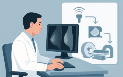
For a male patient with physical exam findings suspicious for breast cancer, the ACR rates both US breast, Mammography diagnostic, and Digital breast tomosynthesis diagnostic as Usually Appropriate. In clinical practice, these studies are often used together to provide a comprehensive initial evaluation.
Breast Ultrasound (US) is an excellent first-line tool in this setting. It uses no ionizing radiation (adult radiation relative level: O, 0 mSv) and is highly effective at characterizing palpable abnormalities. Ultrasound can readily determine if a mass is solid or cystic, define its margins and shape, and assess its vascularity. Critically, it provides real-time visualization of the axilla, allowing for detailed evaluation of lymph node morphology. Perhaps its most important function in this high-suspicion scenario is its ability to guide percutaneous biopsy of both the primary breast mass and any suspicious axillary nodes in the same session, streamlining the path to a definitive pathologic diagnosis.
Diagnostic Mammography, including digital breast tomosynthesis (DBT), is also rated Usually Appropriate and plays a complementary role. While it involves a low dose of ionizing radiation (adult radiation relative level: ☢☢, 0.1-1 mSv), it offers a global view of the entire breast tissue. Mammography is superior to ultrasound for detecting suspicious microcalcifications, which can be a sign of ductal carcinoma in situ (DCIS) that may coexist with an invasive cancer. It also helps define the extent of the mass and screen the remainder of the breast and the contralateral breast for any other occult lesions.
The choice of which to perform first can depend on institutional preference, but a common workflow is to perform both. Often, the patient will undergo mammography first, followed immediately by a targeted ultrasound of the palpable finding and the axilla.
Why Other Studies Are Not Recommended Initially
It is equally important to know which studies to avoid for the initial workup.
- MRI breast without and with IV contrast: This is rated Usually Not Appropriate as a first-line diagnostic tool. While breast MRI is extremely sensitive for detecting cancer, it is not the right initial test for a palpable, suspicious mass. It is more costly, requires IV gadolinium contrast, and its high sensitivity can lead to the detection of incidental, benign findings, potentially resulting in unnecessary follow-up or biopsies. Its primary roles are in preoperative staging after a cancer diagnosis is confirmed, assessing response to neoadjuvant therapy, or as a problem-solving tool for equivocal findings on mammography and ultrasound.
- MRI breast without IV contrast: This is also Usually Not Appropriate. A non-contrast breast MRI has very limited utility in the evaluation of breast cancer, as the key diagnostic information is derived from tissue enhancement patterns after contrast administration.
What Is the Downstream Workflow After Initial Breast Imaging?
The results of the initial ultrasound and mammogram will directly guide the subsequent steps in a clear, protocol-driven manner. The goal is to rapidly obtain a tissue diagnosis.
- If Imaging is Suspicious (BI-RADS 4 or 5): Given the clinical presentation, this is the most anticipated outcome. A finding categorized as suspicious or highly suggestive of malignancy (BI-RADS 4 or 5) requires immediate action. The next step is an image-guided core needle biopsy. Ultrasound is the preferred modality for guiding the biopsy of both the breast mass and any abnormal-appearing axillary lymph nodes. This procedure provides the tissue necessary for a definitive histopathologic diagnosis, which is essential for all subsequent treatment planning.
- If Imaging is Indeterminate (BI-RADS 3): This is a less likely outcome in a patient with clinically suspicious findings. A BI-RADS 3 (probably benign) assessment might be given for a mass with some atypical features that do not meet the threshold for suspicion. However, due to the high-risk physical exam, short-term imaging follow-up is generally inappropriate. In this context, a biopsy is almost always recommended to resolve the discordance between the clinical and imaging findings.
- If Imaging is Negative or Benign (BI-RADS 1 or 2): A completely negative or definitively benign imaging result in the face of a clinically suspicious mass is a red flag for clinical-radiologic discordance. This situation should not lead to reassurance. The palpable finding must be addressed. An immediate referral for surgical consultation for consideration of an excisional biopsy is warranted, as imaging can rarely be falsely negative.
Pitfalls to Avoid (and When to Get Help)
In this high-stakes clinical scenario, avoiding common pitfalls is crucial for ensuring a timely and accurate diagnosis.
- Ignoring Clinical-Radiologic Discordance: Do not be falsely reassured by a “negative” imaging report if your physical exam is highly suspicious. A palpable, fixed mass warrants a tissue diagnosis, even if initial imaging is unremarkable.
- Delaying Biopsy: Once imaging identifies a suspicious lesion (BI-RADS 4 or 5), there is no role for a “watch and wait” approach. Prompt image-guided biopsy is the standard of care.
- Incomplete Axillary Evaluation: The status of the axillary lymph nodes is a critical prognostic factor. Ensure that the ultrasound report explicitly describes the axillary nodes and that any suspicious nodes are biopsied, preferably at the same time as the primary breast mass.
If there is any uncertainty regarding the imaging findings or the appropriate next step, a direct consultation with the interpreting breast radiologist is invaluable. This ensures the clinical and radiologic pictures are aligned and the patient is directed to the most appropriate next step without delay.
Related ACR Topics and Tools
For a comprehensive overview of all clinical variants related to the symptomatic male breast, please see our parent guide. For further exploration of imaging criteria, protocols, and radiation safety, the following GigHz resources are available:
- Evaluation of the Symptomatic Male Breast: ACR Appropriateness Decoded
- Imaging Appropriateness Selector
- Imaging Protocol Library
- Radiation Dose Calculator
Frequently Asked Questions
Should I order a mammogram, an ultrasound, or both for a man with a suspicious breast mass?
According to the ACR, both diagnostic mammography (including tomosynthesis) and breast ultrasound are rated ‘Usually Appropriate.’ In practice, both are typically performed. They provide complementary information: mammography is excellent for detecting microcalcifications and seeing the overall tissue architecture, while ultrasound is superior for characterizing the palpable mass and guiding a biopsy of the mass and any suspicious axillary lymph nodes.
Why isn’t breast MRI recommended as the first imaging test in this scenario?
Breast MRI is rated ‘Usually Not Appropriate’ for the initial evaluation of a suspicious palpable mass. While highly sensitive, it is not the right first step because it is less specific, more expensive, and requires IV contrast. Its primary role is for staging or problem-solving after a cancer diagnosis has already been established by biopsy.
If the ultrasound and mammogram are negative but I can still feel a hard lump, what should I do?
This situation is known as clinical-radiologic discordance and should be taken very seriously. A negative imaging report does not override a suspicious physical exam. The patient should be referred for a surgical consultation to consider an excisional biopsy of the palpable finding to obtain a definitive tissue diagnosis.
Is the imaging workup different if the patient is younger than 25?
For this specific scenario—a clinically suspicious mass with features like skin changes or axillary adenopathy—the age of the patient does not change the initial imaging recommendation. The workup remains the same. Age becomes a differentiating factor only in cases of an ‘indeterminate’ palpable mass without these high-risk features.
Does a history of gynecomastia change the imaging plan if a new, suspicious lump develops?
No. While a patient may have underlying gynecomastia, the development of a new, discrete, hard, or eccentric mass with suspicious features should be investigated according to this high-risk protocol. The new finding should be treated as a distinct clinical problem, and the presence of gynecomastia does not lower the suspicion for malignancy.
Reviewed by Pouyan Golshani, MD, Interventional Radiologist — May 26, 2026