What Is the Best Breast Cancer Screening Strategy for High-Risk Women Over 30?

A 38-year-old woman with a strong family history of breast cancer sits in your exam room. Her mother was diagnosed at age 45 and her maternal aunt at 51. She has no symptoms and a normal clinical breast exam, but she is understandably anxious and wants to establish a proactive screening plan. You know she qualifies as high-risk, but the question is which imaging modalities, and in what combination, offer the most effective surveillance. This scenario requires moving beyond standard screening guidelines to a more intensive, multi-modal approach.
According to the American College of Radiology (ACR) Appropriateness Criteria, for a high-risk female aged 30 or older, both Digital Breast Tomosynthesis screening and supplemental MRI are rated Usually Appropriate.
Who Fits This Clinical Scenario?
This guidance applies specifically to asymptomatic women aged 30 and older who are identified as being at high risk for breast cancer. High risk is generally defined as a lifetime risk of 20% or greater, as calculated by risk assessment models that incorporate family history and other personal risk factors (e.g., Tyrer-Cuzick, BRCAPRO).
This category clinically includes patients with:
- A known pathogenic or likely pathogenic mutation in a breast cancer susceptibility gene (e.g., BRCA1, BRCA2, PALB2, TP53).
- An untested first-degree relative of a known mutation carrier.
- A history of chest wall radiation therapy (e.g., for Hodgkin lymphoma) received between the ages of 10 and 30.
- A personal history of specific breast lesions like lobular carcinoma in situ (LCIS), although this can sometimes fall into an intermediate-risk category depending on other factors.
This workflow is distinct from other common screening scenarios. It does not apply to women at average risk, who typically begin screening later and with mammography alone. It also differs from the workup for women at intermediate risk (e.g., those with dense breasts as their primary risk factor) or high-risk women younger than 30, both of which have their own specific ACR-guided recommendations.
What Diagnoses Are You Working Up in This Scenario?
In a high-risk screening scenario, the primary goal is the early detection of occult breast malignancy before it becomes clinically apparent. The imaging workup is designed to identify subtle architectural changes, microcalcifications, or suspicious enhancing masses that could represent a developing cancer.
The principal target is early-stage invasive ductal carcinoma (IDC), the most common type of breast cancer. Detecting IDC at a small size and before nodal involvement dramatically improves prognosis. Similarly, identifying its precursor, ductal carcinoma in situ (DCIS), allows for treatment that can prevent progression to invasive disease.
A key consideration in high-risk patients, particularly those with genetic mutations, is the potential for more aggressive or mammographically subtle cancers. For instance, invasive lobular carcinoma (ILC) can present as subtle architectural distortion rather than a discrete mass, making it harder to detect on mammography alone. Supplemental imaging with MRI is particularly valuable for identifying these less obvious malignancies. The overarching goal is to find any potential cancer at its most nascent and treatable stage.
Why Is a Multi-Modal Approach with Tomosynthesis and MRI Recommended?
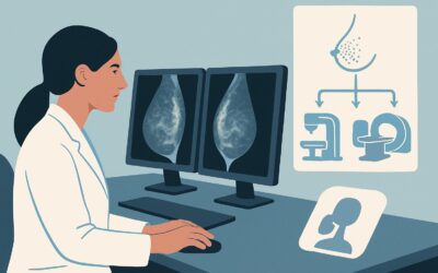
For high-risk women over 30, no single imaging study is sufficient. The ACR designates both Digital Breast Tomosynthesis (DBT) screening and MRI of the breast with and without IV contrast as Usually Appropriate, reflecting the need for a complementary, multi-modal strategy.
Digital Breast Tomosynthesis (DBT), or 3D mammography, serves as the foundational imaging test. By acquiring images from multiple angles to create thin slices, DBT reduces the effect of overlapping breast tissue. This improves cancer detection rates and, just as importantly, decreases the rate of false positives and unnecessary patient recalls compared to standard 2D mammography. For this reason, it is the preferred mammographic technique.
However, mammography’s sensitivity is limited, especially in women with dense breast tissue, which is common in premenopausal women. This is where MRI breast without and with IV contrast plays a critical role. MRI does not use ionizing radiation (Radiation Level: O 0 mSv) and offers superior sensitivity for detecting invasive cancers, including those not visible on mammography. It is recommended as a supplemental screening tool to be performed in addition to, not in place of, annual mammography/DBT.
In contrast, other modalities are rated lower for this specific scenario.
- US breast is rated May be appropriate. While it can detect some mammographically occult cancers, it is highly operator-dependent and has a higher false-positive rate, leading to more benign biopsies than MRI. It is typically reserved for patients with a contraindication to MRI.
- MRI breast without IV contrast is Usually not appropriate. The administration of gadolinium-based contrast is essential for breast MRI, as malignant lesions are typically characterized by their unique pattern and kinetics of enhancement. Without contrast, the study is non-diagnostic for cancer screening.
The standard high-risk screening protocol therefore involves annual DBT and annual supplemental breast MRI. For detailed technical specifications on image acquisition, refer to our guide on the Screening Mammography (with DBT) protocol.
What’s Next After Screening? Downstream Workflow
The results of annual screening with DBT and MRI will guide the subsequent clinical pathway, typically reported using the Breast Imaging Reporting and Data System (BI-RADS) classification.
- Negative or Benign Result (BI-RADS 1 or 2): If both DBT and MRI are negative or show clearly benign findings, the patient continues with her established annual screening schedule. No further immediate action is needed beyond reinforcing the importance of continued adherence to the high-risk surveillance plan.
- Incomplete or Indeterminate Result (BI-RADS 0 or 3): A BI-RADS 0 assessment means the initial imaging is incomplete and requires additional evaluation, such as diagnostic mammographic views or a targeted ultrasound, to clarify a potential finding. A BI-RADS 3 finding is likely benign but requires short-interval follow-up (typically in 6 months) to ensure stability. This is a common outcome, and careful management is crucial to avoid both unnecessary anxiety and missed diagnoses.
- Suspicious Result (BI-RADS 4 or 5): These findings are suspicious for malignancy and require tissue sampling. The next step is an image-guided biopsy (e.g., stereotactic, ultrasound-guided, or MRI-guided) of the suspicious lesion. A positive biopsy result triggers an immediate referral to a multidisciplinary breast cancer team, including a breast surgeon and medical oncologist, to plan treatment.
This structured downstream workflow ensures that every screening result leads to a clear and appropriate next step, from routine follow-up to urgent diagnostic intervention.
Pitfalls to Avoid (and When to Get Help)
Navigating high-risk breast cancer screening requires careful attention to detail to avoid common missteps that can compromise patient care.
First, avoid relying solely on mammography/DBT. For this specific high-risk population, omitting supplemental screening with breast MRI significantly reduces the overall sensitivity of the surveillance program. Second, ensure accurate risk assessment. Misclassifying a patient as average or intermediate risk when they meet high-risk criteria will lead to inadequate screening. Conversely, over-screening a patient not truly at high risk can lead to unnecessary tests and anxiety.
Another pitfall is improper scheduling. Ideally, DBT and MRI should be staggered by six months to provide imaging surveillance at more frequent intervals throughout the year. Finally, do not order a breast MRI without intravenous contrast for screening; it is a non-diagnostic study for this indication. If a patient has a severe contraindication to gadolinium, consultation with a breast radiologist is essential to determine the best alternative, which may be supplemental ultrasound.
Related ACR Topics and Tools
This article covers a single, specific clinical scenario. For a comprehensive overview of all patient presentations and imaging recommendations within this topic, please see our parent guide.
- For breadth across all scenarios in Female Breast Cancer Screening, see our parent guide: Female Breast Cancer Screening: ACR Appropriateness Decoded.
For additional decision support and technical guidance, the following GigHz tools are available:
- ACR Appropriateness Criteria Lookup — for adjacent scenarios
- Imaging Protocol Library — for technique on the recommended study
- Radiation Dose Calculator — for cumulative dose conversations
Frequently Asked Questions
Should high-risk women alternate between mammography and MRI each year?
No, this is a common misconception. Current guidelines recommend that high-risk women receive both an annual mammogram (preferably DBT) AND an annual supplemental breast MRI. These tests are complementary, not interchangeable. To maximize surveillance, many centers schedule the two exams six months apart.
What is the starting age for high-risk screening?
For women with a genetic mutation or a strong family history, screening often begins at age 30 (or sometimes as early as 25), but not before age 25. For women with a history of chest radiation, screening should begin 8 years after the completion of radiation therapy, but not before age 25.
Is an abbreviated breast MRI appropriate for high-risk screening?
Yes, the ACR rates abbreviated breast MRI as ‘Usually Appropriate’ for this scenario. Abbreviated protocols use fewer sequences, reducing scan time and cost while maintaining high diagnostic accuracy for cancer detection. Its availability may vary by institution.
What if a high-risk patient has a contraindication to MRI, such as a non-compatible implanted device or severe renal insufficiency?
If a patient cannot undergo an MRI, supplemental screening with whole-breast ultrasound is rated as ‘May be appropriate’ and is the most common alternative. While not as sensitive as MRI, it can detect some mammographically occult cancers. This decision should be made in consultation with a breast imaging specialist.
Does having dense breasts alone qualify a patient as ‘high-risk’ for this screening protocol?
No. While breast density is an independent risk factor for breast cancer and can mask cancers on mammography, it typically places a woman in the intermediate-risk category, not high-risk. The high-risk protocol discussed here is reserved for those with a lifetime risk of 20% or more due to genetic factors, family history, or prior chest radiation.
Reviewed by Pouyan Golshani, MD, Interventional Radiologist — May 30, 2026