Should You Order MRI for a Suspected Silicone Implant Complication in a Patient Under 30?
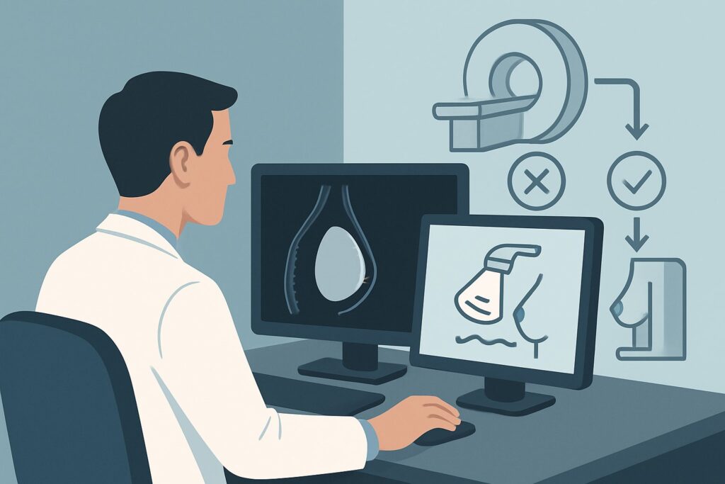
A 28-year-old patient presents to your clinic with new, unilateral breast pain and a palpable change in the shape of her right breast. She underwent bilateral silicone breast augmentation six years ago and is now concerned about a possible implant rupture. As the ordering clinician, you face a critical decision: which imaging study will provide the most accurate diagnosis without unnecessary radiation or contrast exposure? This article provides a detailed workflow for this specific clinical scenario, grounded in the American College of Radiology (ACR) Appropriateness Criteria. For this presentation, an MRI of the breast without IV contrast is rated Usually Appropriate and is the recommended initial study.
Who Fits This Clinical Scenario?
This guidance is specifically for clinicians evaluating a patient who meets all the following criteria:
- Age: Younger than 30 years.
- Patient Identity: Female or transfeminine.
- Implant Type: Silicone gel-filled breast implants.
- Presentation: Suspected implant complication (e.g., new pain, change in breast size or shape, palpable abnormality, firmness).
- Imaging Stage: This is the initial, first-line imaging workup for the new symptoms.
It is crucial to distinguish this scenario from similar but distinct clinical situations that follow different imaging pathways. This advice does not apply if your patient has saline implants, as their rupture is typically diagnosed clinically (deflation) and imaging is less frequently required. It also does not apply to asymptomatic screening for “silent rupture” of silicone implants, which has its own recommended timeline and imaging protocol. Finally, for patients age 40 or older, the role of mammography for concurrent breast cancer screening alongside implant evaluation changes the decision-making process significantly.
What Diagnoses Are You Working Up in This Scenario?
When a younger patient with silicone implants presents with symptoms, your differential diagnosis is focused on complications related to the implant itself. The primary goal of imaging is to assess implant integrity and evaluate for peri-implant issues.
Intracapsular Implant Rupture: This is the most common form of silicone implant failure. The implant shell tears, but the leaked silicone gel is contained by the surrounding fibrous capsule that the body naturally forms. Patients may present with subtle changes in shape or new pain. The classic imaging finding on MRI is the “linguine sign,” representing the collapsed implant shell floating within the silicone gel.
Extracapsular Implant Rupture: A more significant complication where silicone gel breaches both the implant shell and the fibrous capsule, extravasating into the surrounding breast parenchyma or even migrating to nearby lymph nodes. This can lead to a palpable mass or an inflammatory reaction known as a siliconoma. It is a critical diagnosis to make, as it often necessitates more complex surgical management.
Peri-implant Fluid Collection: This includes seromas (clear fluid) or hematomas (blood) that can accumulate around the implant. While small amounts of fluid can be normal, a new or enlarging collection may indicate trauma, inflammation, or, in rare cases, be associated with Breast Implant-Associated Anaplastic Large Cell Lymphoma (BIA-ALCL).
Capsular Contracture: This is a clinical diagnosis characterized by the tightening and hardening of the fibrous capsule around the implant, which can cause pain and cosmetic deformity. While imaging cannot diagnose contracture directly, it is essential for excluding implant rupture as the underlying cause of the patient’s symptoms.
Why Is MRI Breast Without IV Contrast the Recommended Study?
The ACR designates MRI of the breast without IV contrast as Usually Appropriate for this scenario because it offers the highest sensitivity and specificity for detecting silicone implant rupture. This non-contrast protocol is specifically designed to evaluate implant integrity.
The high-resolution, multi-planar images of a dedicated breast MRI allow for clear visualization of the implant shell, the internal silicone gel, and the surrounding fibrous capsule. Silicone-specific sequences can suppress signals from water and fat, making any free silicone from an extracapsular rupture highly conspicuous. This is the most reliable method for identifying the “linguine sign” of intracapsular rupture and distinguishing it from normal radial folds of the implant.
Alternative modalities are rated lower for specific reasons in this context:
- Ultrasound (US) of the breast is rated May be appropriate. While it is non-invasive and uses no radiation, it is highly operator-dependent and significantly less sensitive than MRI, particularly for intracapsular rupture. It may fail to detect subtle signs of shell collapse. The “stepladder sign” on ultrasound is a specific indicator of intracapsular rupture, but its absence does not rule it out. US can be useful for evaluating a focal palpable concern or guiding aspiration of a fluid collection but is not the primary tool for a comprehensive integrity check.
- Mammography and Digital Breast Tomosynthesis (DBT) are both rated Usually not appropriate for the primary purpose of evaluating implant integrity. The dense silicone gel is radiopaque, obscuring the internal structure of the implant and making rupture detection nearly impossible. These studies also involve ionizing radiation (RRL ☢☢ 0.1-1mSv), which should be avoided in a young patient when a non-radiation alternative like MRI (RRL O 0 mSv) is superior for answering the clinical question.
- MRI of the breast with IV contrast is also Usually not appropriate. The addition of gadolinium-based contrast does not improve the visualization of implant rupture and is not necessary for this indication. Omitting contrast avoids potential risks and reactions associated with the agent.
When ordering the study, specifying “non-contrast MRI for silicone implant integrity” ensures the radiology department performs the correct sequences. Once you’ve decided on MRI, our protocol guide covers the technical details and reading principles in depth: MRI Breast With and Without Contrast.
What’s Next After MRI? Downstream Workflow
The results of the non-contrast breast MRI will guide your next steps in management, which almost always involves a consultation with a plastic or breast surgeon.
- If the MRI is positive for rupture (intracapsular or extracapsular): The patient should be referred to a surgeon for consultation. The standard management is surgical, involving implant removal (explantation) with or without replacement, and capsulectomy (removal of the fibrous capsule). For extracapsular rupture, the surgeon may need to excise any associated siliconomas.
- If the MRI is negative for rupture but shows a significant fluid collection: A large or symptomatic seroma may warrant ultrasound-guided aspiration for both diagnostic and therapeutic purposes. The fluid should be sent for cytology, especially if there is any clinical or imaging suspicion for BIA-ALCL. Surgical consultation is still appropriate to discuss management of the collection.
- If the MRI is negative for rupture or other significant findings: If the patient’s symptoms persist despite a normal MRI, the diagnosis is more likely to be clinical, such as capsular contracture or musculoskeletal pain. Management would focus on reassurance and symptomatic relief. If a palpable abnormality was the primary concern and it persists despite a negative MRI, further evaluation with targeted ultrasound may be considered to ensure no subtle lesion was missed.
A negative MRI provides strong evidence against implant rupture, allowing you to confidently shift the focus of your workup and management away from implant failure.
Pitfalls to Avoid (and When to Get Help)
Navigating this clinical scenario requires careful attention to detail to avoid common missteps.
- Ordering the Wrong MRI Protocol: Do not order a standard “MRI breast with and without contrast.” This is a different study designed for cancer detection. Explicitly request a non-contrast MRI for silicone implant evaluation.
- Relying Solely on Ultrasound: While tempting due to its accessibility, a negative ultrasound is not sufficient to rule out a silicone implant rupture. If clinical suspicion is high, a negative US should be followed by an MRI.
- Dismissing Patient Symptoms: A patient’s report of a change in breast shape or feel is a significant clinical finding. Even if imaging is negative for rupture, these symptoms warrant a thorough evaluation and consideration of other causes like capsular contracture.
- Ignoring the Contralateral Breast: While the evaluation is prompted by unilateral symptoms, the MRI protocol should always include imaging of both breasts, as implant rupture can be asymptomatic (“silent”).
If the imaging findings are equivocal or do not align with the clinical picture, a discussion with the interpreting radiologist and a referral to a surgeon specializing in breast reconstruction and revision are the appropriate next steps.
Related ACR Topics and Tools
For a comprehensive overview of all clinical variants related to breast implant imaging, including saline implants and asymptomatic screening, please refer to our parent guide. For other clinical questions, the tools below can help you apply evidence-based guidelines to your practice.
- For breadth across all scenarios in Breast Implant Evaluation, see our parent guide: Breast Implant Evaluation: ACR Appropriateness Decoded.
- ACR Appropriateness Criteria Lookup — for adjacent scenarios
- Imaging Protocol Library — for technique on the recommended study
- Radiation Dose Calculator — for cumulative dose conversations
Frequently Asked Questions
Why is MRI without contrast preferred over MRI with contrast for suspected silicone implant rupture?
MRI with intravenous contrast is not necessary to evaluate the integrity of a silicone implant. The specific silicone-sensitive sequences used in a non-contrast protocol are optimized to visualize the implant shell and detect free silicone gel. Adding contrast provides no additional diagnostic information for rupture, while exposing the patient to the potential risks and costs of a gadolinium-based contrast agent.
If my patient is younger than 30, do I still need to worry about breast cancer?
While the risk of breast cancer is lower in this age group, it is not zero. A thorough clinical breast exam is always warranted. If there is a distinct, palpable lump that is separate from the implant, a diagnostic mammogram and/or targeted ultrasound may be appropriate in addition to the MRI for implant evaluation. However, for symptoms clearly related to the implant itself (e.g., diffuse change in shape), the primary investigation should focus on implant integrity first.
What if my patient has a contraindication to MRI, like a non-compatible device or severe claustrophobia?
In cases where MRI is contraindicated, high-resolution ultrasound performed by an experienced technologist is the next best option. It is rated ‘May be appropriate’ by the ACR. It’s important to communicate the limitations of ultrasound to the patient and acknowledge that it is less sensitive than MRI for detecting intracapsular rupture. The clinical and sonographic findings should be reviewed with a surgeon to decide on the best course of action.
Does the age of the silicone implant affect the choice of imaging?
No, for a symptomatic patient under 30, non-contrast MRI is the recommended initial study regardless of implant age. However, the likelihood of rupture does increase with the age of the implant, which may raise your clinical suspicion. The FDA recommends asymptomatic screening for silent rupture with MRI starting 5-6 years after placement, but for a patient with new symptoms, the workup is diagnostic, not for screening.
How does this guidance change for a transfeminine patient on hormonal therapy?
The imaging recommendation does not change. For the specific question of silicone implant integrity, non-contrast MRI remains the most accurate modality. Hormonal therapy can cause changes in the native breast tissue, but this does not affect the ability of MRI to visualize the implant shell and detect rupture.
Reviewed by Pouyan Golshani, MD, Interventional Radiologist — May 29, 2026