Should You Order Ultrasound for Focal, Noncyclical Breast Pain in Women Over 40?

A 48-year-old woman presents to your clinic with a two-month history of persistent, sharp pain in the upper outer quadrant of her left breast. She can point to the exact spot with one finger. The pain is unrelated to her menstrual cycle and is significant enough that it’s causing her anxiety. On examination, there is tenderness but no discrete palpable mass, skin changes, or nipple discharge. You need to decide on the most appropriate initial imaging study to evaluate her symptoms and rule out an underlying pathology. This article provides a detailed clinical workflow for this specific scenario, guided by the American College of Radiology (ACR) Appropriateness Criteria. For this patient, both breast ultrasound and diagnostic mammography are rated ‘Usually Appropriate’, forming the cornerstone of the initial workup.
Who Fits This Clinical Scenario for Focal Breast Pain?
This guidance is specifically for women aged 40 and older presenting with clinically significant breast pain for initial imaging. To apply this workflow correctly, the patient’s presentation must meet two key criteria:
1. Focal Pain: The pain is localized to a specific, small area, typically one quadrant of the breast or less. The patient can often point to the exact spot. This is distinct from diffuse pain that affects the entire breast or is bilateral.
2. Noncyclical Pain: The pain is constant or intermittent but is not associated with the menstrual cycle. This differentiates it from the more common cyclical mastalgia that fluctuates with hormonal changes.
This workflow is NOT intended for patients with different presentations, which are covered under separate ACR guidelines. Key exclusions include:
- Women under 40: Younger patients have different pre-test probabilities for malignancy and greater breast density, altering the imaging algorithm. Separate guidelines exist for patients under 30 and those aged 30 to 39.
- Clinically insignificant pain: Patients with diffuse, bilateral, or clearly cyclical pain without other findings generally do not require imaging, as the likelihood of identifying a pathologic cause is extremely low.
- Associated clinical findings: If the pain is accompanied by a palpable lump, skin changes (dimpling, erythema), or pathologic nipple discharge, the workup follows the guidelines for those specific findings, which have a higher suspicion for malignancy.
What Diagnoses Are You Working Up with Imaging for Focal Breast Pain?
While breast pain is a common complaint, it is an uncommon presenting symptom of breast cancer. However, its focal and persistent nature in a woman over 40 mandates a thorough evaluation to exclude malignancy and identify benign causes. The imaging workup is designed to differentiate among several potential diagnoses.
The most consequential, though least likely, diagnosis to exclude is an underlying breast malignancy. A small percentage of breast cancers can present with focal pain as the primary symptom, even without a palpable mass. This may be due to perineural invasion or local inflammatory effects. Imaging is critical for identifying suspicious masses, architectural distortion, or calcifications that may represent an occult cancer.
A far more common cause of focal pain is a benign breast cyst. These fluid-filled sacs can develop tension, causing sharp, localized pain. Ultrasound is exceptionally effective at identifying and characterizing cysts, often providing immediate reassurance. A complex cyst with internal debris or wall thickening may also be the source.
Fibrocystic changes can also cause focal, noncyclical pain. While often associated with diffuse, cyclical tenderness, a specific area of prominent fibroglandular tissue or a developing fibroadenoma can be a pain generator.
Less common causes include inflammatory conditions like focal mastitis or duct ectasia, where a milk duct becomes blocked or inflamed, leading to localized pain and tenderness. Finally, imaging helps rule out a breast parenchymal cause, pointing toward an extramammary etiology such as costochondritis (inflammation of rib cartilage), which can mimic focal breast pain.
Why Are Ultrasound and Diagnostic Mammography the Recommended Studies?
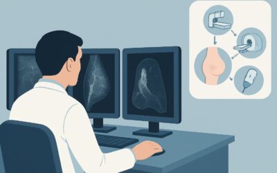
For a woman aged 40 or older with focal, noncyclical breast pain, the ACR rates both US breast, Mammography diagnostic, and Digital breast tomosynthesis diagnostic as ‘Usually Appropriate’. In practice, these studies are complementary and often performed together, starting with the mammogram.
The initial step is typically a diagnostic mammogram (either 2D or tomosynthesis/3D). This provides a comprehensive overview of the entire breast and axilla, serving as the baseline for comparison and detecting findings like suspicious calcifications that ultrasound may miss. Tomosynthesis is particularly useful in women with dense breasts, as it reduces the effect of overlapping tissue.
Following the mammogram, a targeted ultrasound of the area of pain is almost always performed. Ultrasound is the problem-solving tool. Its high spatial resolution is excellent for evaluating the specific symptomatic area identified by the patient. It can readily distinguish solid from cystic lesions, characterize the margins and vascularity of any identified mass, and guide biopsy if needed. For many benign causes, such as a simple cyst, ultrasound is definitive.
This combined approach leverages the strengths of both modalities: the mammogram’s global view for calcifications and architectural distortion, and the ultrasound’s focused, high-resolution characterization of the symptomatic area.
Why are other studies rated lower for this scenario?
- MRI breast without and with IV contrast is rated ‘Usually not appropriate’ for initial evaluation of focal pain. While highly sensitive, its specificity is lower, leading to more false positives and unnecessary biopsies for this indication. MRI is reserved for specific situations, such as problem-solving after inconclusive mammography/ultrasound or for high-risk screening.
- Sestamibi MBI (Molecular Breast Imaging) is also ‘Usually not appropriate’. This nuclear medicine study involves a significant radiation dose (☢☢☢ 1-10 mSv) and is typically used as a secondary tool for problem-solving in specific cases, not as a first-line diagnostic test for pain.
The recommended combination of diagnostic mammography (☢☢ 0.1-1mSv) and ultrasound (O 0 mSv) provides a thorough evaluation with a justifiable radiation level.
What’s Next After Ultrasound and Mammography? Downstream Workflow
The results of the combined mammogram and ultrasound will guide the subsequent steps in the patient’s care. The workflow branches based on the BI-RADS (Breast Imaging Reporting and Data System) assessment.
- Negative or Benign Finding (BI-RADS 1 or 2): If both the mammogram and the targeted ultrasound of the painful area are negative or show a clearly benign finding (e.g., a simple cyst), the patient can be reassured that there is no radiologic evidence of malignancy. The pain is likely due to benign fibrocystic changes or has an extramammary cause (e.g., musculoskeletal). Management focuses on symptom control, and the patient should return to her routine screening mammography schedule.
- Probably Benign Finding (BI-RADS 3): If a finding is identified that has a very high likelihood of being benign (typically <2% risk of malignancy), the standard recommendation is short-interval follow-up imaging, usually with ultrasound in 6 months, to ensure stability. This avoids biopsy for lesions that are overwhelmingly likely to be benign.
- Suspicious or Highly Suggestive of Malignancy (BI-RADS 4 or 5): If imaging reveals a suspicious mass, architectural distortion, or calcification cluster, the definitive next step is a tissue diagnosis. An image-guided core needle biopsy (most often with ultrasound guidance, given its use in characterizing the focal finding) is required to establish a pathologic diagnosis and guide treatment.
- Incomplete (BI-RADS 0): This assessment is often given on the initial mammogram before the targeted ultrasound is complete. The final report after both studies should provide a definitive BI-RADS 1-5 category.
Pitfalls to Avoid (and When to Get Help)
Navigating the workup for focal breast pain requires careful attention to detail to avoid common missteps.
1. Pitfall: Ordering Ultrasound Alone. In women 40 and over, skipping the diagnostic mammogram and ordering only a targeted ultrasound is a significant pitfall. This approach can miss suspicious calcifications or architectural distortion outside the immediate area of pain, which may be the true cause of symptoms or an incidental cancer.
2. Pitfall: Accepting a “Negative” Screening Mammogram. If a patient had a recent screening mammogram that was negative but now presents with new focal pain, a diagnostic workup is still required. A diagnostic mammogram includes special views and is interpreted with the knowledge of the clinical symptom, followed by a targeted ultrasound.
3. Pitfall: Dismissing Pain After Negative Imaging. While negative imaging is reassuring, the patient’s symptom is real. It’s important to consider and evaluate for extramammary causes like costochondritis, rib fracture, or neuralgia if breast imaging is normal.
4. Pitfall: Not Correlating Imaging with the Symptom. The radiologist must be clearly informed of the precise location of the pain. The goal is to find a radiologic correlate for the clinical symptom. If imaging is negative at the site of pain, that is the key finding.
If imaging is negative but the pain persists and worsens, or if a palpable abnormality develops, re-evaluation and consultation with a breast specialist or surgeon are warranted.
Related ACR Topics and Tools
This article focuses on a single clinical scenario. For a comprehensive overview of all variants and patient presentations related to breast pain, please consult our parent guide. You can also use the tools below to explore adjacent scenarios, imaging techniques, and radiation safety considerations.
- For breadth across all scenarios in Breast Pain, see our parent guide: Breast Pain: ACR Appropriateness Decoded.
- To explore other clinical situations, use the ACR Appropriateness Criteria Lookup.
- For details on imaging techniques, see the Imaging Protocol Library.
- For patient conversations about medical radiation, use the Radiation Dose Calculator.
Frequently Asked Questions
If my patient had a normal screening mammogram 3 months ago, do I still need to order a diagnostic workup for new focal pain?
Yes. A screening mammogram is performed on an asymptomatic patient. New, focal, noncyclical pain is a clinical symptom that requires a dedicated diagnostic evaluation. This includes a diagnostic mammogram (which may have different views than the screening study) and a targeted ultrasound of the painful area to ensure no underlying pathology has developed.
Why not just start with an ultrasound and skip the mammogram and its radiation?
In women 40 and older, a mammogram is crucial because it provides a global view of the breast and is superior to ultrasound for detecting certain signs of cancer, such as suspicious microcalcifications. An ultrasound alone could miss a malignancy that is not located at the precise site of pain. The combined approach is the standard of care.
What if the imaging is completely normal but the patient’s pain persists?
If a thorough diagnostic mammogram and targeted ultrasound are negative (BI-RADS 1), you can confidently reassure the patient that there is no imaging evidence of breast cancer or another significant breast abnormality. The next step is to consider non-breast causes of pain, such as musculoskeletal issues (e.g., costochondritis), and to manage the symptoms. The patient should continue with her routine annual screening mammography.
Is tomosynthesis (3D mammography) required for this diagnostic workup?
Digital breast tomosynthesis is rated ‘Usually Appropriate’ by the ACR, alongside standard 2D diagnostic mammography. It is often preferred, especially in women with dense breast tissue, as it can reduce tissue overlap and improve cancer detection. However, a high-quality 2D diagnostic mammogram followed by targeted ultrasound is also an acceptable and appropriate workup.
Does a finding of a simple cyst on ultrasound explain the patient’s focal pain?
Yes, very often. Simple cysts are a common cause of focal breast pain, especially if they are under tension. When the ultrasound identifies a simple cyst that corresponds directly to the site of the patient’s pain, it is considered the likely cause. This is a benign finding that provides a clear explanation for the symptom and allows for reassurance.
Reviewed by Pouyan Golshani, MD, Interventional Radiologist — June 27, 2026