What Imaging Should You Order for Asymptomatic Silicone Breast Implant Screening?
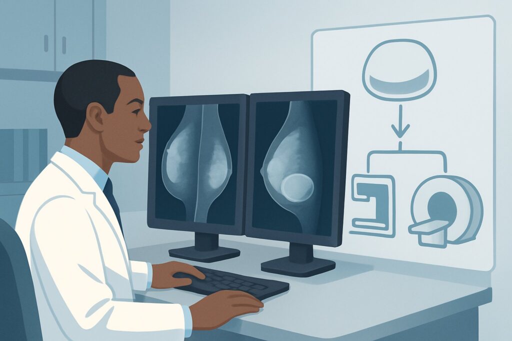
A 46-year-old patient is in your office for a routine annual visit. She underwent bilateral silicone breast augmentation six years ago and feels perfectly well, with no new symptoms, pain, or changes in breast shape. Recalling the FDA recommendations for implant surveillance, you know it’s time to consider imaging to assess for silent rupture. The patient is anxious about radiation and wants the most straightforward, effective option. This raises a common clinical question: what is the appropriate initial imaging study for routine, asymptomatic screening of silicone breast implants?
According to the American College of Radiology (ACR) Appropriateness Criteria, for this specific scenario, a breast ultrasound (US) is rated Usually appropriate. This article provides a detailed workflow for this presentation, explaining the rationale for this choice and the downstream steps based on the imaging results.
Who Fits This Clinical Scenario?
This guidance is specifically for an adult patient of any age, either female or transfeminine, who has silicone breast implants and is asymptomatic. The workflow applies to two distinct timing situations:
- Initial Screening: The patient is between 5 to 6 years from their initial implant placement.
- Follow-up Screening: The patient had a previous negative screening study and is now due for routine follow-up, typically every 2 to 3 years.
The key elements are the implant type (silicone) and the absence of symptoms. Patients who present with new clinical signs or symptoms—such as breast pain, a palpable lump, changes in breast size or shape, or skin changes—fall into a different clinical category requiring a diagnostic workup, not routine screening.
This workflow is distinct from several related scenarios:
- Patients with saline implants: Saline implant rupture is typically diagnosed clinically (deflation) and does not require the same imaging surveillance protocol.
- Patients less than 5 years post-placement: Routine screening for silent rupture is not typically initiated before the 5-year mark.
- Patients with suspected rupture: Any symptomatic patient, regardless of time since placement, should be evaluated under the “suspected implant complication” or “suspected rupture” criteria, which may alter the choice and urgency of imaging.
What Diagnoses Are You Working Up in This Scenario?
The primary goal of imaging in this asymptomatic patient is surveillance for implant integrity. The differential diagnosis, or list of potential findings, is centered on identifying subclinical changes before they become symptomatic problems.
The most critical finding to identify is a silent implant rupture. Silicone gel is cohesive, meaning an implant can rupture its outer shell without an immediate change in the breast’s shape or feel. This can be an intracapsular rupture, where the silicone remains contained by the fibrous capsule the body forms around the implant, or an extracapsular rupture, where silicone has breached both the shell and the fibrous capsule. Early detection allows for planned surgical intervention.
Another consideration is silicone gel bleed. This refers to the microscopic migration of silicone molecules through an intact implant shell. While a minor degree of gel bleed is common, imaging can sometimes detect extracapsular silicone in nearby tissues or lymph nodes, even without a frank rupture.
Imaging may also incidentally identify other conditions. A peri-implant fluid collection, such as a seroma, can be visualized. While often benign, a new or enlarging collection could warrant further investigation. Finally, while not the primary purpose of this specific study, imaging provides a general assessment of the implant’s position and the surrounding breast tissue, though it is not a substitute for dedicated breast cancer screening.
Why Is Breast US the Recommended Study for This Presentation?
The ACR rates both breast ultrasound (US) and non-contrast breast Magnetic Resonance Imaging (MRI) as Usually appropriate for this scenario. However, ultrasound is often the practical and effective first-line choice for routine screening due to its accessibility, lower cost, and lack of ionizing radiation.
Breast US is highly effective at visualizing the implant shell and the surrounding gel. Radiologists look for specific signs of rupture. The “stepladder sign” on ultrasound, which represents the collapsed implant shell floating within the silicone gel, is a reliable indicator of intracapsular rupture. For extracapsular rupture, the “snowstorm sign” or “echogenic noise” signifies free silicone in the breast parenchyma. Because it uses no radiation (0 mSv), it is an ideal modality for repeated surveillance every 2 to 3 years.
While non-contrast breast MRI is also Usually appropriate and is considered the most sensitive and specific imaging test for detecting silicone implant rupture, it is more expensive and less widely available than ultrasound. It is often reserved as a problem-solving tool if the ultrasound results are unclear or equivocal.
Why are other studies rated lower?
- Mammography and Digital Breast Tomosynthesis (DBT): These are rated Usually not appropriate for the specific question of implant integrity. While essential for breast cancer screening, mammography has low sensitivity for detecting intracapsular silicone rupture. The compression required can also, in rare cases, risk damaging an already compromised implant. These studies also involve a small amount of ionizing radiation (☢☢ 0.1-1 mSv).
- MRI Breast with IV Contrast: This is also rated Usually not appropriate. The administration of intravenous gadolinium-based contrast does not improve the visualization of the implant shell or the detection of free silicone. It adds unnecessary cost, time, and the potential for contrast-related adverse events without providing additional diagnostic value for this clinical question.
What’s Next After Breast US? Downstream Workflow
The results of the screening breast ultrasound will guide your next steps in a clear decision tree. The goal is to confirm implant integrity and establish a plan for future surveillance or intervention.
If the ultrasound is negative (implant is intact):
This is the most common outcome. Reassure the patient that the implant appears intact. The recommended next step is to continue routine surveillance, with the next screening ultrasound scheduled in 2 to 3 years. The patient should also continue their standard, age-appropriate breast cancer screening as recommended by their primary care provider or gynecologist.
If the ultrasound is positive for rupture (intracapsular or extracapsular):
A definitive finding of rupture on ultrasound warrants a referral to a plastic or breast surgeon. The surgeon will discuss the findings with the patient and review options, which typically include implant removal with or without replacement. While some surgeons may request a confirmatory non-contrast breast MRI, a high-quality ultrasound with classic signs of rupture is often sufficient to proceed with surgical planning.
If the ultrasound is indeterminate or equivocal:
Sometimes, findings like complex folds, peri-implant fluid, or unclear echotexture can make it difficult to definitively confirm or exclude a rupture. In this situation, the appropriate next step is to order the other Usually appropriate study: a non-contrast breast MRI. MRI can resolve the ambiguity and provide a definitive diagnosis, guiding whether the patient can return to routine screening or needs a surgical consultation.
Pitfalls to Avoid (and When to Get Help)
Navigating asymptomatic implant screening requires attention to a few common pitfalls to ensure an accurate and efficient workup.
First, avoid confusing routine implant integrity screening with diagnostic breast cancer screening. If a patient has a palpable lump, do not simply order an implant screening US; a diagnostic mammogram and/or diagnostic ultrasound focused on the palpable abnormality is required.
Second, be aware that normal radial folds in an implant can sometimes mimic the “stepladder sign” of rupture on ultrasound. This is an area where radiologist expertise is key. Ensure the study is performed at a center experienced in breast imaging.
Third, do not order a contrast-enhanced breast MRI for this indication. It adds no value for assessing implant integrity and introduces unnecessary risk and cost. The correct order is a non-contrast study.
If an ultrasound report is returned as “indeterminate” or “equivocal,” the best course of action is to escalate to the next appropriate study. Do not simply repeat the ultrasound. The definitive problem-solving tool in this workflow is a non-contrast breast MRI.
Related ACR Topics and Tools
For a comprehensive overview of all clinical scenarios related to breast implant imaging, including symptomatic patients and those with saline implants, please consult our parent guide. This article focuses on one specific workflow, while the parent guide provides the broader context.
- For breadth across all scenarios in Breast Implant Evaluation, see our parent guide: Breast Implant Evaluation: ACR Appropriateness Decoded.
To explore other clinical presentations or refine your imaging orders, the following GigHz tools are available:
- ACR Appropriateness Criteria Lookup — for adjacent scenarios
- Imaging Protocol Library — for technique on the recommended study
- Radiation Dose Calculator — for cumulative dose conversations
Frequently Asked Questions
Why not just order a breast MRI for every patient, since it’s the most sensitive test?
While breast MRI is highly sensitive and specific for silicone implant rupture, breast ultrasound is also very effective and is rated equally as ‘Usually appropriate’ by the ACR for this scenario. Ultrasound is more widely available, significantly less expensive, and faster to perform, making it a more practical and efficient first-line screening tool for asymptomatic patients. MRI is best reserved for cases where the ultrasound is inconclusive.
Does this implant screening ultrasound replace my regular mammograms for breast cancer screening?
No, it does not. This ultrasound is specifically designed to evaluate the integrity of the silicone implant. It is not a substitute for routine, age-appropriate breast cancer screening with mammography or digital breast tomosynthesis (DBT). Patients with implants should continue to follow standard guidelines for breast cancer screening.
What if my patient is 7 years post-implant placement and has never had screening? Does this workflow still apply?
Yes. This workflow applies to the initial screening at 5 to 6 years, but it is also the correct pathway for a patient who is presenting for their first screening later than that, as long as they remain asymptomatic. The recommendation would be to proceed with a screening breast ultrasound.
Is there a risk of the ultrasound procedure itself causing a rupture?
No. A breast ultrasound is a non-invasive procedure that uses a handheld transducer and gel on the skin’s surface. It involves gentle pressure but does not use compression or force that would pose a risk to the integrity of the breast implant.
What are the specific ultrasound findings for silicone implant rupture?
The key finding for an intracapsular rupture (the most common type) is the ‘stepladder sign,’ which shows the collapsed implant shell floating within the silicone gel. For an extracapsular rupture, where silicone has leaked into the surrounding tissue, radiologists look for the ‘snowstorm sign,’ which appears as a bright, echogenic area with posterior acoustic shadowing.
Reviewed by Pouyan Golshani, MD, Interventional Radiologist — June 27, 2026