Should You Order Screening Imaging After Prophylactic Mastectomy Without Reconstruction?

A 48-year-old female with a known BRCA1 mutation presents for her annual follow-up. Five years ago, she underwent a bilateral prophylactic mastectomy without reconstruction and has been well since. She has no new complaints, and her surgical scars are well-healed. During the visit, she asks, “Shouldn’t I be getting some kind of yearly scan, like an MRI or something, just to be safe?” You know that routine imaging after mastectomy is a nuanced topic and need to decide on the appropriate surveillance plan. This article details the American College of Radiology (ACR) Appropriateness Criteria for this specific high-risk screening scenario.
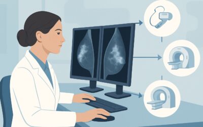
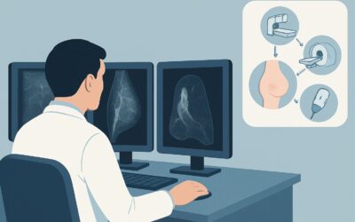
For a high-risk patient with a history of bilateral prophylactic mastectomy and no reconstruction, the ACR has determined that all routine screening imaging modalities, including Ultrasound (US), Mammography, and Magnetic Resonance Imaging (MRI), are Usually Not Appropriate. The following workflow explains the rationale behind this recommendation and outlines the correct surveillance strategy.
Who Fits This Clinical Scenario?
This guidance applies specifically to asymptomatic women presenting for routine breast cancer screening who meet all the following criteria:
- High-Risk Status: The patient has a known high-risk factor for breast cancer, such as a pathogenic variant in BRCA1, BRCA2, or other high-penetrance genes, or a very strong family history that prompted the prophylactic surgery.
- Surgical History: The patient has undergone a bilateral prophylactic (risk-reducing) mastectomy. This is distinct from a therapeutic mastectomy performed to treat an existing cancer.
- No Reconstruction: The patient has not had any form of breast reconstruction, either with implants or autologous tissue flaps. The chest wall is flat.
It is crucial to distinguish this presentation from similar but distinct clinical situations that follow different guidelines. This advice does not apply if the patient:
- Has a new clinical finding: If the patient or clinician discovers a palpable lump, skin change, or focal pain, this is no longer a screening scenario. The workup would instead follow the ACR variant for a symptomatic patient after mastectomy, where targeted imaging is appropriate.
- Had a mastectomy for a cancer diagnosis: Surveillance guidelines for patients with a personal history of breast cancer are different, as the risk of recurrence is higher than the risk of a new primary in this prophylactic setting.
- Has breast reconstruction: The presence of implants or autologous tissue flaps introduces new tissue and interfaces that can obscure clinical exams and may warrant imaging if concerns arise. These scenarios are covered separately by the ACR.
What Are the Theoretical Risks After Prophylactic Mastectomy?
In a screening context where imaging is not recommended, the “differential diagnosis” shifts to a discussion of the residual risks that surveillance aims to mitigate. The purpose of a prophylactic mastectomy is to remove as much breast tissue as possible, which dramatically reduces breast cancer risk by 90-95%. However, the risk is not zero. The ongoing surveillance, primarily through clinical examination, monitors for the following rare events.
Recurrence in Residual Glandular Tissue: It is technically impossible to remove 100% of breast tissue. Microscopic amounts can remain along the chest wall, near the axilla, or under the skin flaps. A new primary cancer could theoretically arise from this tissue, though the incidence is extremely low. These are typically small and superficial, making them amenable to detection by a careful physical exam.
Chest Wall Recurrence: This refers to cancer involving the skin, subcutaneous tissue, or pectoralis muscle. While more common after therapeutic mastectomy for an existing cancer, it is an exceedingly rare event after prophylactic surgery. When it occurs, it often presents as a palpable skin nodule or a visible change.
Axillary Adenopathy: The development of new, firm, or fixed lymph nodes in the axilla could be a sign of a new malignancy. While the primary source is less likely to be occult breast tissue in this setting, it remains a critical component of the physical examination. Other causes of adenopathy (e.g., infectious, inflammatory, other malignancies) must also be considered.
Why Is Routine Imaging Usually Not Appropriate for Screening in This Scenario?
The core principle behind the ACR’s “Usually Not Appropriate” rating for all imaging modalities in this screening context is the extremely low pre-test probability of finding a clinically significant cancer. After a prophylactic mastectomy, the volume of at-risk tissue is so small that the potential harms of routine screening—false positives, patient anxiety, and unnecessary biopsies—far outweigh the minimal potential benefit.
The annual clinical breast examination (CBE), performed by an experienced provider, becomes the primary and most effective surveillance tool. A thorough CBE of the chest wall, scars, and regional lymph node basins (axillary, supraclavicular, and infraclavicular) is highly effective at detecting the superficial nodules that would constitute a new cancer in this setting.
Let’s review why common imaging modalities are not recommended for routine screening here:
- Mammography and Digital Breast Tomosynthesis (DBT): These modalities are rated Usually Not Appropriate. There is insufficient residual tissue to compress effectively, making the examination technically difficult and of very low diagnostic yield. The radiation dose (☢☢ 0.1-1mSv), while small, is not justified given the lack of benefit.
- Breast MRI with and without IV Contrast: This is also rated Usually Not Appropriate for screening. While MRI is a highly sensitive tool for intact high-risk breasts, its utility is diminished post-mastectomy. Normal post-surgical changes, such as scar tissue and fat necrosis, can enhance and lead to a high rate of false-positive findings, prompting unnecessary biopsies and significant patient anxiety. The procedure is also costly and requires IV contrast.
- Breast Ultrasound (US): Ultrasound is rated Usually Not Appropriate as a screening tool. Performing a comprehensive screening ultrasound of the entire chest wall is not standardized and has a low yield for detecting non-palpable cancers in this setting. However, it is important to note that ultrasound becomes the primary diagnostic tool if a new abnormality is found on clinical examination.
What’s Next? The Downstream Workflow After a Clinical Exam
The recommended surveillance strategy for this patient population is centered on the annual clinical breast exam, not imaging. The downstream workflow is straightforward and depends entirely on the exam findings.
If the Clinical Breast Exam is Normal: If the examination of the chest wall, scars, and regional lymph nodes is unremarkable, the patient should be reassured. No imaging is indicated. The plan is to continue with annual clinical exams and encourage breast self-awareness. This is the most common and expected outcome.
If the Clinical Breast Exam is Abnormal: If the exam reveals a new, discrete finding—such as a palpable nodule, a focal area of thickening, skin changes, or suspicious adenopathy—the patient’s status shifts from “asymptomatic screening” to “symptomatic/diagnostic.” At this point, targeted imaging is warranted. The ACR guidelines for a palpable lump after mastectomy would apply, and the first-line imaging study would be a targeted diagnostic breast ultrasound of the specific area of concern. The goal of the ultrasound is to characterize the palpable finding and determine if it has suspicious features requiring a biopsy.
If Findings are Indeterminate: In the rare case that a targeted ultrasound of a palpable finding is indeterminate, further steps may include a short-interval follow-up ultrasound or a biopsy to obtain a definitive tissue diagnosis. MRI is rarely needed in this diagnostic context unless there is a high suspicion of deeper chest wall invasion.
Pitfalls to Avoid (and When to Get Help)
Navigating surveillance for this specific patient group requires avoiding several common pitfalls. The primary goal is to provide effective surveillance without causing undue harm from over-investigation.
- Ordering a “Reassurance Scan”: Avoid ordering screening imaging solely to alleviate patient anxiety. While well-intentioned, the high likelihood of a false-positive finding can ultimately increase anxiety and lead to a cascade of unnecessary interventions. Patient education about the low risk and the rationale for CBE-only surveillance is a more effective strategy.
- Mistaking Post-Surgical Changes for Pathology: Do not misinterpret expected post-operative findings like scar tissue or fat necrosis as suspicious. A thorough understanding of the patient’s surgical history and a careful physical exam are key to differentiating normal healing from a new, concerning lesion.
- Performing an Incomplete Clinical Exam: The effectiveness of this surveillance strategy hinges on a meticulous annual CBE. Ensure you examine the entire chest wall, the full length of the mastectomy scars, and all regional nodal basins (axillary, supraclavicular, infraclavicular).
If a new, definitive, and suspicious palpable finding is identified, this is the primary trigger to escalate care and initiate a diagnostic imaging workup, starting with targeted ultrasound.
Related ACR Topics and Tools
For a comprehensive overview of all clinical variants related to imaging after mastectomy and reconstruction, or to explore the tools used to develop these recommendations, please refer to the following resources.
- For breadth across all scenarios in Imaging after Mastectomy and Breast Reconstruction, see our parent guide: Imaging after Mastectomy and Breast Reconstruction: ACR Appropriateness Decoded.
- To review other clinical scenarios and their corresponding ACR ratings, use the ACR Appropriateness Criteria Lookup tool.
- For details on how specific imaging studies are performed, consult the Imaging Protocol Library.
- To discuss cumulative radiation exposure with patients, the Radiation Dose Calculator can be a helpful aid.
Frequently Asked Questions
Is there ever a role for imaging in a high-risk patient after prophylactic mastectomy?
Yes, but only for diagnostic purposes, not for routine screening. If a new clinical finding, such as a palpable lump, skin change, or suspicious lymph node, is discovered during a clinical breast exam, then targeted imaging (usually starting with ultrasound) is absolutely appropriate to evaluate that specific finding.
How much breast tissue is actually left after a prophylactic mastectomy?
A total or simple mastectomy, typically performed for prophylaxis, aims to remove over 95% of the glandular breast tissue. However, it is impossible to remove every single cell, as some tissue extends into the skin flaps and toward the axilla. The remaining risk of developing breast cancer in this residual tissue is extremely low, which is why routine screening imaging is not beneficial.
What if my patient is extremely anxious and insists on getting a scan for peace of mind?
This is a common clinical challenge. The best approach is patient education. Explain the ACR rationale: the risk of a new cancer is very low, and the risk of a false positive from a scan is comparatively high. A false positive can lead to unnecessary anxiety, biopsies, and procedures. Reinforce that a thorough annual clinical exam is the most effective and recommended surveillance method for her specific situation.
Does the type of genetic mutation (e.g., BRCA1 vs. BRCA2) change this ‘no imaging’ recommendation?
No. The current ACR Appropriateness Criteria do not differentiate surveillance strategies based on the specific type of high-risk genetic mutation for patients who have already undergone bilateral prophylactic mastectomies. The recommendation to rely on annual clinical breast exams for screening applies to the entire high-risk, post-prophylactic mastectomy population without reconstruction.
If no imaging is needed, what should my annual follow-up visit include?
The annual follow-up should consist of a detailed interval history and a meticulous clinical breast examination (CBE). The CBE should include careful palpation of the entire chest wall, the mastectomy scars, and the regional lymph node basins, including the axillae and the supraclavicular areas. This exam is the cornerstone of surveillance for this patient group.
Reviewed by Pouyan Golshani, MD, Interventional Radiologist — May 29, 2026