Palpable Breast Mass in a Woman Under 30: What to Do After a BI-RADS 3 Ultrasound?

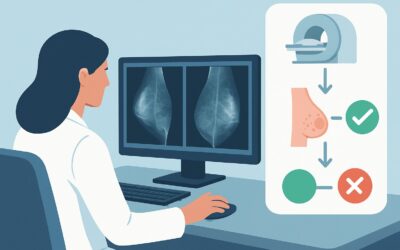
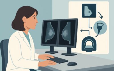
A 28-year-old woman presents to your clinic with a palpable, mobile, rubbery lump she found in her right breast. You appropriately order a targeted breast ultrasound, and the report comes back with a finding classified as probably benign, or BI-RADS 3. The patient is relieved but anxious, asking if a mammogram or MRI is needed to be certain. You are now faced with a common clinical decision point: is more imaging necessary, or is there a different standard of care? This article details the American College of Radiology (ACR) Appropriateness Criteria workflow for this exact scenario, explaining why the next step is typically surveillance, not additional imaging. For a BI-RADS 3 palpable mass in a woman under 30, further studies like a diagnostic digital breast tomosynthesis are considered Usually Not Appropriate.
Who Fits This Clinical Scenario?
This guidance applies to a very specific patient population: an adult female, younger than 30 years of age, who has a palpable breast mass that has been evaluated with ultrasound and categorized as BI-RADS 3 (Probably Benign). A BI-RADS 3 classification implies that the finding has a very high probability (greater than 98%) of being benign, but it is not definitively benign based on imaging features alone. The recommendation is for short-interval follow-up to establish stability.
This workflow is distinct from several similar-sounding but clinically different situations. This guidance does not apply if:
- The patient is 30 years of age or older. Age is a significant risk factor for breast cancer, and the imaging algorithm changes. For women 30-39, diagnostic mammography is often considered alongside ultrasound. For women 40 and older, mammography is the primary initial imaging modality.
- The ultrasound findings are suspicious or malignant (BI-RADS 4 or 5). Any finding with suspicious features warrants immediate consideration for tissue sampling (biopsy), not surveillance.
- The patient has not yet had initial imaging. This article addresses the step after an initial ultrasound. For guidance on the initial imaging workup for a palpable mass in this age group, a different ACR variant applies.
What Diagnoses Are You Working Up in This Scenario?
When an ultrasound of a palpable mass in a woman under 30 yields a BI-RADS 3 result, the differential diagnosis is narrow and heavily weighted toward benign entities. The purpose of the recommended short-term follow-up is to confirm the benign nature by demonstrating stability over time.
Fibroadenoma: This is by far the most common diagnosis for a palpable, solid breast mass in this age group. Fibroadenomas are benign tumors composed of stromal and epithelial tissue. On ultrasound, they typically appear as well-circumscribed, oval, hypoechoic masses that are wider than they are tall, features that often lead to a BI-RADS 3 classification.
Complicated Cyst or Clustered Microcysts: While simple cysts are BI-RADS 2 (Benign), complicated cysts contain internal debris and may not meet all criteria for a simple cyst. They can be palpable and are often classified as BI-RADS 3 to ensure stability, as they can occasionally mimic a solid mass.
Focal Fibrosis or Adenosis: These are benign proliferative changes in the breast tissue that can present as a palpable or imaging-detected mass. They can sometimes have features that overlap with more concerning lesions, warranting a BI-RADS 3 assessment and follow-up.
Malignancy: While breast cancer is rare in women under 30, it is the reason surveillance is necessary. The BI-RADS 3 category exists to detect the small number of malignancies (<2%) that may initially appear benign on imaging. Growth or morphological change on follow-up imaging would be a red flag prompting an upgrade to BI-RADS 4 and a recommendation for biopsy.
Why Is Further Imaging Usually Not Appropriate for a BI-RADS 3 Finding?
In this clinical context, the ACR designates further immediate imaging studies—including diagnostic digital breast tomosynthesis (DBT), mammography, and MRI—as Usually Not Appropriate. The rationale is grounded in balancing the extremely low pre-test probability of malignancy with the risks and limitations of additional tests.
The core principle of managing a BI-RADS 3 lesion is to establish stability over a two-year period via serial ultrasounds. This approach has been shown to be a safe and effective alternative to immediate biopsy for lesions with a very low likelihood of malignancy. Adding other imaging modalities at this stage typically does not add diagnostic value and can introduce new problems.
Here is why alternative studies are not recommended:
- Digital Breast Tomosynthesis (DBT) / Mammography: These studies are rated Usually Not Appropriate. The primary reason is the high breast density typical in women under 30. Dense fibroglandular tissue can obscure underlying masses on mammography, significantly reducing its sensitivity. Furthermore, this involves exposing a young patient to ionizing radiation (Relative Radiation Level ☢☢, 0.1-1 mSv) for a finding that is already well-characterized by ultrasound and has a >98% chance of being benign. The risk of radiation, though small, is not justified by the low potential benefit.
- Breast MRI with and without IV Contrast: This is also rated Usually Not Appropriate. While breast MRI is highly sensitive, its specificity is lower, particularly in premenopausal women where normal hormonal changes can cause benign background enhancement. Ordering an MRI for a BI-RADS 3 finding can lead to the detection of incidental, benign-enhancing foci, resulting in a cascade of unnecessary anxiety, further imaging, and biopsies. It is reserved for high-risk screening or problem-solving for more suspicious (BI-RADS 4 or 5) findings.
The established, evidence-based pathway is to use short-interval follow-up ultrasound as the diagnostic “test of time.”
What’s Next After a BI-RADS 3 Finding? Downstream Workflow
The conclusion that further imaging is “Usually Not Appropriate” does not mean the workup is over. Instead, it shifts from diagnostic imaging to a structured surveillance protocol. Clear communication with the patient about the rationale for this approach is critical for adherence.
The standard downstream workflow for a BI-RADS 3 finding is as follows:
- First Follow-up (6 months): The patient should return in six months for a targeted ultrasound of the known finding. The radiologist will compare the size, shape, and features of the mass to the initial study.
- Subsequent Follow-ups (12 and 24 months): If the finding is stable at the 6-month mark, follow-up ultrasounds are repeated at 12 months and 24 months from the initial exam.
- Interpreting Results:
- If the finding remains stable for two years: After two years of stability (no growth or suspicious change), the lesion is considered definitively benign and is downgraded to BI-RADS 2. No further imaging follow-up for this specific finding is needed.
- If the finding grows or develops suspicious features: If at any follow-up exam the mass has increased in size or developed irregular margins, posterior shadowing, or other concerning features, it is upgraded to BI-RADS 4 (Suspicious). The next step is an image-guided core biopsy.
- If the finding resolves: Occasionally, a finding (like a complicated cyst) may resolve on its own. In this case, no further follow-up is necessary.
This surveillance pathway safely manages these low-risk lesions, minimizing invasive procedures for the vast majority of patients whose masses are benign.
Pitfalls to Avoid (and When to Get Help)
Navigating a BI-RADS 3 recommendation requires careful management and clear communication to avoid common pitfalls.
- Pitfall: Prematurely ordering advanced imaging. Giving in to patient or clinician anxiety by ordering a mammogram or MRI goes against evidence-based guidelines and can lead to a cascade of false positives, unnecessary radiation, and increased healthcare costs.
- Pitfall: Inadequate patient counseling. Failing to explain that “probably benign” means a >98% chance of being benign and that surveillance is the standard of care can lead to poor patient understanding and non-adherence to follow-up.
- Pitfall: Losing the patient to follow-up. The success of this pathway depends on completing the full two-year surveillance. It is essential to have a system in place to track these patients and ensure they return for their scheduled exams.
If a patient’s palpable mass is clearly growing or if follow-up imaging demonstrates suspicious changes, the situation has changed. At that point, you should escalate care by referring the patient for a consultation with a breast surgeon or for an image-guided core needle biopsy.
Related ACR Topics and Tools
This article covers one specific scenario within the broader topic of palpable breast masses. For a comprehensive overview of all clinical variants, including different age groups and imaging findings, please consult our parent guide. For additional decision support, the following GigHz resources are available:
- For breadth across all scenarios in Palpable Breast Masses, see our parent guide: Palpable Breast Masses: ACR Appropriateness Decoded.
- Imaging Appropriateness Selector — for adjacent scenarios
- Imaging Protocol Library — for technique on the recommended study
- Radiation Dose Calculator — for cumulative dose conversations
Frequently Asked Questions
What exactly does a BI-RADS 3 (Probably Benign) assessment mean?
BI-RADS 3 is a category used by radiologists for a finding that is almost certainly benign, but not definitively so. It carries a risk of malignancy of less than 2%. Instead of an immediate biopsy, the standard of care is short-term imaging follow-up (typically with ultrasound at 6, 12, and 24 months) to prove the finding is stable over time.
If there’s any chance it’s cancer, why not just biopsy the mass now?
The decision to biopsy is based on balancing the risk of malignancy against the risks of an invasive procedure (pain, bleeding, infection, scarring). For a BI-RADS 3 finding, the risk of cancer is very low (<2%). Performing biopsies on all of these lesions would result in a large number of unnecessary procedures on benign masses. The surveillance protocol is a well-established, safe, and effective way to monitor these low-risk findings and only proceed to biopsy if there is a change.
Does this guidance change if the patient has a strong family history or a BRCA gene mutation?
Yes, it can. The ACR guidelines discussed here apply to average-risk women. A patient with a known high-risk genetic mutation (like BRCA1 or BRCA2) or other significant risk factors may be managed differently. In such cases, the threshold to proceed with biopsy may be lower, and breast MRI may play a role in screening. These high-risk cases warrant a multidisciplinary discussion and referral to a breast specialist.
How often does a BI-RADS 3 finding turn out to be cancer?
The malignancy rate for BI-RADS 3 lesions is consistently shown in large studies to be less than 2%. This means that for every 100 women with a BI-RADS 3 finding who complete the two-year follow-up protocol, more than 98 will be confirmed to have a benign mass.
What if the patient feels the lump is getting bigger, but the follow-up ultrasound shows it’s stable?
This is a common clinical scenario. A patient’s perception of the size of a lump can be influenced by hormonal changes during the menstrual cycle, which can cause surrounding breast tissue to become more swollen or tender. If high-quality targeted ultrasound performed by an experienced radiologist confirms stability in size and appearance, it is very reassuring. The objective imaging data should be trusted, but it’s important to acknowledge the patient’s concern and explain this discrepancy.
Reviewed by Pouyan Golshani, MD, Interventional Radiologist — June 27, 2026