What Is the Initial Imaging for Pathologic Nipple Discharge in Patients Over 40?
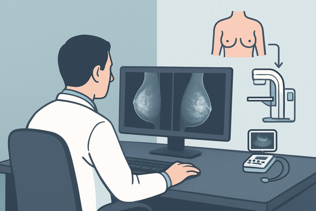
A 52-year-old woman presents to your clinic with a two-month history of spontaneous, bloody discharge from her right nipple. She has no palpable breast mass on examination, and her personal and family histories are unremarkable for breast cancer. You recognize these features as “pathologic” nipple discharge, a presentation that requires a definitive workup to exclude malignancy. The immediate clinical question is which imaging study to order first to evaluate the underlying cause. According to the American College of Radiology (ACR) Appropriateness Criteria, for this specific scenario, both diagnostic mammography (including tomosynthesis) and breast ultrasound are considered Usually Appropriate as the initial imaging steps. This article details the evidence-based workflow for this common and consequential clinical presentation.
Who Fits This Clinical Scenario for Pathologic Nipple Discharge?
This guidance applies to a specific patient population: adult males or females aged 40 years or older presenting with pathologic nipple discharge for initial imaging evaluation.
Inclusion Criteria:
- Age: 40 years or older.
- Presentation: Pathologic nipple discharge. This is defined by discharge that is spontaneous (occurs without manipulation), unilateral (from one breast), and arises from a single duct. The character of the discharge is typically bloody (sanguineous), serosanguinous, clear (serous), or watery.
Exclusion Criteria (These patients follow a different workflow):
- Patients Younger Than 40: The risk of malignancy is lower, and the imaging approach is different. For instance, the workup for a female younger than 30 or a patient aged 30 to 39 with the same symptoms follows a separate ACR variant.
- Physiologic Nipple Discharge: This article does not apply to patients with discharge that is bilateral, involves multiple ducts, is milky (galactorrhea), or only occurs with breast compression or stimulation.
- Concurrent Palpable Mass: If a distinct breast mass is palpable at the time of presentation, the workup is primarily guided by the evaluation of the mass itself, which constitutes a different clinical scenario.
What Diagnoses Are You Working Up in This Scenario?
In a patient over 40 with pathologic nipple discharge, the primary goal of imaging is to identify or exclude an underlying malignancy, though benign causes are more common. The differential diagnosis guides the imaging strategy.
Intraductal Papilloma
This is the most frequent cause of pathologic nipple discharge. Papillomas are benign, wart-like growths within a milk duct. While they are not cancerous, they can sometimes be associated with atypical cells or Ductal Carcinoma In Situ (DCIS), necessitating definitive diagnosis and often surgical excision.
Ductal Carcinoma In Situ (DCIS)
This is a non-invasive form of breast cancer where abnormal cells are confined to the milk ducts. Pathologic nipple discharge can be the sole presenting symptom of DCIS, particularly when it is located in the central ducts behind the nipple. Early detection is critical.
Invasive Ductal Carcinoma (IDC)
While less common to present only with nipple discharge without a palpable mass, an underlying invasive cancer must be considered and ruled out. The cancer can invade from a duct wall and cause bleeding or obstruction, leading to discharge.
Duct Ectasia
This is a benign condition where a milk duct widens and its walls thicken, which can lead to blockage and fluid buildup. The resulting discharge is often thick, sticky, and may be green or black, but it can also be serous or serosanguinous, mimicking a more concerning presentation.
Why Are Diagnostic Mammography and Ultrasound the Recommended Initial Studies?
For a patient aged 40 or older with pathologic nipple discharge, the ACR rates both Diagnostic Mammography (including Digital Breast Tomosynthesis) and US breast as Usually Appropriate. These modalities are used together to provide a comprehensive initial evaluation. The workflow typically begins with the mammogram, followed immediately by a targeted ultrasound.
The rationale for this dual approach is rooted in the complementary strengths of each study. Diagnostic mammography provides a global assessment of the entire breast. It is exceptionally sensitive for detecting suspicious microcalcifications, which are often the only sign of an underlying DCIS. Digital Breast Tomosynthesis (DBT), or 3D mammography, is particularly valuable in this setting, as it reduces the effect of overlapping breast tissue and can better delineate subtle architectural distortions or asymmetries that might indicate a lesion.
Following the mammogram, a targeted breast ultrasound is performed, focusing on the retroareolar region of the affected breast. Ultrasound is superior for visualizing the ducts themselves and identifying small, non-calcified intraductal masses, such as papillomas, that may be invisible on a mammogram. It can precisely characterize ductal dilation, wall thickening, and intraluminal debris or masses. If a suspicious lesion is found, ultrasound provides the ideal modality for real-time guidance of a core needle biopsy.
Why are other studies not recommended for initial workup?
- MRI breast without and with IV contrast: This is rated Usually not appropriate for the initial workup. While highly sensitive, breast MRI has lower specificity and can lead to unnecessary biopsies. Its primary role is as a problem-solving tool if mammography and ultrasound are negative or inconclusive but clinical suspicion remains high, or for preoperative planning if cancer is diagnosed.
- Ductography (Galactography): Also rated Usually not appropriate, this invasive procedure involves cannulating the discharging duct and injecting contrast. It has been largely supplanted by the combination of high-resolution ultrasound and MRI, which provide excellent ductal evaluation without the technical challenges and patient discomfort of ductography.
The recommended combination of mammography (☢☢ 0.1-1mSv) and ultrasound (O 0 mSv) offers a highly effective diagnostic pathway with minimal or no ionizing radiation.
What’s Next After Initial Breast Imaging? Downstream Workflow
The results of the diagnostic mammogram and targeted ultrasound will dictate the subsequent steps in the patient’s management.
- If a suspicious finding is identified: When imaging reveals a suspicious solid mass, a cluster of microcalcifications, or significant architectural distortion (e.g., BI-RADS 4 or 5), the next step is an image-guided biopsy. An ultrasound-guided core needle biopsy is typically performed for solid masses, while a stereotactic-guided biopsy is used for calcifications visible only on mammography. The pathology results from the biopsy will determine the need for surgical consultation and further treatment.
- If imaging is negative (BI-RADS 1 or 2): If both the diagnostic mammogram and targeted retroareolar ultrasound are completely normal, the likelihood of malignancy is low. However, given the “pathologic” nature of the discharge, clinical follow-up is essential. The patient should be counseled on self-monitoring. If the discharge persists, is particularly bothersome, or changes character, a referral to a breast surgeon for consultation is often appropriate. Surgical intervention, such as a terminal duct excision, may be considered for both diagnostic purposes and to resolve symptoms.
- If imaging is indeterminate or shows a likely benign finding (BI-RADS 3): For findings like a solitary dilated duct or a lesion with classic features of a benign papilloma, a short-term imaging follow-up in 6 months may be recommended. Alternatively, depending on the specific finding and clinical concern, a biopsy may be pursued to achieve a definitive diagnosis.
Pitfalls to Avoid (and When to Get Help)
Navigating the workup for pathologic nipple discharge requires careful attention to detail to avoid common missteps.
- Pitfall 1: Ordering a Screening Mammogram. A patient with a clinical symptom like pathologic discharge requires a diagnostic mammogram, not a screening one. This ensures a radiologist is present to review the images in real-time and prescribe additional views or a targeted ultrasound as needed during the same visit.
- Pitfall 2: Stopping with a Negative Mammogram. In this age group, a negative mammogram alone is insufficient to rule out a central ductal lesion. A targeted retroareolar ultrasound is a mandatory component of the initial workup.
- Pitfall 3: Dismissing Persistent Discharge After Negative Imaging. While the risk is low, a small percentage of malignancies can be occult on initial imaging. If a patient’s pathologic discharge continues despite a negative workup, do not ignore it. Escalation to a breast surgeon for consideration of further evaluation, which may include MRI or surgical duct excision, is the appropriate next step.
Related ACR Topics and Tools
For a comprehensive overview of all clinical scenarios related to nipple discharge and for further exploration of imaging guidelines, the following resources are available. For breadth across all scenarios in Evaluation of Nipple Discharge, see our parent guide: Evaluation of Nipple Discharge: ACR Appropriateness Decoded.
- Imaging Appropriateness Selector — for adjacent and alternative clinical scenarios
- Imaging Protocol Library — for detailed technique on recommended studies
- Radiation Dose Calculator — for discussing cumulative radiation exposure with patients
Frequently Asked Questions
Is a breast MRI a better first test for pathologic nipple discharge since it’s more sensitive?
No, for the initial evaluation of pathologic nipple discharge in a patient over 40, breast MRI is rated as ‘Usually not appropriate’ by the ACR. While highly sensitive, it has lower specificity, which can lead to false positives and unnecessary biopsies. The standard, evidence-based approach starts with diagnostic mammography and targeted ultrasound, reserving MRI for problem-solving if the initial workup is negative but clinical suspicion remains high.
What if the patient is a male over 40 with pathologic nipple discharge?
The initial imaging workup is the same for both males and females in this age group. A diagnostic mammogram and a targeted ultrasound are the recommended first steps. While breast cancer is much less common in men, it does occur, and pathologic nipple discharge is a concerning symptom that warrants a full evaluation.
If the mammogram is negative, is an ultrasound still necessary?
Yes, absolutely. A targeted retroareolar ultrasound is a critical component of the workup, even if the mammogram is negative. Small, non-calcified intraductal lesions, such as papillomas or some forms of DCIS, can be occult on mammography but visible on ultrasound. Omitting the ultrasound would be an incomplete evaluation.
Does the color of the nipple discharge change the imaging plan?
For this scenario, the initial imaging plan (diagnostic mammogram and targeted ultrasound) remains the same regardless of whether the pathologic discharge is bloody, serosanguinous, or clear/serous. All of these are considered suspicious and require the same thorough initial workup. A greenish or milky discharge is more suggestive of a benign or physiologic process and may be managed differently.
What is the role of ductography in 2026?
Ductography (or galactography) is now rated ‘Usually not appropriate’ by the ACR for this indication. It is an invasive procedure that has been largely replaced by high-resolution ultrasound and, when needed, breast MRI. These non-invasive modalities provide excellent visualization of the ductal system without the need to cannulate the duct and inject contrast.
Reviewed by Pouyan Golshani, MD, Interventional Radiologist — August 4, 2026