What Imaging Should You Order for a New Axillary Lump During Neoadjuvant Chemo?
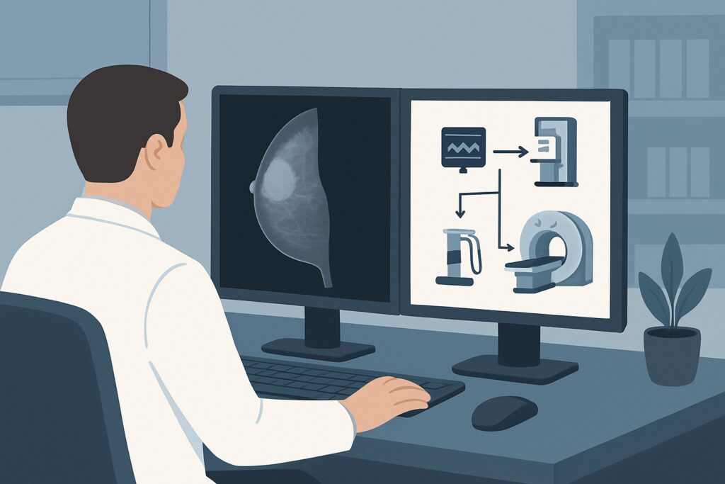
A 48-year-old woman is midway through her neoadjuvant chemotherapy (NAC) regimen for a 3 cm, clinically node-negative invasive ductal carcinoma of the right breast. During a self-exam, she discovers a new, firm, palpable lump in her right axilla. Her oncologist now faces a critical decision: Is this a sign of disease progression, a benign reactive node, or something else? Choosing the right imaging study is essential to answer this question promptly and accurately, as the finding could significantly alter her surgical and therapeutic plan. This article provides a detailed clinical workflow for this specific scenario, explaining why the American College of Radiology (ACR) designates axillary ultrasound as the most appropriate initial imaging test.
Who Fits This Clinical Scenario?
This guidance is specifically for a female patient with a known diagnosis of breast cancer who meets all of the following criteria:
- The primary breast tumor was greater than 2 cm in size at diagnosis.
- She was clinically node-negative (cN0) at the time of her initial staging.
- She is currently undergoing neoadjuvant chemotherapy.
- She presents with a new palpable lump in the ipsilateral axilla that was not present before starting treatment.
It is crucial to distinguish this situation from similar but distinct clinical presentations. This workflow does not apply to:
- Patients at initial diagnosis: The workup for axillary staging before any treatment begins follows a different pathway. This includes scenarios for newly diagnosed breast cancer, whether clinically node-negative or node-positive.
- Patients with a new axillary lump but no history of breast cancer: This presentation falls under the workup for a new palpable, unilateral axillary lump, where the differential diagnosis is broader.
- Patients with known positive nodes being monitored for treatment response: Evaluating the response of a biopsy-proven metastatic node to chemotherapy is a different clinical question than investigating a newly appeared node.
What Diagnoses Are You Working Up in This Scenario?
The appearance of a new axillary mass during neoadjuvant chemotherapy raises several possibilities, ranging from the most concerning to the benign. The primary goal of imaging is to differentiate between these potential causes.
Metastatic Lymph Node: This is the most significant concern. The development of a new metastatic node suggests that the cancer is not responding adequately to the current chemotherapy regimen or that micrometastatic disease, invisible at initial staging, has now grown to a palpable size. Confirming this would change the patient’s nodal status from cN0 to ycN+ (post-treatment clinical node positive), directly impacting prognosis and dictating a more aggressive surgical approach, such as an axillary lymph node dissection rather than a sentinel node biopsy.
Reactive Lymphadenopathy: A more common and benign cause. Systemic chemotherapy can induce an inflammatory response, causing normal lymph nodes to enlarge and become palpable. These reactive nodes are typically a sign of the body’s immune response and do not represent cancer progression. Imaging helps characterize the node’s morphology to distinguish it from a malignant one.
Treatment-Related Inflammatory Changes: Less frequently, other non-nodal changes can occur in the axilla. These can include fat necrosis, especially if a prior axillary procedure was performed, or a seroma. While less likely to present as a firm, distinct lump, they remain on the differential.
Infection or Abscess: In a patient who may be neutropenic from chemotherapy, an infection is a possibility, though it is usually accompanied by other signs like erythema, warmth, or systemic fever. An abscess could present as a tender, palpable mass.
Why Is Axillary Ultrasound the Recommended Study for This Presentation?
For a patient presenting with a new palpable axillary lump mid-chemotherapy, the ACR Appropriateness Criteria rate US axilla as Usually appropriate. It is the definitive first-line imaging study for this specific clinical question.
The primary strength of ultrasound is its high spatial resolution, which allows for detailed morphologic assessment of lymph nodes. Radiologists can evaluate key features that help distinguish benign from malignant nodes. Suspicious features include a rounded shape (as opposed to the normal oval or reniform shape), cortical thickening (often asymmetric or focal), and the partial or complete effacement of the normal fatty hilum. Color Doppler imaging can further assess for abnormal vascular patterns. Critically, ultrasound uses no ionizing radiation (0 mSv) and does not require intravenous contrast, making it a safe and readily accessible tool.
Perhaps most importantly, ultrasound provides real-time guidance for percutaneous biopsy. If a suspicious node is identified, a US-guided fine-needle aspiration (FNA) or core needle biopsy can be performed in the same session or scheduled promptly. This ability to move directly from diagnosis to tissue sampling is a major advantage, providing the cytological or histological proof needed to alter management.
Other imaging modalities are considered Usually not appropriate for this focused clinical question:
- MRI breast without and with IV contrast: While breast MRI is the primary tool for assessing the response of the main breast tumor to NAC, it is not the ideal first step for evaluating a new, focal axillary lump. It is more resource-intensive, and its ability to characterize nodal morphology is not superior to a dedicated, high-resolution ultrasound.
- FDG-PET/CT skull base to mid-thigh: This is a powerful tool for systemic staging but is considered overkill for this specific problem. It involves a significant radiation dose (☢☢☢☢ 10-30 mSv) and is not necessary for the initial characterization of a single palpable finding. The goal here is targeted evaluation and tissue sampling, not a full-body restaging, unless there are other signs of widespread disease progression.
What’s Next After Axillary US? Downstream Workflow
The results of the axillary ultrasound will guide the subsequent steps in the patient’s care. The workflow branches based on the imaging findings.
If the US reveals a suspicious-appearing lymph node (e.g., BI-RADS 4 or 5): The immediate next step is a US-guided biopsy (FNA or core needle biopsy) to obtain a tissue diagnosis. If the biopsy confirms metastatic carcinoma, the patient’s nodal status is upstaged. This information is critical for the multidisciplinary tumor board to discuss potential changes to the systemic therapy and to plan for definitive axillary surgery (typically an axillary lymph node dissection) after completion of NAC.
If the US shows only benign-appearing or reactive lymph nodes (e.g., BI-RADS 2): This finding is highly reassuring. It suggests the palpable lump is due to a benign process. The patient can typically continue with her planned neoadjuvant chemotherapy regimen. Close clinical follow-up of the axilla is warranted to ensure the lump resolves or remains stable.
If the US is negative or indeterminate, but a definite lump remains palpable: This situation represents a clinical-radiologic discordance and requires careful consideration. A direct conversation between the ordering clinician and the radiologist is essential. The radiologist may be able to perform a US-guided biopsy of the palpable abnormality even if a discrete, suspicious node is not clearly visible. If suspicion remains high despite a negative targeted US, escalation to a surgical consultation for consideration of an excisional biopsy may be necessary.
Pitfalls to Avoid (and When to Get Help)
Navigating this scenario requires vigilance to avoid common missteps. Here are key pitfalls to watch for:
- Dismissing the finding: Never assume a new palpable lump during NAC is benign or “just reactive” without an objective imaging evaluation. It must be worked up.
- Stopping at imaging: A suspicious ultrasound is an indication for biopsy, not a final diagnosis. Management changes should be based on pathology, not just imaging appearance.
- Incomplete examination: Ensure the ultrasound report documents a thorough evaluation of all three axillary levels and explicitly correlates the imaging findings with the site of the palpable lump.
- Ignoring clinical-radiologic discordance: If you can feel a distinct, hard mass but the ultrasound is reported as normal, do not stop. This is a red flag that requires escalation. If a confident diagnosis cannot be made with imaging and percutaneous biopsy, a multidisciplinary discussion or surgical consultation is the appropriate next step.
Related ACR Topics and Tools
This article focuses on a single, specific clinical scenario. For a comprehensive overview of all variants and recommendations for axillary imaging, or to explore the tools used to develop these guidelines, please refer to the following resources.
- For breadth across all scenarios in Imaging of the Axilla, see our parent guide: Imaging of the Axilla: ACR Appropriateness Decoded.
- To explore other clinical situations, consult the ACR Appropriateness Criteria Lookup tool.
- For details on imaging techniques, see the Imaging Protocol Library.
- To discuss radiation exposure with patients, use the Radiation Dose Calculator.
Frequently Asked Questions
Why not just go straight to a PET/CT to restage the patient completely?
A PET/CT is a systemic staging tool and is considered ‘Usually not appropriate’ by the ACR for this specific, focal question. The immediate goal is to diagnose the palpable lump with minimal radiation and cost. An axillary ultrasound is more sensitive for nodal morphology and directly enables biopsy. A full restaging with PET/CT would only be considered if there were other signs of systemic disease progression.
If the ultrasound is suspicious, is a fine-needle aspiration (FNA) or a core needle biopsy better?
Both are valid options, and the choice often depends on institutional preference and pathologist expertise. A core needle biopsy provides tissue architecture and may allow for receptor testing if needed, which can be advantageous. An FNA provides cytology and is often faster and less invasive. The key is to obtain a definitive pathologic diagnosis.
What if the patient had a suspicious node at diagnosis that was biopsied and confirmed positive (cN1)?
That is a different clinical scenario. In that case, the goal of mid-treatment imaging would be to assess treatment response in a known positive node, not to diagnose a new finding. This might involve placing a clip in the positive node before starting chemotherapy to ensure it can be identified for surgical removal later.
Does the type of breast cancer (e.g., triple-negative vs. ER-positive) change this recommendation?
No, the initial imaging recommendation for a new palpable axillary lump remains the same regardless of the breast cancer subtype. The clinical urgency may be higher with more aggressive subtypes like triple-negative or HER2-positive breast cancer, but the diagnostic algorithm starting with axillary ultrasound is unchanged.
Can I order a diagnostic mammogram or tomosynthesis of the axilla instead of an ultrasound?
No, both diagnostic mammography and digital breast tomosynthesis are rated as ‘Usually not appropriate’ for this indication. While they can visualize the axilla, their ability to characterize lymph node morphology is significantly inferior to ultrasound. Ultrasound is the superior modality for this specific clinical task.
Reviewed by Pouyan Golshani, MD, Interventional Radiologist — May 29, 2026