Which Supplemental Screening Is Best for Average-Risk Women with Extremely Dense Breasts?
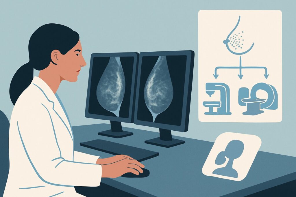
A 48-year-old woman with no significant family history of breast cancer comes to your office to review her annual screening mammogram report. The findings are negative for malignancy (BI-RADS 1), but the report notes BI-RADS density category D, or “extremely dense breasts.” The radiologist’s comment suggests that this density may obscure small cancers and recommends discussing supplemental screening. You now face the clinical question: What is the most appropriate imaging study to order next to improve cancer detection for this average-risk patient? According to the American College of Radiology (ACR) Appropriateness Criteria, multiple modalities are rated as Usually Appropriate, including Digital Breast Tomosynthesis (DBT) as the foundational screening tool, with Breast MRI being a key consideration for additional imaging.
Who Fits This Clinical Scenario for Supplemental Screening?
This guidance applies specifically to adult women who meet a precise set of criteria. The patient must be at average risk for breast cancer. This generally means she has no personal history of breast cancer, no known high-risk genetic mutations (e.g., BRCA1, BRCA2), no strong family history suggesting a hereditary syndrome, and no history of high-dose radiation to the chest before age 30.
The central feature of this scenario is the mammographic finding of extremely dense breasts, corresponding to Breast Imaging Reporting and Data System (BI-RADS) density category D. This indicates that the breast is composed of almost all fibroglandular tissue, which can significantly lower the sensitivity of mammography by masking underlying lesions.
It is critical to distinguish this patient from those in similar but distinct clinical situations:
- Women with heterogeneously dense breasts (BI-RADS C): While still considered “dense,” the degree of masking is less severe, and the risk-benefit calculation for supplemental screening may differ.
- Women at intermediate or high risk: Patients with a personal history of breast cancer, a lifetime risk of 20% or greater, or known genetic mutations fall into a different risk category. For these women, supplemental screening with MRI is often standard practice, regardless of breast density.
- Symptomatic patients: A woman presenting with a palpable lump, nipple discharge, or focal pain is not undergoing screening. She requires a diagnostic workup, which follows a separate clinical pathway, typically starting with diagnostic mammography and targeted ultrasound.
What Diagnoses Are You Working Up in This Scenario?
Supplemental screening is not prompted by a specific symptom but by the known limitations of mammography in a particular patient population. The goal is to detect occult, or hidden, malignancies that are not visible on a standard 2D or even 3D mammogram due to the masking effect of dense tissue.
The primary target is occult invasive ductal carcinoma (IDC). These are the most common type of invasive breast cancers. In dense breasts, a small, developing IDC may not form a distinct spiculated mass or may be indistinguishable from the surrounding glandular tissue on a mammogram, delaying diagnosis until it is larger and potentially at a more advanced stage.
Another key consideration is occult invasive lobular carcinoma (ILC). This subtype is notoriously difficult to detect mammographically, even in non-dense breasts, because it often grows in a diffuse, single-file pattern without forming a discrete lump. The architectural distortion it creates can be exceptionally subtle and easily masked by dense tissue, making supplemental imaging particularly valuable for its detection.
Supplemental screening also aims to find high-grade ductal carcinoma in situ (DCIS). While many cases of DCIS are identified by suspicious microcalcifications on mammography, some present as non-calcified masses or subtle distortions. These non-calcified forms of DCIS are more likely to be obscured in women with extremely dense breasts.
Finally, it is important to acknowledge that these advanced screening methods will inevitably identify a range of benign findings, such as cysts, fibroadenomas, and intramammary lymph nodes. A key part of the clinical workflow is managing these findings appropriately to confirm their benign nature without subjecting the patient to unnecessary anxiety or invasive procedures.
Why Is Digital Breast Tomosynthesis the Foundational Study, with MRI as a Key Supplement?
For an average-risk woman with extremely dense breasts, the American College of Radiology (ACR) rates several imaging modalities as Usually Appropriate. The clinical workflow involves establishing Digital Breast Tomosynthesis (DBT) as the primary screening method and then considering additional modalities like Magnetic Resonance Imaging (MRI) for further sensitivity.
Digital Breast Tomosynthesis (DBT), or 3D mammography, is now the standard of care for all screening, especially in women with dense breasts. Unlike 2D mammography, which creates a single flat image, DBT acquires multiple low-dose images from different angles. A computer reconstructs these into thin slices, allowing the radiologist to scroll through the breast tissue layer by layer. This process significantly reduces the problem of overlapping tissue, which is the primary cause of masking. DBT has been shown to increase cancer detection rates while simultaneously decreasing the recall rate for false positives. For this reason, it should be considered the baseline screening test for this patient population.
For true supplemental screening—that is, adding another test to enhance detection beyond DBT—the ACR rates MRI breast without and with IV contrast as Usually Appropriate. Breast MRI is the most sensitive imaging modality for detecting invasive breast cancer, with a sensitivity that is not limited by breast density. It is particularly effective at identifying small, otherwise occult invasive ductal and lobular cancers. The ACR also rates MRI breast without and with IV contrast abbreviated as Usually Appropriate. This faster protocol can improve accessibility and reduce costs while retaining high diagnostic accuracy for cancer detection.
Ultrasound (US) breast is rated as May be appropriate. While handheld or automated whole-breast ultrasound can detect additional cancers not seen on mammography, it has a significantly higher rate of false positives compared to MRI, leading to more benign biopsies. It is also highly operator-dependent. For these reasons, while it can be considered, MRI is often preferred if available.
Modalities like Mammography with IV contrast and MRI breast without IV contrast are rated Usually not appropriate. Contrast-enhanced mammography is an emerging technology but is not yet established for screening. Non-contrast MRI lacks the functional data from gadolinium enhancement that is crucial for characterizing and detecting suspicious lesions.
For detailed technical specifications on performing a screening mammogram, see our guide on the Screening Mammography (with DBT) protocol.
What’s Next After Supplemental Screening? Downstream Workflow
The results of supplemental screening will guide the subsequent steps in the patient’s care. The workflow branches based on the findings reported using the BI-RADS classification system.
- Negative or Benign Result (BI-RADS 1 or 2): If the supplemental study (e.g., breast MRI) is negative or shows only clearly benign findings, the patient can return to her routine annual screening schedule, which should include DBT. No further immediate action is required.
- Probably Benign Result (BI-RADS 3): If the study identifies a finding that is almost certainly benign (e.g., a classic fibroadenoma on MRI), the standard recommendation is a short-interval follow-up imaging study, typically in six months, to ensure stability. This avoids unnecessary biopsy while maintaining a high level of safety.
- Suspicious Result (BI-RADS 4 or 5): If the supplemental screening reveals a suspicious lesion, the next step is an image-guided biopsy to obtain a tissue diagnosis. The modality used for the biopsy (ultrasound, stereotactic, or MRI) will depend on which imaging test best visualizes the lesion. A positive biopsy for malignancy will trigger a referral to a breast surgeon and oncologist for treatment planning.
- Incomplete Result (BI-RADS 0): Occasionally, a finding may be indeterminate, requiring further imaging for characterization. For example, a finding on MRI may prompt a targeted second-look ultrasound to see if a sonographic correlate can be identified for an easier biopsy.
This structured approach ensures that positive findings are acted upon promptly while minimizing intervention for benign or probably benign results.
Pitfalls to Avoid (and When to Get Help)
Navigating supplemental screening requires careful consideration to avoid common pitfalls.
First, do not order supplemental screening for a patient who is not at least average risk. The potential harms of false positives and unnecessary biopsies are more likely to outweigh the benefits in very low-risk individuals. Second, avoid ordering breast MRI without intravenous contrast for cancer screening. The non-contrast portion of the exam is insufficient for detecting or characterizing suspicious enhancing lesions. Third, be mindful of the higher false-positive rate of whole-breast ultrasound. While it is an option, it is crucial to counsel the patient beforehand about the increased likelihood of being called back for benign findings. Finally, ensure the patient has no contraindications to the chosen study, such as renal insufficiency for gadolinium-based contrast agents or non-compatible metallic implants for MRI.
If a screening study returns a complex or equivocal finding (BI-RADS 0), or if the biopsy results are discordant with the imaging findings, it is appropriate to escalate care by recommending consultation at a multidisciplinary breast center.
Related ACR Topics and Tools
For a comprehensive overview of all clinical variants and imaging modalities related to this topic, please refer to our parent guide. Additional GigHz resources can help you navigate adjacent clinical questions and technical specifications.
- For breadth across all scenarios in Supplemental Breast Cancer Screening Based on Breast Density, see our parent guide: Supplemental Breast Cancer Screening Based on Breast Density: ACR Appropriateness Decoded.
- To explore other clinical scenarios and their ACR-recommended workups, use our ACR Appropriateness Criteria Lookup.
- For detailed procedural techniques, consult the Imaging Protocol Library.
- To discuss cumulative radiation exposure with your patients, our Radiation Dose Calculator can be a useful tool.
Frequently Asked Questions
Does every woman with extremely dense breasts need supplemental screening?
Not necessarily. For average-risk women, it is a shared decision-making process. While supplemental screening with modalities like MRI can increase cancer detection, it also increases the chance of false positives, benign biopsies, and patient anxiety. The discussion should weigh the potential benefit of detecting an early cancer against these potential harms.
Why is breast MRI considered ‘Usually Appropriate’ if Digital Breast Tomosynthesis (DBT) is also recommended?
DBT is the foundational screening test that should replace 2D mammography for all women, especially those with dense breasts. Breast MRI is considered a supplemental tool to be used *in addition* to mammography for those who desire the highest sensitivity for cancer detection. MRI is not limited by breast density and detects more cancers than mammography and ultrasound combined, but it is more costly and has a higher false-positive rate.
If my patient is average risk with extremely dense breasts, should I order an MRI every year?
The optimal frequency of supplemental screening with MRI for average-risk women is still an area of active research. Some guidelines suggest alternating annual mammography with annual MRI, while others may recommend both in the same year or MRI every 2-3 years. The decision should be individualized based on patient preference, risk tolerance, and insurance coverage.
What is the difference between standard breast MRI and abbreviated breast MRI?
Abbreviated breast MRI is a significantly shorter version of the full protocol, typically taking less than 10 minutes. It acquires fewer image sequences but includes the most critical pre- and post-contrast images needed to detect suspicious enhancement. Studies have shown it maintains a cancer detection rate comparable to the full protocol, making it a more accessible and cost-effective option for screening.
Is insurance coverage a barrier for supplemental screening in this scenario?
Coverage can be a significant issue. While many states have passed legislation requiring insurers to cover supplemental screening for women with dense breasts, the specifics vary widely. Some policies may cover ultrasound but not MRI, or they may require high deductibles. It is essential to consider the patient’s financial responsibility and verify insurance coverage before ordering the test.
Reviewed by Pouyan Golshani, MD, Interventional Radiologist — May 30, 2026