What Imaging Is Best for Axillary Adenopathy with Silicone Implants?
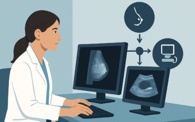
A 35-year-old patient with a history of silicone breast augmentation presents to your clinic with a new, palpable, non-tender lump in her left axilla. On examination, you confirm a firm, mobile lymph node. The breast examination is unremarkable, with no palpable masses or skin changes. You need to determine the most appropriate initial imaging study to evaluate this unexplained axillary adenopathy, considering her implant history. This article details the clinical workflow for this specific scenario, guiding you through the differential diagnosis and the rationale for the recommended imaging. According to the American College of Radiology (ACR) Appropriateness Criteria, for this presentation, a breast ultrasound is rated Usually appropriate.
Who Fits This Clinical Scenario for Axillary Adenopathy?
This workflow is specifically for patients matching a precise set of criteria. Applying this guidance to a different clinical presentation may lead to a suboptimal diagnostic pathway.
Inclusion Criteria:
- Age: 30 to 39 years of age.
- Patient Identity: Female or transfeminine.
- Presentation: Unexplained axillary adenopathy (a palpable or incidentally discovered enlarged lymph node in the armpit) without a known breast mass.
- Implant History: Current or prior history of silicone breast implants.
- Imaging Stage: This is the initial, first-line imaging workup.
Exclusion Criteria (These Route to Different Workflows):
- Patients with Saline Implants: The differential diagnosis and imaging priorities differ, particularly regarding silicone-specific complications.
- Patients with a Palpable Breast Mass: If a breast mass is present in addition to adenopathy, the workup prioritizes characterizing the breast lesion, which follows a different diagnostic algorithm.
- Asymptomatic Patients: This guidance does not apply to routine, asymptomatic implant screening or surveillance.
- Suspected Implant Rupture as the Primary Symptom: Patients presenting with breast pain, contour changes, or other signs of rupture are evaluated under a different ACR scenario.
What Diagnoses Are You Working Up in This Scenario?
Unexplained axillary adenopathy in a patient with silicone implants requires consideration of a specific differential diagnosis that bridges benign, implant-related, and malignant causes. The initial imaging choice is designed to efficiently differentiate among these possibilities.
Silicone-Induced Adenopathy (Silicone Granuloma)
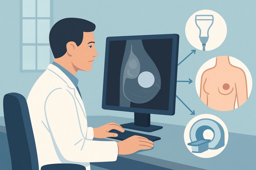
This is a key consideration unique to this patient population. Even intact silicone implants can have microscopic “gel bleed,” and frank rupture can release free silicone. This silicone can migrate to regional lymph nodes, provoking a foreign body giant cell reaction. The result is enlarged, firm lymph nodes that are clinically indistinguishable from malignancy. Imaging is crucial for identifying signs of silicone within the node itself.
Reactive or Inflammatory Adenopathy
The most common cause of enlarged lymph nodes in the general population, this remains a frequent cause in patients with implants. It can result from local skin infections, dermatologic conditions, or systemic viral or bacterial illnesses. These nodes typically have a benign appearance on imaging, with a preserved fatty hilum.
Occult Breast Carcinoma
Axillary lymph node metastasis can be the first and only sign of an underlying breast cancer that is too small or diffuse to be detected on clinical examination. This is a critical diagnosis to exclude, as the presence of nodal metastasis immediately upstages the cancer and dictates treatment.
Breast Implant-Associated Anaplastic Large Cell Lymphoma (BIA-ALCL)
A rare T-cell lymphoma that can develop in the scar tissue capsule and fluid surrounding breast implants (particularly textured-surface implants). While it most commonly presents as a delayed seroma (fluid collection), it can also manifest with capsular masses or lymph node involvement. It must be considered in the differential for any new axillary finding in a patient with implants.
Systemic Malignancy
Less commonly, the axillary adenopathy may represent a systemic process like non-Hodgkin lymphoma, leukemia, or metastasis from a non-breast primary cancer (e.g., melanoma, lung cancer). Imaging features may suggest a systemic cause, but biopsy is often required for definitive diagnosis.
Why Is Breast Ultrasound the Recommended First Step for This Presentation?
The ACR designates breast ultrasound as Usually appropriate for this scenario because it directly and effectively addresses the primary clinical question: characterizing the abnormal axillary lymph node. It provides high-resolution detail of nodal architecture without exposing the patient to ionizing radiation.
Ultrasound excels at evaluating key nodal features. A radiologist can assess the size of the node, the thickness of its cortex, and the presence or absence of a normal fatty hilum. A thickened cortex or an effaced, absent hilum are suspicious features for malignancy. Conversely, a prominent but preserved hilum suggests a benign reactive process. Most importantly for this scenario, ultrasound can often directly visualize free silicone within the lymph node. This appears as multiple, intensely echogenic (bright) spots without posterior acoustic shadowing, a pattern often described as a “snowstorm” appearance. Identifying this is highly specific for silicone-induced adenopathy and can often obviate the need for more invasive workups.
The ACR also rates diagnostic mammography and digital breast tomosynthesis (DBT) as Usually appropriate. These are often performed in conjunction with the targeted axillary ultrasound. While ultrasound is superior for evaluating the node itself, mammography/DBT is essential for examining the ipsilateral breast tissue to search for an occult primary cancer that may be the source of a metastatic node. However, in patients under 40, dense breast tissue can limit the sensitivity of mammography, making ultrasound a critical first step.
In contrast, breast MRI (with or without contrast) is rated Usually not appropriate as the initial imaging test. While highly sensitive, MRI is less specific, more costly, and less accessible than ultrasound. It is not the right tool for the initial characterization of a lymph node. Its role is reserved for problem-solving, such as when initial imaging is indeterminate or for preoperative staging if cancer is diagnosed.
The recommended initial study, ultrasound, involves no ionizing radiation (0 mSv), a key advantage in this younger patient population who may require future imaging.
What’s Next After Ultrasound? Downstream Workflow
The results of the initial ultrasound (and concurrent diagnostic mammogram) will guide the subsequent steps in the patient’s management. The workflow branches based on whether the findings are benign, suspicious, or specific for an implant-related complication.
If the Findings Suggest Silicone Adenopathy:
If ultrasound demonstrates the classic “snowstorm” appearance of silicone within an otherwise morphologically normal-appearing lymph node, and the mammogram is negative, the likely diagnosis is silicone-induced adenopathy. The next step is often clinical correlation and short-term imaging follow-up (e.g., a 6-month follow-up ultrasound) to ensure stability. Biopsy may be considered if there are any atypical features but can often be avoided.
If the Findings Are Suspicious for Malignancy:
If a lymph node shows suspicious features—such as a thickened cortex, effaced hilum, or rounded shape—the next step is an image-guided biopsy. Ultrasound-guided core needle biopsy is the standard of care. This allows for histopathologic diagnosis to differentiate between breast cancer metastasis, lymphoma (including BIA-ALCL), or another malignancy. The results of this biopsy will determine the need for further systemic staging and treatment planning.
If the Findings Are Benign/Reactive:
If the lymph nodes are mildly enlarged but maintain a normal, benign appearance (preserved fatty hilum) and the mammogram is negative, the likely diagnosis is reactive adenopathy. The next step is typically clinical management. This involves a careful history and physical exam to look for a potential inflammatory cause (e.g., skin infection, recent vaccination) and a short-term clinical or imaging follow-up to ensure resolution or stability.
If the Findings Are Indeterminate:
If the initial imaging is inconclusive, further evaluation may be warranted. This could involve a biopsy of the indeterminate node or, in select cases, a problem-solving breast MRI to better evaluate the breast parenchyma for an occult primary and assess the extent of adenopathy.
Pitfalls to Avoid (and When to Get Help)
Navigating this clinical scenario requires careful attention to the patient’s specific history and imaging findings. Here are several common pitfalls to avoid:
- Dismissing Adenopathy as “Just a Reactive Node”: In any patient, but especially one with breast implants, new axillary adenopathy must be fully worked up to exclude malignancy.
- Attributing All Adenopathy to Implants: While silicone-induced adenopathy is a key consideration, it is a diagnosis of exclusion after malignancy has been ruled out. Do not assume a node is benign without appropriate imaging characterization.
- Forgetting to Order a Diagnostic Mammogram: While ultrasound is the primary tool for the node, a concurrent diagnostic mammogram/tomosynthesis is crucial for evaluating the breast as a potential source of metastasis.
- Performing an Excisional Biopsy First: An image-guided core needle biopsy is the preferred initial diagnostic step for a suspicious node. A premature surgical excision can disrupt lymphatic drainage and complicate subsequent surgical planning if malignancy is found.
If imaging reveals a highly suspicious node or if a biopsy confirms malignancy, immediate escalation to a multidisciplinary breast care team, including a breast surgeon and oncologist, is essential.
Related ACR Topics and Tools
The ACR Appropriateness Criteria are extensive, covering thousands of clinical variants. For a comprehensive overview of imaging for all implant-related concerns, from rupture to asymptomatic screening, please see our parent guide. For tools to help you apply these criteria in your practice, see the resources below.
- For breadth across all scenarios in Breast Implant Evaluation, see our parent guide: Breast Implant Evaluation: ACR Appropriateness Decoded.
- ACR Appropriateness Criteria Lookup — for adjacent scenarios
- Imaging Protocol Library — for technique on the recommended study
- Radiation Dose Calculator — for cumulative dose conversations
Frequently Asked Questions
Why is breast MRI not recommended as the first imaging test for this scenario?
Breast MRI is rated ‘Usually not appropriate’ as the initial test because it is a high-sensitivity but lower-specificity tool that is more expensive and less accessible than ultrasound. Ultrasound is superior for the primary task of characterizing the lymph node’s internal architecture and detecting signs of silicone. MRI is reserved for problem-solving if initial imaging is inconclusive or for staging after a cancer diagnosis.
If the ultrasound shows silicone in the lymph node, is a biopsy always unnecessary?
Not always. While the presence of silicone is strong evidence for silicone-induced adenopathy, a biopsy may still be necessary if the lymph node also has morphologically suspicious features (e.g., a focal, markedly thickened cortex). Malignancy can coexist with silicone deposits. The decision is based on the overall imaging appearance of the node, not just the presence of silicone.
Does this guidance change if the patient had textured implants versus smooth implants?
The initial imaging workup (ultrasound and mammogram) remains the same regardless of implant surface type. However, the index of suspicion for Breast Implant-Associated Anaplastic Large Cell Lymphoma (BIA-ALCL) is higher in patients with a history of textured implants. This clinical context is important for the radiologist and pathologist to know, especially if a biopsy is performed.
What if the patient is 42 years old instead of 38?
The ACR criteria are often grouped by age brackets. While the fundamental workup for unexplained axillary adenopathy is similar, patients aged 40 and older are in a different screening mammography cohort. The emphasis on performing a diagnostic mammogram/tomosynthesis in conjunction with the ultrasound is even greater due to the increased age-related risk of breast cancer.
Can ultrasound reliably distinguish between silicone adenopathy and metastatic cancer?
In many cases, yes. The ‘snowstorm’ appearance of free silicone is very specific and distinct from the typical appearance of a metastatic lymph node, which usually shows cortical thickening and loss of the fatty hilum. However, in cases where both features might be present or the findings are atypical, an ultrasound-guided biopsy is the definitive next step to establish a diagnosis.
Reviewed by Pouyan Golshani, MD, Interventional Radiologist — May 29, 2026