Should You Order Brain Imaging for Asymptomatic Stage I Breast Cancer?

A 58-year-old woman sits in your clinic, newly diagnosed with a 1.2 cm, ER-positive, HER2-negative, node-negative breast cancer. She is otherwise healthy and has no neurologic complaints, but she read online that breast cancer can spread to the brain and is anxious for a “full body scan” to be sure she is clear. You are now faced with the decision of whether to order imaging to rule out brain metastases as part of her initial staging. This article details the clinical workflow for this specific scenario, explaining why routine brain imaging is not indicated.
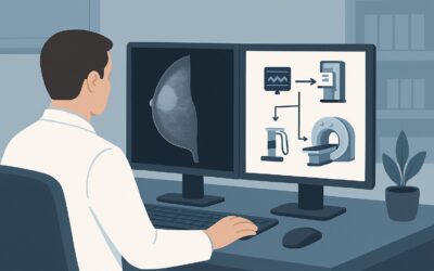
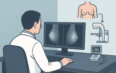
For the initial staging of an asymptomatic patient with newly diagnosed Stage I breast cancer, the American College of Radiology (ACR) Appropriateness Criteria rate MRI head without and with IV contrast as Usually not appropriate.
Who Fits This Clinical Scenario for Brain Metastasis Screening?
This guidance applies to a very specific patient population: those with newly diagnosed, biopsy-proven Stage I breast cancer who are completely asymptomatic from a neurologic standpoint. The key inclusion criteria are:
- Newly Diagnosed: The imaging is being considered for initial staging, not for surveillance after treatment has been completed.
- Stage I Disease: The tumor is small (≤2 cm) and there is no evidence of spread to the axillary lymph nodes (N0) or distant sites (M0) based on initial clinical and pathologic evaluation.
- Asymptomatic: The patient has no new, persistent, or progressive neurologic signs or symptoms. This includes the absence of headaches, seizures, focal weakness or numbness, changes in vision or speech, or new-onset balance problems.
It is crucial to distinguish this scenario from others that may seem similar. This workflow does not apply if:
- The patient has neurologic symptoms: Any new, unexplained neurologic deficit warrants a dedicated workup, which would typically involve brain imaging. That patient is no longer asymptomatic.
- The patient has higher-stage disease: Patients with Stage II or III breast cancer have a higher risk of distant metastases, and the recommendations for systemic staging are different.
- The workup is for other sites: This guidance is specific to screening for brain metastases. The decision to image for bone, thoracic, or abdominal metastases follows different logic and criteria.
What Diagnoses Are You Working Up in This Scenario?
In this clinical context, the primary and often sole concern driving the consideration for imaging is the presence of occult brain metastases. However, the decision to order a test must be weighed against the pre-test probability of the disease and the potential for incidental findings.
Brain Metastasis: This is the principal diagnosis to be excluded. For patients with Stage I breast cancer, however, the incidence of brain metastases at initial diagnosis is exceedingly low. Major studies and society guidelines consistently find the risk to be less than 1%. Ordering a high-sensitivity test in a low-prevalence population dramatically increases the likelihood that a positive finding is a false positive.
Benign Incidentaloma: A far more likely outcome of screening an asymptomatic patient is the discovery of an incidental, benign finding. These can include small meningiomas, developmental venous anomalies, arachnoid cysts, or other non-neoplastic lesions. While clinically insignificant, their discovery often triggers a cascade of follow-up imaging, specialist consultations (e.g., neurosurgery), and significant patient anxiety, without providing any clinical benefit.
Primary Central Nervous System (CNS) Neoplasm: While theoretically possible, discovering an unrelated primary brain tumor (like a glioma or meningioma) in an asymptomatic patient during a breast cancer workup is a rare event. It is not a primary target of this screening consideration.
Why Is Routine Brain Imaging ‘Usually Not Appropriate’ for Asymptomatic Stage I Breast Cancer?
The ACR panel’s designation of all brain imaging modalities as “Usually not appropriate” for this scenario is rooted in a clear risk-benefit analysis: the potential harms of screening far outweigh the potential benefits in this low-risk population. The rationale is built on the very low prevalence of disease and the high potential for iatrogenic harm from over-investigation.
No imaging study is recommended. The core principle is that screening is not indicated. Let’s review the specific modalities and why they are rated poorly for this task:
- MRI head without and with IV contrast (0 mSv): While MRI is the most sensitive modality for detecting brain metastases, its use for screening in this asymptomatic, early-stage population is rated Usually not appropriate. The extremely low pre-test probability means the positive predictive value of the test is poor. The risk of uncovering incidental, non-pathologic findings that lead to further workup and anxiety is high.
- CT head with or without IV contrast (☢☢☢ 1-10 mSv): CT of the head is also rated Usually not appropriate. It is less sensitive than MRI for detecting small metastases, particularly in the posterior fossa. Furthermore, it exposes the patient to ionizing radiation without a clear clinical benefit. Given the low likelihood of finding true metastatic disease, the added radiation exposure is not justified.
- FDG-PET/CT whole body (☢☢☢☢ 10-30 mSv): This modality is also rated Usually not appropriate for screening for brain metastases. While PET/CT is used for systemic staging in more advanced disease, it has poor sensitivity for small brain lesions. Standard PET/CT protocols often show high background glucose uptake in the brain’s gray matter, which can obscure small metastatic deposits. It also carries a significant radiation dose.
In summary, the consensus from the ACR and other major oncology guidelines (like NCCN and ASCO) is that for asymptomatic patients with Stage I breast cancer, the risk of brain metastases is too low to justify routine screening with any imaging modality. The proper course is clinical vigilance.
What’s Next? The Correct Downstream Workflow Instead of Imaging
Since imaging is not recommended at initial staging, the appropriate downstream workflow is based on clinical monitoring and proceeding with standard-of-care treatment for the primary cancer. The decision tree is straightforward.
If the patient remains asymptomatic:
Proceed with the planned local and systemic therapy for the Stage I breast cancer. This typically involves surgery (lumpectomy or mastectomy), possibly radiation, and endocrine therapy or chemotherapy based on the tumor’s biologic subtype. No brain imaging is performed. The focus should be on patient education regarding symptoms that warrant reporting.
If the patient develops new neurologic symptoms:
This fundamentally changes the clinical scenario. The patient is no longer asymptomatic, and the pre-test probability of an intracranial process has increased significantly. The next step is to order diagnostic brain imaging. In this new context, MRI head without and with IV contrast becomes the appropriate study to evaluate for metastases or other causes of the patient’s symptoms. This would be followed by a referral to neurology or neuro-oncology if the imaging is positive.
If the patient has a high-risk subtype (e.g., triple-negative):
While certain subtypes have a higher predilection for CNS metastasis over the course of the disease, guidelines do not recommend routine brain imaging at initial Stage I presentation even for these subtypes if the patient is asymptomatic. The risk at this very early stage remains low. The strategy remains vigilant clinical follow-up.
Pitfalls to Avoid (and When to Get Help)
Navigating the initial workup for early-stage breast cancer requires avoiding common pitfalls that can lead to over-testing and patient anxiety.
- Pitfall 1: Imaging to alleviate patient anxiety. While patient concerns are valid, ordering low-yield tests can cause more harm by uncovering incidental findings that require a stressful and unnecessary workup. A clear conversation about the extremely low risk is more beneficial than a scan.
- Pitfall 2: Overlooking subtle neurologic symptoms. Be thorough in the review of systems. A vague complaint of “fuzziness” might be stress, but a new, persistent, unilateral headache or subtle word-finding difficulty should not be dismissed. Misclassifying a symptomatic patient as asymptomatic is a critical error.
- Pitfall 3: Applying advanced-stage guidelines to early-stage disease. The imaging workup for Stage III or metastatic breast cancer is completely different. It is essential to apply the correct guidelines for the patient’s specific stage.
If a patient develops clear, objective neurologic signs or symptoms at any point, escalate immediately by ordering the appropriate diagnostic imaging (brain MRI) and consulting with neurology or medical oncology.
Related ACR Topics and Tools
This article covers one specific decision point in early-stage breast cancer. For a comprehensive overview of all imaging decisions in this disease stage, from initial workup to surveillance, please consult our parent guide. For other tools to help with imaging decisions, see the resources below.
- For breadth across all scenarios in Stage I Breast Cancer: Initial Workup and Surveillance for Local Recurrence and Distant Metastases in Asymptomatic Women, see our parent guide: Stage I Breast Cancer: Initial Workup and Surveillance for Local Recurrence and Distant Metastases in Asymptomatic Women: ACR Appropriateness Decoded.
- Imaging Appropriateness Selector — for adjacent scenarios
- Imaging Protocol Library — for technique on the recommended study
- Radiation Dose Calculator — for cumulative dose conversations
Frequently Asked Questions
Why not get a baseline brain MRI ‘just in case’ for a Stage I breast cancer patient?
A ‘baseline’ scan is not recommended because the incidence of brain metastases in asymptomatic, Stage I breast cancer is extremely low (typically <1%). The potential harms, such as discovering a benign incidental finding that leads to anxiety and further unnecessary tests, outweigh the negligible chance of finding a clinically significant metastasis. Major oncology guidelines agree that this type of screening provides no survival benefit.
What if my patient has a high-risk subtype like triple-negative or HER2-positive breast cancer?
While these subtypes do have a higher propensity for brain metastases over the entire course of the disease, the risk at initial presentation as Stage I disease is still very low. Therefore, major guidelines, including the ACR Appropriateness Criteria, do not recommend routine brain imaging for asymptomatic patients even with these subtypes. The standard of care remains clinical surveillance for neurologic symptoms.
If my patient develops a headache after her diagnosis, should I order an MRI?
This requires careful clinical judgment. A new, persistent, and progressive headache, or one associated with other neurologic signs (like nausea, vomiting, or focal deficits), warrants imaging. However, a tension-type headache in the context of a stressful new cancer diagnosis may not. A thorough history and neurologic exam are key to distinguishing between a symptom that requires investigation and a benign one.
What is the harm of a ‘false positive’ or incidental finding on a brain MRI?
Discovering an incidentaloma (e.g., a small meningioma or an arachnoid cyst) can trigger a ‘workup cascade.’ This may involve serial follow-up MRIs for years, costly consultations with neurosurgeons, and significant, prolonged anxiety for the patient, all for a finding that is benign and would never have caused a problem. This creates iatrogenic harm without improving the patient’s cancer outcome.
If not brain imaging, what other staging scans are needed for Stage I breast cancer?
For most patients with clinical Stage I breast cancer, no distant staging imaging (e.g., CT chest/abdomen/pelvis, bone scan, or PET/CT) is recommended. The risk of finding distant metastases is very low, and routine staging is not considered cost-effective or beneficial. Staging is primarily clinical and pathologic, based on the breast imaging, physical exam, and surgical pathology.
Reviewed by Pouyan Golshani, MD, Interventional Radiologist — May 26, 2026