Which Imaging Is Best for Locoregional Staging of Stage IIB-III Breast Cancer?
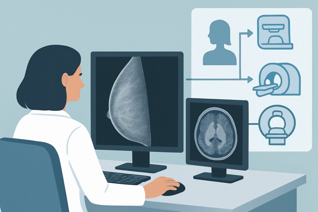
A 58-year-old woman presents to your clinic with a palpable 4 cm mass in her left breast and a firm, non-tender lymph node in her axilla. A core biopsy has already confirmed invasive ductal carcinoma. Based on the tumor size and clinically positive node, you estimate her clinical stage to be at least IIB. Before the multidisciplinary tumor board can finalize a plan for neoadjuvant therapy, you need to accurately assess the full extent of her locoregional disease. This article details the ACR-guided imaging workflow for evaluating locoregional disease in newly diagnosed, clinically advanced (Stage IIB-III) breast cancer. According to the ACR Appropriateness Criteria, an axillary ultrasound (US axilla) is a Usually Appropriate initial study in this setting.
Who Fits This Clinical Scenario for Locoregional Staging?
This guidance applies specifically to patients with a new, biopsy-proven diagnosis of invasive breast cancer—including invasive ductal carcinoma (IDC), invasive lobular carcinoma (ILC), or not otherwise specified (NOS)—who present with clinically advanced disease. This corresponds to clinical stages IIB, IIIA, IIIB, or IIIC. These stages are typically defined by a combination of a large primary tumor (e.g., >5 cm) or significant lymph node involvement identified on physical examination (e.g., palpable, matted, or fixed axillary nodes).
This workflow is distinct from other common clinical situations. It is crucial to differentiate this scenario from:
- Early-Stage Breast Cancer (Stage I-IIA): Patients with smaller tumors and clinically negative lymph nodes follow a different diagnostic algorithm, which may begin with sentinel lymph node biopsy rather than upfront axillary imaging.
- Evaluation for Distant Disease: This article focuses on locoregional staging (the breast, chest wall, and regional lymph nodes). The workup to rule out distant metastases in the bones, liver, lungs, or brain is a separate, though often concurrent, clinical question with its own set of recommended studies.
- Inflammatory Breast Cancer: This aggressive presentation has a unique clinical and imaging workup and is not covered by this specific scenario.
Applying this guidance to the correct patient population ensures appropriate resource utilization and accurate staging, which is foundational for effective treatment planning.
What Diagnoses Are You Working Up in This Locoregional Evaluation?
In this scenario, the primary diagnosis of invasive breast cancer is already established. The purpose of imaging is not to find the cancer, but to define its precise extent. The key questions you are trying to answer involve the T (tumor), N (node), and M (metastasis) staging components, specifically focusing on the locoregional environment.
Axillary Lymph Node Metastases: This is the most critical determination. Identifying the presence and burden of cancer in the axillary lymph nodes is a primary driver of staging, prognosis, and treatment. Imaging seeks to identify nodes with suspicious features that warrant biopsy, which can confirm nodal involvement and potentially allow the patient to proceed directly to axillary lymph node dissection, bypassing a sentinel node procedure.
Extra-Axillary Nodal Disease: In advanced disease, cancer can spread to other regional lymph node basins. Imaging aims to detect involvement of the internal mammary, supraclavicular, or infraclavicular nodes. The presence of disease in these areas significantly upstages the cancer (to N2 or N3) and mandates changes to radiation therapy fields and systemic treatment regimens.
Extent of Primary Tumor and Chest Wall Invasion: It is essential to determine if there are additional, clinically occult cancer foci in the breast (multifocal or multicentric disease) or if the known tumor extends into the pectoralis muscle or chest wall. These findings influence the feasibility of breast-conserving surgery (lumpectomy) versus mastectomy and are critical for radiation planning.
Why Is Axillary Ultrasound the Recommended First Step for Locoregional Staging?
For a patient with clinically suspicious axillary nodes, the ACR designates axillary ultrasound (US axilla) as a Usually Appropriate initial imaging study. Its role is pivotal for confirming nodal involvement and guiding the subsequent therapeutic pathway.
The primary strength of axillary ultrasound is its ability to characterize lymph node morphology with high resolution. It can readily identify features suspicious for metastasis, such as cortical thickening, a rounded shape, and the loss of the normal fatty hilum. More importantly, ultrasound provides real-time guidance for fine-needle aspiration (FNA) or core needle biopsy of any abnormal-appearing nodes. A positive biopsy confirms nodal metastasis, providing crucial staging information that directly influences decisions about neoadjuvant chemotherapy and axillary surgery. Furthermore, US axilla is non-invasive, widely accessible, and uses no ionizing radiation (0 mSv).
While axillary US is the key first step for the nodes, other imaging modalities are also rated Usually Appropriate for a complete locoregional workup in this advanced stage:
- MRI of the breast without and with IV contrast: This study is highly sensitive for defining the extent of the primary tumor within the breast. It excels at detecting additional cancer sites (multifocal/multicentric disease) and assessing for chest wall invasion, particularly for invasive lobular carcinoma or in women with dense breast tissue.
- FDG-PET/CT from skull base to mid-thigh: This whole-body functional imaging study is also Usually Appropriate. It is excellent for detecting metastatic disease in regional lymph nodes that are difficult to assess with other methods, such as the internal mammary and supraclavicular chains. It simultaneously provides information about distant metastatic disease, combining locoregional and distant staging into a single examination, albeit with a significant radiation dose (☢☢☢☢ 10-30 mSv).
In contrast, a whole-body bone scan is rated Usually Not Appropriate for this specific question of locoregional staging. While it is used to evaluate for bony metastases (a question of distant disease), it provides no information about the primary tumor or soft tissue lymph nodes, making it unsuitable for this purpose. Similarly, a dedicated contrast-enhanced CT of the chest, abdomen, and pelvis is also Usually Not Appropriate for the primary goal of locoregional staging, as its soft tissue resolution in the breast and axilla is inferior to MRI and ultrasound.
What’s Next After Axillary Ultrasound? Downstream Workflow
The results of the initial axillary ultrasound and subsequent biopsy create a clear decision-making pathway for the multidisciplinary team.
- If the axillary US is positive (and biopsy confirms metastasis): The patient is confirmed to have node-positive disease. This information is used to complete the clinical staging. The patient will typically proceed to neoadjuvant (pre-operative) chemotherapy. The confirmed nodal involvement means they are generally not a candidate for a sentinel lymph node biopsy and will likely require an axillary lymph node dissection after chemotherapy.
- If the axillary US is negative (no suspicious nodes seen): In a patient with clinically advanced disease (e.g., a very large primary tumor), a negative ultrasound may not be sufficient to rule out microscopic nodal disease. The patient may still proceed to a sentinel lymph node biopsy at the time of surgery to pathologically assess the nodes. However, in the context of planned neoadjuvant therapy, treatment may begin based on the high pre-test probability of nodal involvement from the primary tumor size alone.
- If the axillary US is indeterminate (nodes are equivocal): If nodes are seen that do not meet classic criteria for malignancy but are not clearly benign, an ultrasound-guided biopsy is still the recommended next step to resolve the ambiguity. The results of the biopsy will then guide the workflow as described above.
Following the complete locoregional staging (using US, MRI, and/or PET/CT), the case is typically discussed at a multidisciplinary tumor board to finalize a comprehensive treatment plan, which usually involves neoadjuvant systemic therapy, followed by surgery and radiation.
Pitfalls to Avoid (and When to Get Help)
Navigating the workup for advanced breast cancer requires careful coordination. Here are several common pitfalls to avoid:
- Skipping the Axillary US-Guided Biopsy: In a patient with clinically palpable nodes, proceeding directly to surgery without a pre-operative tissue diagnosis of the axilla can complicate treatment planning. A positive biopsy confirms the need for neoadjuvant therapy and more extensive axillary surgery.
- Relying Solely on Mammography for Extent of Disease: In patients with large tumors, dense breasts, or invasive lobular carcinoma, mammography can significantly underestimate the true extent of the cancer. Breast MRI is often necessary for accurate surgical planning.
- Misinterpreting the Role of PET/CT: While excellent for staging, a negative PET/CT in the axilla does not have sufficient negative predictive value to rule out microscopic nodal disease. It is a staging tool, not a substitute for pathologic nodal evaluation.
- Delaying the Workup: Stage IIB-III breast cancer is aggressive. The diagnostic and staging workup should be completed efficiently to allow for the timely initiation of neoadjuvant therapy, which can improve outcomes.
If imaging findings are complex or discordant with the clinical picture, consultation with a breast radiologist or discussion at a multidisciplinary tumor board is essential to formulate the best path forward.
Related ACR Topics and Tools
For a comprehensive overview of imaging across all clinical scenarios related to invasive breast cancer, please refer to our parent guide. For tools to assist in ordering the correct study and understanding its technical aspects, the following resources are available.
- For breadth across all scenarios in Imaging of Invasive Breast Cancer, see our parent guide: Imaging of Invasive Breast Cancer: ACR Appropriateness Decoded.
- To look up appropriateness criteria for adjacent or alternative clinical scenarios, use the ACR Appropriateness Criteria Lookup.
- For detailed technical parameters on how imaging studies are performed, consult the Imaging Protocol Library.
- To discuss cumulative radiation exposure with your patients, especially when considering PET/CT, refer to the Radiation Dose Calculator.
Frequently Asked Questions
Why is axillary ultrasound performed before a sentinel lymph node biopsy in this stage?
In clinically advanced (Stage IIB-III) breast cancer, there is a high pre-test probability of axillary node involvement. An axillary ultrasound with biopsy can confirm this non-surgically. If positive, it establishes N1 (or greater) disease, and the patient can proceed directly to neoadjuvant chemotherapy and a planned axillary dissection, making a sentinel node biopsy unnecessary.
Is a breast MRI always necessary for Stage IIB-III cancer?
While rated ‘Usually Appropriate’ by the ACR, it is not mandatory for every patient. However, it is strongly recommended for patients with invasive lobular carcinoma (which is often underestimated by mammography), those with dense breast tissue, or when there is a discrepancy between clinical exam and mammogram findings. It is crucial for planning breast-conserving surgery.
When should I choose FDG-PET/CT over separate CT scans and a bone scan?
FDG-PET/CT is rated ‘Usually Appropriate’ for this scenario because it provides comprehensive locoregional and distant staging in a single exam. It is particularly useful for detecting metastases in extra-axillary nodal basins (internal mammary, supraclavicular) and distant sites. In contrast, separate CTs and a bone scan are rated ‘Usually Not Appropriate’ for the specific question of locoregional staging, though they may be considered for distant staging if PET/CT is unavailable.
What if the patient has a contraindication to MRI contrast (e.g., severe renal failure)?
If a patient cannot receive gadolinium-based contrast for a breast MRI, the utility of the exam for assessing disease extent is significantly reduced (MRI without contrast is rated ‘Usually Not Appropriate’). In such cases, clinicians must rely more heavily on high-quality diagnostic mammography (including tomosynthesis) and breast ultrasound. Contrast-enhanced mammography, where available, is a ‘May be appropriate’ alternative.
Does this guidance apply to male breast cancer?
Yes, the general principles of staging invasive breast cancer apply to all patients. Men with Stage IIB-III breast cancer would undergo a similar locoregional staging workup, including evaluation of the axilla, to determine the extent of disease and guide therapy.
Reviewed by Pouyan Golshani, MD, Interventional Radiologist — May 30, 2026