Should You Order Screening Imaging for Transmasculine Patients After Top Surgery?

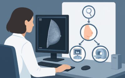
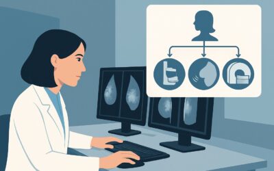
A 32-year-old transmasculine patient, who underwent bilateral mastectomies five years ago, presents for his annual primary care visit. He is otherwise healthy but mentions a family history of post-menopausal breast cancer in his mother. He asks, “Do I still need mammograms or any other kind of breast cancer screening?” This common and important question places you at a clinical decision point: how do you appropriately screen for breast cancer in a patient with a surgically altered chest? This article provides a detailed workflow for this specific scenario, grounded in the American College of Radiology (ACR) Appropriateness Criteria. For a transmasculine patient with prior bilateral mastectomies, routine screening with imaging modalities like mammography, digital breast tomosynthesis, and ultrasound is rated as Usually not appropriate.
Who Fits This Clinical Scenario?
This guidance applies specifically to transmasculine (female-to-male) individuals of any age and any underlying risk level who have undergone gender-affirming bilateral mastectomies, often referred to as “top surgery.” The key inclusion criterion is the history of this specific surgical procedure, which removes the vast majority of breast glandular tissue.
It is critical to distinguish this situation from related but distinct clinical scenarios that require a different imaging approach. This workflow does not apply to:
- Transmasculine patients with intact breast tissue: Individuals who have not had chest surgery, or who have only had a reduction mammoplasty, retain significant glandular tissue and follow different screening guidelines based on age and risk factors.
- Transfeminine (male-to-female) patients: Individuals on gender-affirming hormone therapy (estrogen) have different risk profiles and screening recommendations, which typically begin after a certain duration of hormone use and at a specific age.
- Patients with a new palpable lump: This article addresses screening in an asymptomatic patient. The presence of a new, palpable chest wall or axillary abnormality shifts the indication from screening to diagnostic, which has a separate and distinct workup.
Applying this screening guidance to the wrong patient population can lead to missed diagnoses or unnecessary procedures. Always confirm the patient’s surgical history and current symptoms before making a decision.
What Diagnoses Are You Working Up in This Scenario?
In a screening context, the goal is not to work up a specific symptom but to assess the baseline risk of malignancy and determine if routine imaging is justified. For transmasculine individuals post-mastectomy, the primary consideration is the residual risk of developing breast cancer in the small amount of tissue that may remain.
Residual Breast Cancer Risk: Bilateral mastectomy, especially procedures that are not “total” or “radical” mastectomies in the oncologic sense, can leave behind small amounts of breast glandular tissue. This tissue may be located along the chest wall, near the pectoralis muscle, in the axillary tail (near the armpit), or preserved in nipple-areolar complexes if a nipple-sparing technique was used. While the surgery reduces breast cancer risk by over 95%, the risk is not zero. The clinical question is whether this very low absolute risk is high enough to warrant routine imaging surveillance.
Fat Necrosis or Scar Tissue: Post-surgical changes are the most common cause of palpable findings in this population. Scar tissue, suture granulomas, and fat necrosis can all present as firm nodules. While benign, these can cause patient anxiety and may be difficult to distinguish from a malignancy on palpation alone, underscoring the importance of a clear diagnostic pathway if a lump does appear.
Chest Wall Lesions: Though less common, other non-breast pathologies of the chest wall must be considered. This includes benign entities like lipomas or sebaceous cysts, as well as rare primary or metastatic malignancies of the skin, muscle, or bone. The focus of breast-specific screening, however, remains on cancer arising from residual glandular elements.
Why Is Routine Imaging Usually Not Appropriate for Screening?
The ACR designates all standard imaging modalities as “Usually not appropriate” for routine screening in this patient population. The rationale is based on a careful balance of the very low incidence of cancer against the limitations and potential harms of the imaging tests themselves in this specific anatomical context.
Mammography and Digital Breast Tomosynthesis (DBT) are both rated Usually not appropriate. After top surgery, there is typically insufficient mobile tissue to position on the mammography detector and apply adequate compression. Attempting to do so is often technically difficult, painful for the patient, and results in suboptimal images with very low diagnostic yield. The radiation exposure (ACR RRL® ☢☢, 0.1-1 mSv), while small, is not justified given the low probability of detecting a clinically significant cancer in this setting.
US breast (Ultrasound) is also rated Usually not appropriate for screening. While ultrasound does not use ionizing radiation (ACR RRL® O, 0 mSv), its use as a screening tool in a low-prevalence population is limited by a high rate of false-positive findings. Normal post-surgical changes like scarring and fat necrosis can be difficult to distinguish from suspicious lesions, potentially leading to unnecessary anxiety and biopsies. However, it is crucial to note that ultrasound becomes the primary, recommended modality for a diagnostic workup if a new palpable abnormality is discovered.
MRI breast without and with IV contrast is also Usually not appropriate for routine screening. While highly sensitive, breast MRI has the highest false-positive rate of all modalities and is significantly more expensive and less accessible. Its use is reserved for very specific high-risk scenarios (e.g., known pathogenic variants like BRCA1/2) and is not indicated for routine surveillance in the general post-mastectomy transmasculine population.
What’s Next? The Downstream Workflow
If routine imaging is not recommended, what is the appropriate surveillance strategy? The workflow shifts from imaging to physical examination and patient education.
- If the patient is asymptomatic: The recommended course is to forego screening imaging. Instead, the focus should be on performing a thorough clinical chest wall examination as part of the annual physical. This includes careful palpation of the entire chest wall, any reconstructed nipple-areolar complexes, and the axillae. Counsel the patient on the importance of chest wall self-awareness and instruct them on how to perform a monthly self-exam, encouraging them to report any new or changing lumps, bumps, or skin changes promptly.
- If a new palpable abnormality is found (by patient or clinician): The clinical scenario immediately changes from screening to diagnostic. At this point, imaging is indicated. The appropriate next step is a targeted diagnostic breast ultrasound of the specific area of concern. The radiologist can correlate the imaging findings directly with the palpable lump.
- If ultrasound is indeterminate or suspicious: Based on the ultrasound findings (e.g., a BI-RADS 4 or 5 lesion), the next step is typically an ultrasound-guided core needle biopsy to obtain a tissue diagnosis. This ensures that any suspicious finding is appropriately evaluated histologically.
This pathway ensures that imaging is used judiciously and effectively, reserved for situations where there is a specific clinical question to answer rather than being used for low-yield screening.
Pitfalls to Avoid (and When to Get Help)
Navigating care for transmasculine patients post-mastectomy requires awareness of several potential pitfalls:
- Conflating Screening with Diagnostic Workup: Do not order screening mammography for a patient with a new palpable lump. The correct order is a diagnostic ultrasound targeted to the finding.
- Ignoring Patient Anxiety: A patient’s request for screening may be driven by significant anxiety, especially with a family history. Dismissing their concerns is a pitfall; instead, use it as an opportunity to educate them on why clinical and self-exams are the more effective strategy in their specific case.
- Incomplete Physical Exam: Ensure the clinical exam covers the entire area where residual tissue may be present, including the full chest wall, incision lines, and deep into the axilla.
- Assuming No Risk: While the risk is very low, it is not zero. Avoid telling patients they have “no risk” of breast cancer. Frame it accurately as a “dramatically reduced risk” that no longer warrants the harms of routine imaging.
If a palpable lesion is identified and the initial diagnostic ultrasound is equivocal, or if there is a high clinical suspicion despite negative imaging, consultation with a breast surgeon or radiologist specializing in breast imaging is the appropriate escalation.
Related ACR Topics and Tools
The ACR Appropriateness Criteria are a comprehensive resource for evidence-based imaging decisions. For this and related scenarios, the following GigHz tools can streamline your workflow and provide additional context for patient conversations.
- For breadth across all scenarios in Transgender Breast Cancer Screening, see our parent guide: Transgender Breast Cancer Screening: ACR Appropriateness Decoded.
- To review other clinical presentations, use the ACR Appropriateness Criteria Lookup.
- For details on how specific studies are performed, consult the Imaging Protocol Library.
- To discuss radiation exposure from alternative (but not recommended) studies, the Radiation Dose Calculator can be a useful aid.
Frequently Asked Questions
Does a family history of breast cancer change the screening recommendation for a transmasculine patient after top surgery?
For most family histories, the recommendation does not change; routine imaging is still ‘Usually not appropriate.’ The dramatic risk reduction from mastectomy generally outweighs the increased baseline risk from family history. However, for patients with known high-penetrance genetic mutations (e.g., BRCA1/2), a specialized high-risk consultation is warranted, as surveillance strategies may differ and could involve MRI, although this is outside the scope of routine screening.
If a patient had a nipple-sparing mastectomy, is there more risk?
Nipple-sparing techniques intentionally leave more tissue behind at the nipple-areolar complex compared to free nipple grafts. While this may slightly increase the amount of residual glandular tissue, it does not currently change the overarching ACR recommendation to forgo routine screening imaging. The emphasis remains on diligent clinical exams and patient self-awareness, with particular attention to the nipple and subareolar regions.
What is the recommended follow-up if a patient reports a new lump but a diagnostic ultrasound is negative (BI-RADS 1 or 2)?
If a targeted diagnostic ultrasound of a palpable lump is definitively benign (e.g., a simple cyst or classic post-surgical scar), the standard of care is typically clinical follow-up. Reassure the patient and recommend continued self-monitoring. A short-term follow-up clinical exam in 3-6 months can provide additional reassurance for both the patient and provider.
Should I stop testosterone therapy before any diagnostic breast imaging?
No, there is no evidence to suggest that testosterone therapy interferes with the accuracy of diagnostic breast ultrasound or that it needs to be stopped for this purpose. Patients should continue their prescribed gender-affirming hormone therapy.
Why is ultrasound recommended for a palpable lump if it’s not recommended for screening?
This highlights the critical difference between a screening test and a diagnostic test. A screening test is used on an asymptomatic population to find occult disease and requires high performance with low false positives. A diagnostic test is used to evaluate a specific problem, like a lump. Ultrasound is excellent for characterizing a palpable finding (e.g., is it cystic or solid?) and guiding a biopsy if needed. Its focused, problem-solving nature makes it the ideal tool for a diagnostic workup, even though it is not suitable for broad, asymptomatic screening in this population.
Reviewed by Pouyan Golshani, MD, Interventional Radiologist — May 29, 2026