Should You Order Imaging for Routine Surveillance After Mastectomy for DCIS?

A 62-year-old woman, status post left mastectomy for Ductal Carcinoma in Situ (DCIS) four years ago, presents for her annual wellness visit. She is asymptomatic and reports no concerns with the reconstructed breast or chest wall. Her physical examination is unremarkable, with no palpable nodules, skin changes, or axillary adenopathy. As you consider her ongoing cancer surveillance plan, you question whether routine imaging of the ipsilateral chest wall is indicated. This article details the American College of Radiology (ACR) Appropriateness Criteria for this specific scenario: routine surveillance for local recurrence in an adult patient with a history of mastectomy for DCIS.
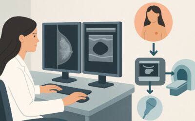
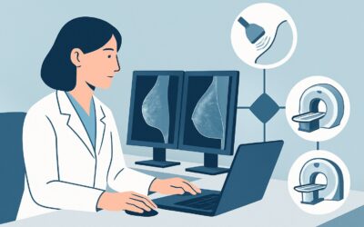
For this presentation, imaging studies such as breast ultrasound are designated as Usually not appropriate. This guide will explore the clinical reasoning behind this recommendation, the appropriate surveillance strategy, and the differential diagnosis to keep in mind.
Who Fits This Clinical Scenario?
This guidance applies to a very specific patient population: asymptomatic adults undergoing routine, scheduled surveillance after a total mastectomy for Ductal Carcinoma in Situ (DCIS). The key inclusion criteria are:
- History: Confirmed history of DCIS treated with total mastectomy on the ipsilateral side.
- Presentation: The patient is completely asymptomatic. There are no new lumps, pain, skin changes (such as erythema, thickening, or retraction), or nipple discharge.
- Intent: The goal is routine surveillance, not a diagnostic workup for a new finding.
It is critical to distinguish this scenario from similar but distinct clinical situations that require a different imaging approach. This guidance does not apply if the patient:
- Is symptomatic: If the patient or clinician detects a new palpable lump, skin change, or other abnormality on physical exam, the situation shifts from surveillance to a diagnostic evaluation. This falls under the ACR scenario for suspected local recurrence, where imaging is indicated.
- Had breast conservation therapy (BCT): Patients who underwent lumpectomy followed by radiation have a different risk profile and surveillance protocol, which includes annual mammography of the treated breast.
- Has a new diagnosis of DCIS: This article does not cover the initial workup of newly diagnosed DCIS prior to treatment.
What Diagnoses Are You Working Up in This Scenario?
While routine imaging is not recommended, the purpose of ongoing clinical surveillance is to detect potential local recurrence. The differential diagnosis for a new finding on the chest wall after mastectomy includes both malignant and benign etiologies. Understanding these possibilities informs the rationale for relying on physical examination as the primary surveillance tool.
Local Recurrence of DCIS or Invasive Carcinoma
This is the primary concern. Recurrence after mastectomy for pure DCIS is uncommon, but it can occur. It typically presents as a subcutaneous or cutaneous nodule on the chest wall, near the mastectomy scar. The goal of surveillance is to detect such a recurrence at an early, treatable stage.
Fat Necrosis
A common benign consequence of surgery and/or reconstruction, fat necrosis can present as a firm, sometimes tender, palpable mass. On physical exam, it can be difficult to distinguish from a malignant recurrence, which is why any new palpable finding warrants a diagnostic imaging workup, even if the underlying cause is likely benign.
Suture Granuloma
This is a benign inflammatory reaction to suture material left behind during surgery. Suture granulomas can form palpable nodules along the scar line weeks, months, or even years after the procedure. They are often firm and fixed, mimicking a recurrence on palpation.
Post-Surgical Scar Tissue (Fibrosis)
Normal healing involves the formation of scar tissue. Sometimes this can be thicker or more nodular than expected, creating a palpable area of concern. While a normal part of the healing process, any change in a stable scar should be evaluated.
Why Is Imaging Usually Not Appropriate for This Presentation?
The American College of Radiology (ACR) rates all imaging modalities as Usually not appropriate for routine surveillance in an asymptomatic patient following mastectomy for DCIS. This includes modalities with and without ionizing radiation.
The core rationale is based on a risk-benefit analysis. The rate of local recurrence after mastectomy for pure DCIS is very low, often cited in the range of 1-2% over 10 years. Given this low pre-test probability, the potential yield of any routine screening imaging test is minimal. The potential harms—including false-positive findings leading to patient anxiety, unnecessary biopsies of benign post-surgical changes like fat necrosis or suture granulomas, and increased healthcare costs—outweigh the potential benefits in an asymptomatic individual.
Let’s examine the specific ratings for common modalities:
- US breast: Rated Usually not appropriate. While ultrasound uses no ionizing radiation (0 mSv), its routine use in this setting is hampered by a high rate of false positives. Normal post-surgical changes, such as seromas, fat necrosis, and scar tissue, can have an ambiguous appearance on ultrasound, often prompting unnecessary follow-up and biopsies.
- Mammography screening / Digital breast tomosynthesis screening: Rated Usually not appropriate. After a total mastectomy, there is insufficient breast tissue for effective mammographic evaluation. While imaging of the chest wall is possible, it has a very low yield for detecting recurrence and exposes the patient to a small amount of ionizing radiation (☢☢ 0.1-1 mSv).
- MRI breast without and with IV contrast: Rated Usually not appropriate. Although MRI is a highly sensitive modality for detecting breast cancer, its specificity can be lower, particularly in the post-operative setting where inflammation and scar tissue can enhance. Its routine use for surveillance in this low-risk population is not justified due to high cost and the potential for false-positive findings.
The standard of care for surveillance in this scenario is a diligent annual clinical breast examination and patient education on breast self-awareness. Imaging is reserved for the diagnostic workup of new signs or symptoms.
What’s Next After a Routine Exam? Downstream Workflow
The downstream workflow for a patient in this scenario is guided entirely by the findings of the physical examination and patient-reported symptoms.
If the Clinical Exam is Normal and the Patient is Asymptomatic:
The appropriate next step is to continue with routine annual clinical surveillance. No imaging is indicated. The patient should be reassured and reminded of the importance of self-monitoring and reporting any new changes they notice between appointments. Schedule a follow-up for the next annual examination.
If the Clinical Exam is Abnormal or the Patient Reports a New Symptom:
The workflow changes immediately from surveillance to diagnostic. If a new nodule, skin thickening, or other concerning finding is identified, the patient no longer fits this “routine surveillance” scenario. At this point, targeted diagnostic imaging is warranted. The clinical question has changed to evaluating a specific finding, which is a different ACR variant (“Suspected local recurrence based on symptoms…”). Typically, a targeted breast ultrasound of the area of concern would be the first imaging step. Depending on the findings, this may be followed by ultrasound-guided biopsy.
In summary, a negative exam leads to continued observation, while a positive exam triggers a new diagnostic cascade.
Pitfalls to Avoid (and When to Get Help)
Navigating surveillance for post-mastectomy DCIS patients requires careful adherence to guidelines to avoid common errors.
- Applying BCT Guidelines to Mastectomy Patients: Do not order annual mammograms for a post-mastectomy patient. The surveillance protocols for breast conservation therapy (lumpectomy) and mastectomy are fundamentally different.
- Dismissing a Palpable Finding: While routine imaging is not recommended for asymptomatic patients, any new palpable finding must be taken seriously and worked up with diagnostic imaging. Do not assume a new lump is just scar tissue without evaluation.
- Underestimating Patient Anxiety: Some patients may feel anxious without the “reassurance” of an annual imaging test. It’s crucial to explain the evidence-based rationale for relying on physical exams, highlighting that this approach avoids the harms of false positives from low-yield tests.
- Neglecting Patient Education: Since clinical and self-exams are the primary surveillance tools, ensure the patient understands what to look for and the importance of promptly reporting any changes.
If a new, concerning finding is identified on physical exam, the patient should be promptly referred for diagnostic breast imaging.
Related ACR Topics and Tools
For a comprehensive overview of imaging recommendations across all DCIS-related clinical presentations, from initial diagnosis to other surveillance scenarios, please consult our parent guide. For further exploration of adjacent criteria or imaging techniques, the following resources are available.
- For breadth across all scenarios in Imaging of Ductal Carcinoma in Situ (DCIS), see our parent guide: Imaging of Ductal Carcinoma in Situ (DCIS): ACR Appropriateness Decoded.
- To look up other clinical scenarios, use the ACR Appropriateness Criteria Lookup tool.
- To review technical details for various imaging studies, visit the Imaging Protocol Library.
- To discuss cumulative radiation exposure with patients, the Radiation Dose Calculator can be a helpful aid.
Frequently Asked Questions
Why is routine imaging not recommended after mastectomy for DCIS, but it is after a lumpectomy (BCT)?
After a lumpectomy (breast conservation therapy), a significant amount of breast tissue remains, which carries a risk of new or recurrent cancer. Annual mammography is effective for screening this remaining tissue. After a total mastectomy, nearly all breast tissue is removed, making the risk of local recurrence very low (around 1-2%). Routine imaging of the chest wall has a very low yield and a high rate of false positives in this setting, so the risks are considered to outweigh the benefits.
Does this ‘no imaging’ recommendation change if the patient has a high-risk genetic mutation like BRCA1 or BRCA2?
This is a complex area, and guidelines may vary. While the ACR criteria for this specific scenario do not differentiate based on genetic risk, high-risk patients are often managed in specialized clinics. Some guidelines may recommend annual chest wall MRI for high-risk patients after mastectomy, though the evidence for this is still evolving. Consultation with a breast specialist or high-risk clinic is advisable for these patients.
How often should a clinical breast exam be performed for surveillance in this scenario?
Most major guidelines, including those from the National Comprehensive Cancer Network (NCCN), recommend a clinical breast examination every 6 to 12 months for the first 5 years after treatment, and annually thereafter. This frequency, combined with patient self-awareness, is the cornerstone of surveillance.
What if my patient is very anxious and insists on getting an imaging study for reassurance?
This situation requires a careful conversation about the risks and benefits. Acknowledge the patient’s anxiety and explain the rationale behind the guidelines: routine imaging is more likely to lead to a false alarm (and an unnecessary biopsy) than to find a true recurrence. Discussing the very low recurrence rate and the effectiveness of physical examination can be reassuring. Shared decision-making is key, but ordering low-value tests should generally be discouraged.
Does this recommendation apply if the original DCIS had microinvasion?
The presence of microinvasion (DCIS-MI) can place the patient in a slightly higher risk category than pure DCIS. While the ACR criteria group them together for some scenarios, management may be nuanced. However, for an asymptomatic patient after mastectomy, the principle of low recurrence risk still largely applies, and routine imaging is generally not performed. The primary surveillance method remains clinical examination. If there is any uncertainty, consultation with a breast surgical oncologist or medical oncologist is recommended.
Reviewed by Pouyan Golshani, MD, Interventional Radiologist — May 30, 2026