What Is the Right Imaging for Surveillance After Breast Conservation Therapy?

A 58-year-old female presents for her annual follow-up. Two years ago, she completed breast conservation therapy (BCT), including lumpectomy and radiation, for early-stage estrogen-receptor-positive breast cancer. Her surgical margins were negative, and she has been asymptomatic since, with no new lumps, pain, or skin changes. You are preparing to order her annual surveillance imaging and must decide on the most appropriate modality to detect potential recurrence while minimizing false positives in the surgically altered breast. This article details the clinical workflow for this specific scenario, explaining why the American College of Radiology (ACR) finds that Digital breast tomosynthesis diagnostic is Usually Appropriate.
## Who Fits This Clinical Scenario for Post-BCT Surveillance?
This guidance applies to a specific patient population: an adult female who has successfully completed breast conservation therapy for breast cancer. The key inclusion criteria are:
- History: Prior diagnosis of breast cancer treated with BCT (lumpectomy).
- Surgical Status: Confirmed negative (clear) surgical margins on final pathology.
- Clinical Status: Asymptomatic, with no new breast-related symptoms like a palpable mass, focal pain, skin changes, or nipple discharge.
- Timing: The patient is presenting for routine, scheduled surveillance imaging following the completion of her primary treatment (which may or may not have included radiation therapy).
It is critical to distinguish this surveillance scenario from other clinical presentations. This workflow does not apply if:
- The patient has positive surgical margins. This situation requires a different approach focused on evaluating for residual disease, often detailed in the ACR variant for Postsurgical excision for breast cancer. Positive margins.
- The patient presents with new symptoms. A new lump, skin thickening, or nipple discharge shifts the indication from surveillance to a diagnostic workup, which follows a different algorithm.
- The imaging is for a nonmalignant finding. This guidance is specific to post-cancer surveillance, not for follow-up after an excision of a benign lesion like a fibroadenoma.
## What Diagnoses Are You Working Up in This Scenario?
Surveillance imaging after BCT is primarily focused on the early detection of cancer recurrence, while also differentiating it from the expected and benign changes related to treatment. The differential diagnosis includes several key considerations.
The most critical diagnosis to identify is an Ipsilateral Breast Tumor Recurrence (IBTR). This is a new cancer that develops in the same breast that was previously treated. Recurrence can manifest as a new mass, a cluster of suspicious microcalcifications, or architectural distortion. The architectural changes from prior surgery and radiation can make detecting a subtle recurrence challenging, which is why the choice of imaging modality is so important.
Second, the patient remains at risk for a new primary breast cancer, either in the contralateral (untreated) breast or a new, distinct cancer in the ipsilateral (treated) breast. Surveillance imaging must comprehensively evaluate both breasts for any new, unrelated malignancies.
Finally, a significant portion of findings will be benign post-treatment changes. Surgery and radiation therapy create predictable alterations in the breast tissue, including scarring, seromas (fluid collections), fat necrosis, and skin thickening. These changes can evolve over the first few years post-treatment and can sometimes mimic the appearance of a malignancy. A primary goal of surveillance imaging is to confidently characterize these findings as benign and stable, avoiding unnecessary patient anxiety and biopsies.
## Why Is Digital Breast Tomosynthesis the Recommended Study for Surveillance After BCT?
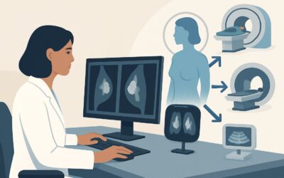
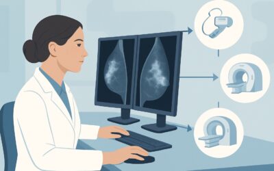
For an asymptomatic female undergoing surveillance after breast conservation therapy with negative margins, the ACR designates Digital breast tomosynthesis diagnostic as Usually Appropriate. Standard 2D mammography (both screening and diagnostic) and screening tomosynthesis also receive this rating, reflecting the central role of mammographic techniques in this setting.
The primary advantage of Digital Breast Tomosynthesis (DBT), or 3D mammography, is its ability to reduce the effect of overlapping breast tissue. This is particularly valuable in the post-treatment breast, where scarring and architectural changes from surgery and radiation can obscure or mimic a true lesion on standard 2D images. By acquiring multiple thin-slice images and reconstructing them, DBT allows the radiologist to scroll through the tissue, improving the conspicuity of masses and architectural distortion. This leads to higher cancer detection rates and, importantly, a reduction in recall rates for benign findings.
### How Do Alternative Studies Compare?
- MRI breast without and with IV contrast is rated as May be appropriate. While breast MRI has the highest sensitivity for detecting invasive breast cancer, its specificity is lower than mammography. This can lead to more false-positive findings, resulting in additional imaging and often unnecessary biopsies. For this reason, it is not recommended for routine annual surveillance in every post-BCT patient. Its use is typically reserved for select cases, such as patients with extremely dense breasts where mammography is limited, or for further evaluation of an indeterminate mammographic finding.
- US breast is also rated as May be appropriate. Ultrasound is an excellent tool for targeted evaluation of a specific palpable or mammographic finding. However, when used as a primary surveillance tool for the entire breast, it is highly operator-dependent and has a lower sensitivity for detecting microcalcifications, which can be an early sign of ductal carcinoma in situ (DCIS) recurrence. It is often used as a supplemental imaging tool but not as the standalone primary modality for routine surveillance.
- MRI breast without IV contrast is Usually not appropriate. Without the use of gadolinium-based contrast, breast MRI cannot reliably differentiate between benign and malignant enhancing lesions, rendering it inadequate for cancer surveillance.
The recommended study, DBT, involves a low radiation dose (ACR Relative Radiation Level ☢☢, 0.1-1 mSv), which is considered a safe and acceptable level for annual imaging. This avoids the need for IV contrast agents required for MRI. When ordering, it is crucial to provide a clear clinical history, including the date of surgery, type of cancer, and radiation history, and to ensure all prior imaging studies are available for comparison.
## What’s the Next Step After Post-BCT Surveillance Tomosynthesis?
The results of the surveillance DBT, reported using the BI-RADS classification system, will guide the subsequent workflow.
- Negative or Benign Finding (BI-RADS 1 or 2): If the examination shows only expected stable post-surgical changes or clearly benign findings, the patient can return to routine annual surveillance. No further immediate action is needed.
- Probably Benign Finding (BI-RADS 3): This category is used for findings that have a very high likelihood of being benign (>98%) but are not definitively so. Common examples in the post-BCT setting include developing focal asymmetry or a new round mass that appears benign. The standard recommendation is a short-interval follow-up imaging study, typically in six months, to ensure stability.
- Suspicious or Highly Suggestive of Malignancy (BI-RADS 4 or 5): These findings warrant tissue sampling to establish a definitive diagnosis. The next step is typically an image-guided core needle biopsy. The modality for guidance (ultrasound, stereotactic/tomosynthesis, or MRI) depends on which technique best visualizes the lesion. A positive biopsy confirming recurrence would trigger a multidisciplinary tumor board discussion to plan further treatment, which could include re-excision, mastectomy, or systemic therapy.
- Incomplete (BI-RADS 0): If the tomosynthesis is inconclusive, additional imaging is required for a final assessment. This almost always involves diagnostic mammographic views (e.g., spot compression, magnification) and a targeted breast ultrasound of the area in question.
## Pitfalls to Avoid (and When to Get Help)
1. Mistaking Scar for Recurrence: Post-surgical scarring can be dense and spiculated, mimicking a malignancy. Comparing with prior studies is essential to confirm stability. A scar should retract and become less prominent over time, whereas a recurrence typically grows.
2. Dismissing Subtle Changes: In a breast with architectural distortion from prior treatment, a subtle new area of distortion or a small cluster of microcalcifications can be the only sign of recurrence. A low threshold for recommending magnification views or biopsy for new, evolving findings is critical.
3. Inadequate Clinical History: Failing to provide the radiologist with the date of surgery, location of the primary tumor, and radiation history can lead to misinterpretation. This context is vital for correctly interpreting post-treatment changes.
4. Over-reliance on a Single Modality: While DBT is the primary tool, an indeterminate finding should prompt the use of supplemental imaging like ultrasound or even MRI. Do not hesitate to order additional studies if the initial images are inconclusive.
If a new finding is suspicious (BI-RADS 4 or 5) or if imaging remains indeterminate after a full diagnostic workup, the appropriate next step is to refer the patient to a breast surgeon or for an image-guided biopsy.
## Related ACR Topics and Tools
This article covers one specific workflow within the broader topic of post-surgical breast imaging. For a comprehensive overview of all related scenarios, from post-benign excision to follow-up for positive margins, please see our parent guide.
- For breadth across all scenarios in Imaging after Breast Surgery, see our parent guide: Imaging after Breast Surgery: ACR Appropriateness Decoded.
For additional decision support and technical details, the following GigHz resources are available:
- ACR Appropriateness Criteria Lookup — for adjacent scenarios
- Imaging Protocol Library — for technique on the recommended study
- Radiation Dose Calculator — for cumulative dose conversations
Frequently Asked Questions
How soon after completing radiation should the first surveillance mammogram be performed?
The general consensus is to perform the first post-treatment surveillance mammogram 6 to 12 months after the completion of radiation therapy. This allows acute radiation-induced inflammation and skin edema to subside, which can otherwise obscure the breast tissue and mimic suspicious findings.
Should I order a ‘screening’ or ‘diagnostic’ mammogram for this patient?
For the first one to two years following BCT, it is often preferable to order a ‘diagnostic’ mammogram/tomosynthesis. This designation allows the radiologist the flexibility to obtain additional views, such as spot compression or magnification, at the time of the initial appointment if needed, without requiring the patient to return for a separate visit.
Does the recommendation for DBT change if the patient has dense breasts?
No, Digital Breast Tomosynthesis (DBT) remains the ‘Usually Appropriate’ study. In fact, the benefits of DBT over standard 2D mammography are often more pronounced in women with dense breasts because it excels at reducing the masking effect of overlapping tissue. In some cases of extremely dense breasts with indeterminate mammographic findings, a supplemental MRI (‘May be appropriate’) may be considered, but DBT is still the primary surveillance modality.
If a patient had a mastectomy on one side and BCT on the other, does this surveillance plan apply?
Yes, this guidance applies to the breast that underwent conservation therapy. The mastectomy side does not typically require routine imaging surveillance unless there is a clinical concern in the chest wall or axilla, or if the patient has an implant that requires monitoring.
What is the role of a physical exam in this surveillance scenario?
A clinical breast examination remains a cornerstone of surveillance alongside imaging. It should be performed regularly by a clinician. While imaging is highly sensitive for non-palpable recurrences, a physical exam can detect palpable abnormalities that may arise between imaging intervals, prompting a diagnostic workup.
Reviewed by Pouyan Golshani, MD, Interventional Radiologist — May 30, 2026