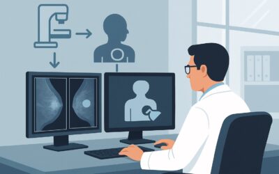
Which Breast Cancer Screening Is Best for Transfeminine Patients on Estrogen for Over 5 Years?

A 42-year-old transfeminine patient presents for her annual physical. She has been on consistent gender-affirming hormone therapy with estrogen for the past seven years and has developed breast tissue. She feels well and has no specific breast complaints but asks, “Should I be getting mammograms now, like my cisgender female friends?” You know that long-term estrogen use is a key factor in breast cancer risk, but the guidelines for this specific population are nuanced. This article provides a clinical workflow for this exact scenario, clarifying which imaging study to order for an average-risk transfeminine patient over 40 with five or more years of hormone exposure.
Based on the American College of Radiology (ACR) Appropriateness Criteria®, the recommended approach, Digital breast tomosynthesis screening, is rated as May be appropriate.
Who Fits This Clinical Scenario?
This guidance applies specifically to transfeminine (male-to-female) individuals who meet all of the following criteria:
- Are 40 years of age or older.
- Have a history of gender-affirming hormone use (typically including estrogen) for a cumulative duration of five years or more.
- Are considered at average risk for breast cancer. This means they do not have a personal history of breast cancer, a known genetic mutation (e.g., BRCA1/2), a history of chest radiation therapy before age 30, or a strong family history suggesting a hereditary cancer syndrome.
It is crucial to distinguish this patient from other similar, but distinct, clinical presentations. This workflow does not apply to:
- Transfeminine patients with less than 5 years of hormone use: The risk profile and screening recommendations differ for individuals with shorter durations of hormone exposure.
- Patients with high-risk factors: Any patient with a known BRCA mutation, significant family history, or prior mantle radiation would be considered high-risk and requires a different screening protocol, often involving MRI.
- Transmasculine (female-to-male) patients: Screening guidelines for transmasculine individuals depend on whether they have had chest surgery (mastectomy) and are addressed in a separate ACR variant.
What Diagnoses Are You Working Up in This Scenario?
The primary goal of screening is the early detection of asymptomatic breast cancer. While the overall incidence in the transfeminine population is still being studied, risk is understood to increase with long-term estrogen exposure, which promotes the development of lobular and ductal breast tissue.
The key diagnosis to identify is invasive breast carcinoma, including both invasive ductal carcinoma (IDC) and invasive lobular carcinoma (ILC), as well as their precursor, ductal carcinoma in situ (DCIS). Early detection of these malignancies when they are small and non-palpable is the fundamental purpose of screening and is associated with improved treatment outcomes.
While screening for malignancy, imaging will also characterize benign findings that are common in hormonally stimulated breast tissue. These can include gynecomastia, which represents benign proliferation of glandular tissue, as well as cysts, fibroadenomas, and other benign proliferative changes. Differentiating these expected benign findings from suspicious malignant lesions is a core function of breast imaging. The rationale for screening in this population is that after 5-10 years of feminizing hormones, the breast tissue architecture and associated cancer risk begin to more closely approximate that of cisgender women.
Why Is Digital Breast Tomosynthesis Screening the Recommended Study for This Presentation?
For this specific scenario, the ACR rates both Digital Breast Tomosynthesis (DBT) screening and standard 2D Mammography screening as May be appropriate. However, DBT is often the preferred modality in modern breast imaging centers due to its superior performance in certain situations.
The rationale for this recommendation is grounded in balancing detection efficacy with the risks of radiation and false positives.
- Superior Visualization with DBT: Digital breast tomosynthesis, or “3D mammography,” acquires images of the breast in thin slices, which are reconstructed into a three-dimensional view. This technique reduces the effect of overlapping breast tissue, a common issue that can hide cancers on standard 2D mammograms. This is particularly valuable in individuals with dense breast tissue, which can be induced by estrogen therapy. By minimizing tissue overlap, DBT has been shown to increase cancer detection rates and decrease recall rates (the need for additional imaging due to an uncertain finding).
- Radiation and Risk Profile: Both DBT and standard 2D mammography use a low dose of ionizing radiation, categorized by the ACR with a relative radiation level of ☢☢ (0.1-1 mSv). This is considered a very small and acceptable risk in the context of the life-saving benefit of early cancer detection.
Why Alternatives Are Rated Lower
Other imaging modalities are considered Usually not appropriate for screening in this average-risk population:
- Breast Ultrasound (US): While excellent for diagnostic workups (e.g., evaluating a palpable lump or a mammographic finding), ultrasound is not recommended as a primary screening tool for average-risk individuals. It has a higher rate of false positives and has not been shown to have a mortality benefit as a standalone screening test.
- Breast MRI (with or without contrast): Breast MRI is a highly sensitive test but is reserved for screening high-risk individuals (e.g., BRCA carriers). For average-risk patients, its use for screening would lead to a significant number of false positives, resulting in unnecessary anxiety, additional imaging, and biopsies. Its higher cost and need for IV contrast also make it unsuitable for routine screening in this context.
For detailed technical specifications on performing this study, see the GigHz guide on the Screening Mammography (with DBT) protocol.
What’s Next After Digital Breast Tomosynthesis Screening? Downstream Workflow
The results of a screening mammogram are reported using the Breast Imaging Reporting and Data System (BI-RADS) score, which dictates the next steps.
- BI-RADS 0: Incomplete. This means the radiologist needs more information to make a final assessment. The patient will be called back for additional imaging, known as a diagnostic workup. This typically includes diagnostic mammographic views (e.g., spot compression, magnification) and often a targeted breast ultrasound of the area in question.
- BI-RADS 1 (Negative) or 2 (Benign Finding): These are normal results. The patient has no evidence of malignancy, and the recommendation is to return to routine annual screening.
- BI-RADS 3: Probably Benign. This finding has a very low (≤2%) chance of being cancer. The standard recommendation is a short-interval follow-up, typically with a mammogram of the affected breast in six months, to ensure the finding is stable.
- BI-RADS 4 (Suspicious) or 5 (Highly Suggestive of Malignancy): These findings warrant a biopsy to establish a definitive diagnosis. The patient should be referred to a breast surgeon or radiologist for a tissue sampling procedure, most commonly a core needle biopsy.
If the workup is ultimately negative, the patient returns to their recommended screening interval. If a malignancy is diagnosed, the patient is referred for oncologic consultation and treatment planning.
Pitfalls to Avoid (and When to Get Help)
When ordering breast cancer screening for transfeminine patients, be mindful of several potential pitfalls to ensure appropriate and effective care.
- Misclassifying Risk: Do not assume a patient is average-risk without taking a thorough personal and family history. The presence of a first-degree relative with premenopausal breast cancer, for example, could shift the patient into a high-risk category requiring a different screening approach.
- Ignoring Palpable Findings: Screening mammography is for asymptomatic patients. If a patient presents with a palpable lump, nipple discharge, or focal pain, the correct order is a diagnostic mammogram and ultrasound, not a screening study.
- Inaccurate Hormone History: The 5-year hormone use threshold is a key decision point. Ensure you have an accurate and cumulative history of all gender-affirming hormone therapies, as intermittent or past use still contributes to the total duration.
If you are uncertain about a patient’s risk level or if the imaging results are complex or discordant with clinical findings, escalation to a breast imaging specialist or a multidisciplinary breast center is the appropriate next step.
Related ACR Topics and Tools
For a comprehensive overview of all clinical variants and patient populations within this topic, please consult our parent guide. Additional tools can help you navigate adjacent scenarios and communicate with your patients about imaging specifics.
- For breadth across all scenarios in Transgender Breast Cancer Screening, see our parent guide: Transgender Breast Cancer Screening: ACR Appropriateness Decoded.
- To explore other clinical scenarios, use the ACR Appropriateness Criteria Lookup.
- For detailed procedural techniques, see the Imaging Protocol Library.
- To discuss radiation exposure with patients, use the Radiation Dose Calculator.
Frequently Asked Questions
Why is the guideline to start screening after 5 years of hormone use at age 40?
This recommendation is based on expert consensus that aims to balance the time required for estrogen to induce sufficient glandular breast tissue development (making screening meaningful) with the age at which breast cancer risk begins to increase. After about 5-10 years of feminizing hormone therapy, the breast cancer risk for transfeminine individuals is thought to approach that of cisgender women, for whom screening typically begins at age 40.
Is the breast cancer risk for a transfeminine patient on long-term hormones the same as for a cisgender woman?
The risk is not identical but approaches that of cisgender women with long-term hormone use. While the absolute risk is lower than for cisgender women, it is significantly higher than for cisgender men. Therefore, dedicated screening is warranted after a sufficient duration of hormone exposure.
How does the presence of breast implants affect screening for a transfeminine patient?
Breast implants, whether saline or silicone, can obscure breast tissue on a mammogram. For patients with implants, mammography should always be performed with specialized implant displacement (Eklund) views to maximize visualization of the native breast tissue. Digital breast tomosynthesis is particularly helpful in this population. The fundamental recommendation to screen does not change, but the technique is modified.
Does the type of estrogen or anti-androgen used affect the screening recommendation?
Currently, clinical guidelines do not differentiate screening recommendations based on the specific type of estrogen (e.g., estradiol valerate, patches) or anti-androgen (e.g., spironolactone, bicalutamide) used. The primary driver for the screening decision is the cumulative duration of estrogen exposure, which promotes breast tissue development, rather than the specific formulation.
What if my patient has a palpable lump? Should I still order a screening DBT?
No. A palpable lump or any other new symptom (like skin changes or nipple discharge) requires a diagnostic workup, not a screening one. You should order a ‘diagnostic mammogram’ and a ‘diagnostic breast ultrasound’ targeted to the area of concern. Screening studies are strictly for asymptomatic individuals.
Reviewed by Pouyan Golshani, MD, Interventional Radiologist — May 29, 2026