Which Imaging Is Best for High-Risk Transfeminine Breast Cancer Screening?
A 28-year-old transfeminine patient is in your clinic for a routine visit. She has been on gender-affirming estrogen therapy for six years and is feeling well. During the visit, she mentions that her mother was diagnosed with breast cancer at age 45 and asks when she should begin her own screening. You know that her long-term hormone use and significant family history place her in a higher-than-average risk category, but the standard screening age of 40 for average-risk individuals may not apply. This article details the American College of Radiology (ACR) Appropriateness Criteria workflow for this specific clinical scenario, helping you select the right initial imaging study. For this patient, the ACR rates ‘Digital breast tomosynthesis screening’ as Usually appropriate.
Who Fits This Clinical Scenario?
This guidance applies to a specific patient population: transfeminine (male-to-female) individuals who are 25 to 30 years of age or older and have a history of gender-affirming hormone use for five or more years. Critically, this workflow is for those considered to have a higher-than-average risk for breast cancer. This includes patients with one or more of the following risk factors:
- A personal history of breast cancer
- A history of chest irradiation between the ages of 10 and 30
- A known genetic predisposition to breast cancer (e.g., BRCA1/2 mutation)
- A significant family history of breast or ovarian cancer
- An untested patient with a first-degree relative who has a known genetic predisposition
This guidance should not be applied to patients who fall outside these criteria. For example, a transfeminine patient with similar risk factors but less than five years of hormone use has a different risk profile and follows a separate ACR pathway. Similarly, a transfeminine patient over 40 with five or more years of hormone use but at average risk follows different screening initiation guidelines. This workflow is also distinct from that for transmasculine individuals, whose screening needs depend on whether they have undergone chest surgery.
What Diagnoses Are You Working Up in This Scenario?
The primary goal of screening in this high-risk population is the early detection of breast malignancy before it becomes clinically apparent. The differential diagnosis in a screening context is focused on identifying occult cancers and distinguishing them from benign changes.
Ductal Carcinoma (Invasive or In Situ): This is the most common type of breast cancer and the principal target of screening. Long-term estrogen exposure promotes the development of ductal and lobular breast tissue, creating the substrate where malignancy can arise. In high-risk individuals, the likelihood of these cancers developing at a younger age is increased, justifying earlier screening initiation.
Lobular Carcinoma (Invasive or In Situ): While less common than ductal carcinoma, lobular carcinoma is another important consideration. The development of lobules is stimulated by hormonal therapy, making this a relevant, albeit less frequent, potential finding on screening imaging.
Benign Proliferative Changes: Conditions like fibrocystic changes, adenosis, or papillomas are common in hormonally stimulated breast tissue. While benign, some proliferative lesions can mimic or occasionally harbor malignancy. A key function of screening is to differentiate these common benign findings from suspicious lesions that require further workup.
Why Is Digital Breast Tomosynthesis Screening the Recommended Study?
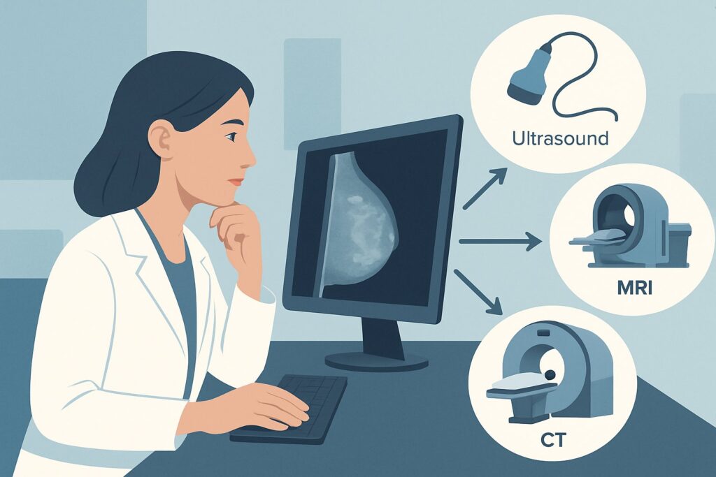
The ACR designates ‘Digital breast tomosynthesis screening’ as Usually appropriate for this high-risk transfeminine patient. Standard 2D ‘Mammography screening’ is also rated Usually appropriate, but tomosynthesis (DBT or “3D mammography”) is often preferred for its distinct advantages in this population.
Gender-affirming hormone therapy induces the growth of fibroglandular tissue, which can result in dense breasts on mammography. Breast density can obscure underlying cancers on traditional 2D mammograms. Digital breast tomosynthesis acquires images from multiple angles to create a three-dimensional reconstruction of the breast. This technique significantly reduces the effect of overlapping tissue, which improves cancer detection rates and lowers the recall rate for false positives—a crucial benefit in any screening program.
The radiation dose for DBT (ACR Relative Radiation Level ☢☢, 0.1-1 mSv) is comparable to that of standard 2D mammography and is considered a low and acceptable level for annual screening. The benefit of early cancer detection in this high-risk group far outweighs the minimal radiation risk.
Why are other studies not recommended for initial screening?
- Breast Ultrasound (US): This modality is rated Usually not appropriate as a primary screening tool. While invaluable for diagnostic workups of palpable lumps or mammographic abnormalities, ultrasound is not sensitive enough to detect microcalcifications, which are often the earliest sign of Ductal Carcinoma in Situ (DCIS). Using it for screening alone would miss a significant number of early-stage cancers.
- Breast MRI with and without contrast: This is also rated Usually not appropriate for initial screening in this specific scenario. While contrast-enhanced MRI is a powerful tool used for supplemental screening in some very high-risk cisgender women, the evidence base and guidelines for its routine use in the transgender population are still evolving. At present, mammography-based screening remains the foundational approach.
When ordering, specifying “screening mammogram with tomosynthesis” is sufficient. For detailed technical parameters used by technologists and radiologists to perform the study, see our guide on the Screening Mammography (with DBT) protocol.
What’s Next After Digital Breast Tomosynthesis Screening? Downstream Workflow
The results of the screening DBT, reported using the Breast Imaging Reporting and Data System (BI-RADS) score, will dictate the next steps in the clinical workflow.
- Negative or Benign Result (BI-RADS 1 or 2): If the study shows no evidence of malignancy, the patient should be advised to continue with annual screening mammography. No further immediate action is needed.
- Probably Benign Result (BI-RADS 3): This indicates a finding that has a very high probability of being benign (<2% risk of malignancy). The standard recommendation is a short-interval follow-up, typically a diagnostic mammogram in six months, to ensure stability. Biopsy is not immediately warranted.
- Suspicious or Highly Suggestive of Malignancy (BI-RADS 4 or 5): These findings require immediate diagnostic workup. The patient should be recalled for diagnostic mammography (which may include magnification and spot compression views) and a targeted breast ultrasound of the area of concern. If the suspicious finding is confirmed on diagnostic imaging, the next step is typically an image-guided core needle biopsy to obtain a tissue diagnosis.
- Incomplete Result (BI-RADS 0): This means additional imaging is needed for a final assessment. This is a common outcome, especially on a first (baseline) mammogram. The patient should be recalled for diagnostic mammography and/or ultrasound to clarify the initial findings.
Pitfalls to Avoid (and When to Get Help)
Navigating breast cancer screening in this specific population requires careful attention to detail. Here are a few common pitfalls to avoid:
- Delaying Screening Initiation: Do not wait until age 40. For a transfeminine individual with over five years of hormone use and high-risk factors, screening should begin earlier, as early as age 25-30, based on shared decision-making and risk assessment.
- Ordering Ultrasound as a Standalone Screen: Avoid ordering a screening breast ultrasound in place of a mammogram. It is not an equivalent or acceptable substitute for primary screening.
- Misinterpreting Breast Density: Be aware that breast tissue in transfeminine patients on hormones is often dense. This is an expected finding, not necessarily a pathologic one, and reinforces the rationale for using DBT over 2D mammography.
- Failing to Act on a BI-RADS 0 Report: An “incomplete” report is not negative. It is crucial to ensure the patient completes the recommended additional diagnostic imaging promptly.
If a biopsy confirms malignancy, immediate referral to a multidisciplinary breast cancer team, including surgical oncology, medical oncology, and radiation oncology, is the essential next step.
Related ACR Topics and Tools
The guidance for this scenario is part of a larger set of recommendations for transgender individuals. For a comprehensive overview of all related clinical variants, from average-risk patients to transmasculine individuals, please see our parent guide. The following GigHz tools can also support your clinical workflow.
- For breadth across all scenarios in Transgender Breast Cancer Screening, see our parent guide: Transgender Breast Cancer Screening: ACR Appropriateness Decoded.
- ACR Appropriateness Criteria Lookup — for adjacent scenarios
- Imaging Protocol Library — for technique on the recommended study
- Radiation Dose Calculator — for cumulative dose conversations
Frequently Asked Questions
Why does screening start so much earlier (age 25-30) in this high-risk group compared to average-risk individuals?
Screening starts earlier because the combination of long-term estrogen exposure (≥5 years) and a high-risk factor (like a BRCA mutation or strong family history) significantly accelerates the potential timeline for breast cancer development. The guidelines aim to begin surveillance at an age where early detection can make a meaningful difference, similar to high-risk screening protocols for cisgender women.
If my patient has breast implants, is digital breast tomosynthesis still the right test?
Yes, digital breast tomosynthesis (DBT) is still the appropriate screening modality for patients with breast implants. Special techniques, known as implant-displaced (Eklund) views, are used to move the implant out of the way and better visualize the native breast tissue. It is crucial to inform the imaging center that the patient has implants when scheduling the exam.
Should I order a screening breast MRI in addition to the mammogram for my high-risk transfeminine patient?
According to the current ACR Appropriateness Criteria for this specific scenario, a screening breast MRI is rated ‘Usually Not Appropriate’ as the initial study. While supplemental MRI is used in some high-risk cisgender populations, the current recommendation for this transfeminine population is to start with mammography-based screening. This recommendation may evolve as more data becomes available.
What if my patient is 32 years old, has been on hormones for 7 years, but has no family history or other risk factors?
That patient would be considered at average risk and would fall into a different clinical scenario. The current ACR guidelines for average-risk transfeminine individuals with ≥5 years of hormone use recommend initiating screening at age 40, not in their early 30s. This article’s guidance is specifically for the high-risk cohort.
Does the type of estrogen or anti-androgen used in hormone therapy affect the screening recommendation?
The current ACR guidelines do not differentiate screening recommendations based on the specific type of estrogen (e.g., estradiol valerate, patches) or anti-androgen (e.g., spironolactone, bicalutamide) used. The primary factors driving the recommendation are the duration of hormone therapy (≥5 years) and the presence of high-risk factors for breast cancer.
Reviewed by Pouyan Golshani, MD, Interventional Radiologist — May 29, 2026