What Is the Best Imaging for Suspected DCIS Recurrence After Mastectomy?
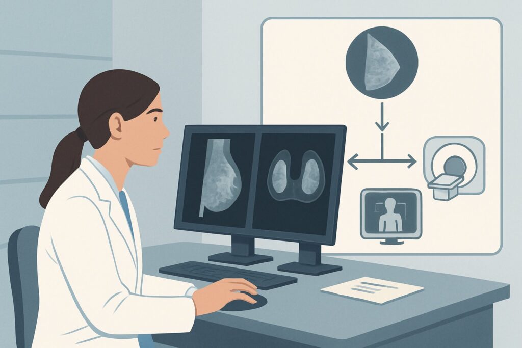
A 58-year-old woman presents to your clinic for follow-up. Six years ago, she underwent a left total mastectomy for extensive, high-grade Ductal Carcinoma in Situ (DCIS). Today, she reports a new, firm, non-tender nodule she can feel along the superior aspect of her mastectomy scar. On examination, you confirm a palpable 1-cm firmness in the subcutaneous tissue. You need to evaluate for local recurrence, but what is the most appropriate initial imaging study in a patient with no remaining breast tissue? This article provides a clinical workflow for this specific scenario, grounded in the American College of Radiology (ACR) Appropriateness Criteria, which rate breast ultrasound as ‘Usually Appropriate’ for this initial workup.
Who Fits This Clinical Scenario?
This guidance is for an adult patient with a prior history of mastectomy for Ductal Carcinoma in Situ (DCIS) who now presents with a new clinical concern for local recurrence. This concern may be based on symptoms (e.g., a new palpable lump, skin changes like erythema or dimpling, nipple discharge if nipple-sparing), a specific finding on physical examination, or, less commonly, an abnormal laboratory value.
This workflow specifically applies to the initial imaging evaluation of a symptomatic patient post-mastectomy. It is crucial to distinguish this from other similar, but distinct, clinical situations:
- Exclusion 1: Routine Surveillance After Mastectomy. This article does not apply to asymptomatic patients undergoing routine surveillance imaging. That scenario, covered in a separate ACR variant, relies primarily on physical examination, as routine imaging is not typically recommended.
- Exclusion 2: History of Breast Conservation Therapy (Lumpectomy). This guidance is for patients post-mastectomy. Patients who underwent lumpectomy and radiation have remaining breast tissue, and the imaging workup for suspected recurrence is different, typically involving diagnostic mammography and/or tomosynthesis.
- Exclusion 3: Newly Diagnosed DCIS. This workflow is for evaluating recurrence, not for the initial workup of a new DCIS diagnosis before treatment.
Correctly identifying your patient’s specific clinical context is the first step to selecting the most effective and appropriate imaging test.
What Diagnoses Are You Working Up in This Scenario?
When a patient presents with a new palpable finding at a mastectomy site, the primary clinical concern is ruling out a local recurrence of cancer. However, several benign conditions can mimic this presentation, forming a key differential diagnosis that the imaging study aims to clarify.
Local Recurrence: This is the most consequential diagnosis to exclude. Recurrence after mastectomy for DCIS is uncommon, but it can occur. It typically presents as a nodule in the skin, subcutaneous tissue, or chest wall musculature (e.g., pectoralis major) at or near the mastectomy scar. The goal of imaging is to identify any suspicious solid mass that would warrant a biopsy.
Fat Necrosis: A very common benign finding, fat necrosis is a result of the surgical trauma and subsequent healing process. It can present as a firm, sometimes irregular, palpable mass that can be clinically indistinguishable from a recurrence. On imaging, it has a characteristic appearance that can often confirm its benign nature without a biopsy.
Suture Granuloma: This is a benign inflammatory reaction to suture material left behind during surgery. It typically presents as a firm nodule directly along the scar line. Like fat necrosis, it can be a convincing clinical mimic of recurrence, but ultrasound can often identify the suture material at the center of the inflammatory mass.
Post-Surgical Fluid Collection (Seroma/Hematoma): While more common in the immediate post-operative period, chronic or organized seromas and hematomas can persist and present as palpable lumps months or even years after surgery. Ultrasound is highly effective at characterizing these collections as fluid-filled rather than solid tissue.
Why Is Breast Ultrasound the Recommended Initial Study for This Presentation?
For a patient with a suspected local recurrence after mastectomy, the ACR designates breast ultrasound as Usually Appropriate. This recommendation is based on its high diagnostic yield, safety profile, and ability to directly evaluate the specific area of clinical concern.
The primary rationale for ultrasound is its excellent spatial resolution for evaluating superficial tissues. After a mastectomy, there is no longer a breast mound to image with mammography. The area of concern is typically within the skin, subcutaneous fat, or the underlying chest wall muscle. Ultrasound is the ideal modality for characterizing a palpable abnormality in these locations. It can readily distinguish solid masses from simple cysts or complex fluid collections, assess the morphology and vascularity of a solid lesion, and precisely guide a biopsy if one is needed.
Furthermore, ultrasound involves no ionizing radiation (0 mSv) and does not require intravenous contrast, making it a safe and accessible first-line test.
Why are other studies rated lower for this initial workup?
- Mammography and Digital Breast Tomosynthesis (DBT) are both rated Usually Not Appropriate. The reason is straightforward: these techniques are designed to compress and image breast tissue. In a post-mastectomy patient, there is no breast tissue to compress, making the examination technically challenging and of extremely low diagnostic value for evaluating the chest wall.
- Breast MRI with and without IV contrast is also rated Usually Not Appropriate as the initial imaging modality. While MRI is a very sensitive test for detecting cancer, it is not the best first step for a focal, palpable finding. Ultrasound provides a targeted, real-time evaluation that can often definitively characterize the lesion or guide a biopsy. MRI is more resource-intensive and may be considered a problem-solving tool if the ultrasound is inconclusive or if there is a strong clinical suspicion for recurrence despite negative ultrasound findings.
What’s Next After Breast Ultrasound? Downstream Workflow
The results of the breast ultrasound will guide the subsequent steps in the patient’s management. The workflow typically follows one of three paths:
1. Positive for a Suspicious Finding: If the ultrasound identifies a solid mass with suspicious features (e.g., irregular shape, spiculated margins, posterior acoustic shadowing), the definitive next step is an ultrasound-guided core needle biopsy. This allows for a histologic diagnosis to confirm or exclude recurrence. If the biopsy confirms malignancy, the patient should be referred to a surgical oncologist and medical oncologist to discuss treatment options, which may include surgical excision, radiation, and/or systemic therapy.
2. Negative or Clearly Benign Finding: If the ultrasound reveals a finding classic for a benign entity, such as a simple cyst, uncomplicated seroma, or typical fat necrosis, no further immediate action is usually required. The imaging report provides reassurance, and the patient can be managed with clinical follow-up. The palpable finding has been correlated with a benign imaging appearance.
3. Indeterminate or Equivocal Finding: Occasionally, the ultrasound findings may be indeterminate. In these cases, or if the ultrasound is negative but the clinical suspicion for recurrence remains very high, a discussion between the referring clinician and the breast radiologist is essential. The next step might be a short-interval follow-up ultrasound to ensure stability or an escalation to a problem-solving modality like contrast-enhanced breast MRI to further evaluate the area of concern.
Pitfalls to Avoid (and When to Get Help)
In evaluating suspected DCIS recurrence post-mastectomy, several common pitfalls can complicate diagnosis. First, avoid ordering a mammogram out of habit; it is not an appropriate test for the post-mastectomy chest wall and provides no useful information. Second, ensure the radiologist is clearly informed of the exact location of the palpable concern so they can perform a targeted ultrasound examination. A “whole chest” ultrasound is not as effective as a focused look at the area of concern. Finally, do not dismiss a palpable finding solely because of a negative ultrasound. While ultrasound is highly sensitive, a very strong clinical suspicion may warrant further discussion or follow-up. If the clinical exam and imaging findings are discordant, escalate by consulting with your breast imaging radiologist to determine the best next step.
Related ACR Topics and Tools
For a comprehensive overview of imaging recommendations across all clinical presentations of Ductal Carcinoma in Situ, from initial diagnosis to surveillance, please see our parent guide. For other tools to assist in ordering the right imaging study, see the resources below.
- For breadth across all scenarios in Imaging of Ductal Carcinoma in Situ (DCIS), see our parent guide: Imaging of Ductal Carcinoma in Situ (DCIS): ACR Appropriateness Decoded.
- To look up appropriateness criteria for other clinical scenarios, use the ACR Appropriateness Criteria Lookup.
- To review imaging techniques and parameters, browse the Imaging Protocol Library.
- To discuss radiation exposure with patients, consult the Radiation Dose Calculator.
Frequently Asked Questions
Why not start with a breast MRI if it’s considered more sensitive for detecting cancer?
While breast MRI is highly sensitive, the ACR rates it ‘Usually Not Appropriate’ as the initial test in this specific scenario. A targeted breast ultrasound is highly effective for evaluating a palpable lump on the chest wall, is more accessible, faster, and does not require IV contrast. Ultrasound can often provide a definitive answer or guide a biopsy directly. MRI is typically reserved as a second-line, problem-solving tool if the ultrasound is negative or inconclusive despite high clinical suspicion.
Is a mammogram ever appropriate for evaluating recurrence after a mastectomy?
No, for a patient who has had a total mastectomy, mammography or tomosynthesis is rated ‘Usually Not Appropriate.’ These imaging techniques require compression of breast tissue, which is absent after a mastectomy. Attempting a mammogram would be technically difficult and would not provide useful diagnostic information about the skin, subcutaneous tissue, or chest wall where a recurrence would occur.
What if the palpable lump feels very deep, close to the ribs?
Breast ultrasound is still the recommended initial imaging study. Modern ultrasound equipment provides excellent visualization of the chest wall, including the intercostal muscles and the surface of the ribs. It is the best initial modality to determine if a deep palpable finding is related to the musculature, a rib, or another structure.
Does the grade of the original DCIS affect the choice of imaging for a suspected recurrence?
No, the imaging approach for a new, palpable finding after mastectomy does not change based on the grade of the original DCIS. The clinical question is the same: to characterize the new abnormality. Breast ultrasound is the most appropriate first step regardless of whether the initial DCIS was low, intermediate, or high grade.
If the ultrasound is negative, can I confidently rule out a recurrence?
A negative targeted ultrasound of a palpable finding is highly reassuring and has a very high negative predictive value. In most cases, it effectively rules out a suspicious underlying cause. However, if there is a strong discordance—meaning the clinical examination is highly suspicious for malignancy despite a normal ultrasound—a discussion with the breast radiologist is recommended to decide between short-term clinical follow-up or proceeding to a more sensitive test like an MRI.
Reviewed by Pouyan Golshani, MD, Interventional Radiologist — May 30, 2026